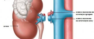
The kidneys are paired bean-shaped organs that perform regulatory functions in relation to chemical homeostasis in the human body and belong to the urinary system. The kidneys remove excess organic molecules (such as glucose molecules) from the blood and play an important role in the process of urine formation by removing organic waste, including urea, from the body.
Performing a homeostatic function, the kidneys play an important role in the urinary system, regulating the acid-base balance and the amount of electrolytes in the human body. By maintaining the body's salt and water balance, the kidneys regulate blood pressure. The kidneys are a natural filter for the blood, removing soluble waste and excess water and sending them to the bladder.
During urine formation, the kidneys excrete waste products such as urea and ammonium and are responsible for the reabsorption of water, amino acids and glucose.
It should also be noted that the kidneys are involved in the formation of certain hormones, including: erythropoietin, calcitriol. This organ also secretes the enzyme renin, which acts in a negative feedback loop on the kidneys, playing a role in regulating blood pressure.
Structure of organs
Thanks to the research carried out, you can be absolutely sure that the anatomy of the human kidneys has been studied
Bud
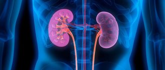
These paired organs are located symmetrically to each other relative to the spine. Only the right kidney in the human body is slightly smaller in size and is located below the left one, since the liver is located above it.
The human kidney is a bean-shaped organ. The outer surface of the human kidneys is dense and smooth; it is covered by a fibrous capsule, which is a thin but very strong film of connective tissue.
In addition, both kidneys are enclosed in a fatty membrane, thanks to which they can be kept in the human body in one place, which is predetermined by anatomy.
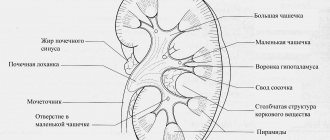
Kidney tissue, called parenchyma, has two layers. The internal structure of the kidney is quite complex, the parenchyma acts as the main filtering instrument, and the pelvis acts as a mechanism for removing harmful substances.
The renal pelvis is formed from the small and large calyces of the kidneys.
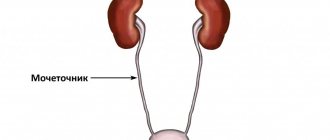
The ureter emerges from the pelvis, which connects it to the bladder and ensures the removal of urine through it.
The nephron is a structural unit of the human kidney, in other words, it is the main filtering element. The nephron consists of renal tubules and corpuscles.
The human kidney tubules externally resemble a ball consisting of blood vessels, surrounded on all sides by a capsule. It is in it that the filtration of blood plasma occurs under a certain pressure.
The liquid formed during this filtration process is primary urine.
Primary urine is not excreted outside, but is directed through long tubules, heading into the collecting duct. As they move through the tubules, beneficial substances (water and electrolytes) are absorbed, and the remaining liquid is excreted.
It is this that is secondary urine, which enters the calyces of the kidneys, then into the pelvis, subsequently into the ureter and is finally excreted from the human body.
Human organs: location in pictures.
Brain
The human brain is the most complex and least studied human organ. He controls all other organs and coordinates their work. In fact, our consciousness is the brain. Despite little knowledge, we still know the location of its main sections. This picture describes in detail the anatomy of the human brain.
Larynx
The larynx allows us to make sounds, speech, singing. The structure of this cunning organ is shown in the picture.
Major organs, chest and abdominal organs
This picture shows the location of the 31 organs of the human body from the thyroid cartilage to the rectum. If you urgently need to look at the location of any organ in order to win an argument with a friend or take an exam, this picture will help.
The picture shows the location of the larynx, thyroid gland, trachea, pulmonary veins and arteries, bronchi, heart and pulmonary lobes. Not much, but very clear.
A schematic arrangement of the human internal organs from the trochea to the bladder is shown in this picture. Due to its small size, it loads quickly, saving you time for peeking during the exam. But we hope that if you are studying to become a doctor, then you do not need the help of our materials.
A picture showing the location of human internal organs, which also shows the system of blood vessels and veins. The organs are beautifully depicted from an artistic point of view, some of them are signed. We hope that among those signed there are those you need.
A picture that details the location of the organs of the human digestive system and the pelvis. If you have a stomach ache, this picture will help you locate the source while the activated charcoal is working, or while you are easing your digestive system in comfort.
Location of the pelvic organs
If you need to know the location of the superior adrenal artery, bladder, psoas major muscle or any other abdominal organ, then this picture will help you. It describes in detail the location of all organs of this cavity.
Tasks of the body
Understanding what the kidneys look like and realizing that the human kidneys have several functions, it is easy to understand how important this organ is for the full functioning of a person. The filtering and excretory function is the main function that nature has endowed the kidneys with.
But besides these tasks, the kidney organs perform several other important functions. In particular, maintaining the water-salt balance in the body, which is important for human life.
And it is the kidneys that monitor such an important ratio, since with a sharp increase in salts, dehydration occurs in the cells, and with a decrease in the natural level of salts, on the contrary, an excessive amount of water is concentrated in them, which provokes swelling.
Kidney functions
Consequently, the osmoregulatory function of the kidneys that occurs in the body is as important and necessary as the excretory function.
The ion-regulating function is also aimed at regulating the ratio, but only the acid-base ratio. Anatomy determines the release of excess hydrogen ions or bicarbonate ions.
Metabolic processes taking place in the human body are also of great importance. The kidney organs also perform metabolic functions, due to which harmful toxins, drug residues, and proteins are eliminated.
The endocrine function performs the task of producing substances that regulate blood pressure, as well as hormones by the adrenal glands. Red blood cells are formed in the body only due to endocrine function.
Kidney functions
The kidneys are involved in homeostasis throughout the body. They regulate electrolyte concentrations and acid-base balance, and also regulate blood pressure and extracellular fluid volume. These functions of the kidney are performed both independently and in interaction with other organs, mainly with the organs of the endocrine system. Endocrine hormones regulate some of the functions of the kidneys. These include renin, angiotensin 2, antidiuretic hormone, aldosterone, natriuretic peptide.
Most of the functions of the kidneys are simple filtration, reabsorption and secretion, occurring in the nephrons of the kidneys. Filtration that occurs in the renal carpuscules is the process by which cells and large proteins are filtered out of the blood, thereby producing an ultrafiltrate, which then becomes urine. The kidneys produce approximately 180 liters of filtrate per day, but produce only about 2 liters of urine. Reabsorption is the transport of molecules from the ultrafiltrate into the blood. Secretion in the kidneys involves the reverse process where molecules from the blood are transported into the urine.
The kidneys excrete various waste products that are produced by metabolizing substances into urine. Such wastes include uric acid, urea, and nitrogenous wastes of protein catabolism.
It should be noted that the organs that maintain the acid-base balance of the body include two paired organs - the lungs and the kidneys. The lungs regulate acid-base homeostasis by regulating the concentration of carbon dioxide in the blood. The role of the kidneys in regulating acid-base balance is to reduce bicarbonate from urine and release fixed acids and hydrogen ions into the urine.
Causes and symptoms of diseases
Kidney diseases are pathologies that provoke disruptions in the functioning of the organ, and also lead to serious damage to the kidney tissue. As a result of such pathologies, kidney function in the human body is noticeably impaired.
Kidney cancer
Most often, all kinds of bacteria and infections have a negative impact on the performance of organs. They are the ones who can provoke stagnation of urine of varying duration, which leads to more serious problems.
The anatomy of the renal organs can be disrupted due to the formation of cysts and tumors of various etymologies.
Metabolic disorders negatively affect many internal processes, and the kidneys are no exception. As a result of a decrease in the performance of the parenchyma, kidney diseases occur.
Pathologies can also be congenital; patients experience various anomalies in the internal structure of the organ itself or in the defective performance of intended functions.
The formation of stones in the kidney organs also causes serious disturbances in their functioning.
Visit doctor
Any pathologies can be detected initially by the patient himself. Symptoms are conventionally divided into general and characteristic.
General symptoms should alert the patient and “refer” him to a medical facility for examination, since such symptoms can only suggest the presence of renal pathology.
But the same symptoms can be accompanied by other diseases. Common symptoms include increased body temperature, chills, fatigue, and increased blood pressure.
Characteristic symptoms include those that are characteristic only of the kidneys. Increased swelling, polyuria, oliguria, pain and burning during urination are all signs indicating obvious problems with the urinary system.
Another characteristic symptom is a change in the color of urine.
If at a certain stage an altered kidney anatomy was discovered, accompanied by characteristic symptoms of pathologies, it is important to immediately begin treatment to prevent a decrease in their functioning or, in complex diseases, their complete loss.
Human organs: location in pictures. Anatomy of body parts
Has it ever seemed strange to you that you have been living for decades, but know absolutely nothing about your own body? Or that you found yourself taking an exam on human anatomy, but did not prepare for it at all. In both cases, you need to catch up on lost knowledge and get to know the human organs better. It is better to see their location in pictures - clarity is very important. Therefore, we have collected pictures for you in which the location of human organs is easily traced and labeled.
If you like games with human internal organs, be sure to try the flash game Amateur Surgeon on our website.
To enlarge any picture, click on it and it will open in full size. This way you can read the fine print. So let's start at the top and work our way down.
Pathologies
Any person's kidneys can be susceptible to many diseases that require emergency treatment. Such diseases can be acquired due to non-compliance with a healthy lifestyle, the basics of proper nutrition, and also hereditary.
Any disease of the kidney organs goes into a chronic stage if the necessary treatment is not carried out.
Glomerulonephritis
Glomerulonephritis is an inflammatory disease that is accompanied by damage to the glomeruli and tubules of the kidneys. The culprits of this complex pathology in most cases are streptococci.
Although medicine knows cases where glomerulonephritis occurred against the background of tuberculosis or malaria. Treatment of glomerulonephritis is long and scrupulous.
Pyelonephritis is another inflammatory disease, the anatomy of which involves damage to the parenchyma, calyces and pelvis of the kidneys. This pathology is provoked by streptococcus, staphylococcus, and E. coli.
The basis for the occurrence of this pathology is a violation of the outflow of urine.
Treatment of pyelonephritis is accompanied by the use of antibiotics, as well as medications that strengthen the body's defenses.
Nephroptosis consists of depletion of the fat capsule, as a result of which the kidney becomes wandering, since there is nothing left to hold it in one place.
Treatment involves normalizing nutrition and wearing a special bandage to hold the kidney in its anatomical location. Full treatment should be accompanied by a complex of physical therapy.
Urolithiasis is characterized by the formation of kidney stones that differ in their chemical composition. Treatment of this pathology is the use of drugs that help dissolve stones and remove them out.
In some cases, operations have to be performed.
Hydronephrosis is characterized by dilation of the kidney cavities due to stagnation of urine. Treatment is primarily aimed at eliminating the root cause.
Kidney failure is the most serious pathology, as it can lead to death. Therefore, it is important to begin proper treatment to prevent such consequences.
The human kidney is the main component of the human genitourinary system. The structure of the human kidney and the physiology of the kidneys are quite complex and specific, but it is they that allow these organs to perform vital functions and have a huge impact on the homeostasis of all other organs in the human body.
Regulation of blood pressure by the kidneys
Although the kidneys cannot directly affect the blood, they play an important role in regulating blood pressure. This occurs by controlling extracellular fluid cells, the size of which depends on plasma sodium concentration. Among the important chemical elements that make up the renin-angiotensin system, renin plays an important role. Changes in renin alter the release of hormones such as aldosterone and angiotensin 2 from this system. Each hormone acts through several mechanisms to increase renal reabsorption of sodium chloride, dilate extracellular fluid cells, and increase blood pressure. The relationship with renin levels is direct. When renin levels increase, the levels of aldosterone and angiotensin 2 increase, which, according to the above scheme, leads to an increase in blood pressure. At low renin levels the process is opposite.
A little about the origin
During their development, the kidneys go through three stages: pronephros, mesonephros and metanephros. The pronephros is a kind of preference, which is a rudiment that does not function in humans. There are no glomeruli in it, and the tubules are not connected to blood vessels. The kidney is completely reduced in the fetus at the 4th week of development. At the same time, at 3-4 weeks, the embryo develops a primary kidney, or mesonephros, the main excretory organ of the fetus in the first half of intrauterine development. It already has glomeruli and tubules connecting to two pairs of ducts: the Wolffian duct and the Müllerian duct, which in the future give rise to the male and female genital organs. The mesonephros actively functions in the fetus until about 4-5 months of development.
The final kidney, or metanephros, is formed in the fetus at 1–2 months, is fully formed at the 4th month of development and subsequently functions as the main excretory organ.
Return to contents
In relation to bones
Where are the human kidneys located? If we take bones as a starting point as relatively immobile and dense formations, then the kidneys are located on both sides of the spine at the level of the lumbar region. The right kidney, as a rule, is located a couple of centimeters lower than its left paired sister. Conventionally, the twelfth rib crosses the left kidney in half, and separates only a third from the right.
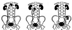
The location in relation to the bones can be high, medium and low. The high position suggests that the kidneys are behind the eleventh and twelfth ribs. When low, both kidneys drop below the twelfth rib. The middle position, as the most typical, was described above.
Knowing the position of the kidneys in each specific clinical case is extremely important for surgeons, as they need to decide which approach to use in order to reach the affected organ during surgery.
Topography
The right kidney is lower than the left due to the location of the liver.
There are two kidneys in the human body. These organs are located behind the peritoneum on both sides of the ridge. Their shapes are a bit like beans. The height of their projection on the lower back in both an adult and a child corresponds to the 11th and 12th thoracic vertebrae and the 1st and 2nd lumbar vertebrae, but the right one, due to its position close to the liver, is placed slightly lower than the left one. In these organs, two surfaces are described - posterior and anterior, two edges - median and lateral, two poles - lower and upper. The upper poles are placed slightly closer to one another than the lower ones, as they are slightly inclined towards the spine.
The hilum is located at the median edge - the area where the ureter and renal vein leave and where the renal artery enters. In addition to the liver, the right kidney is in close proximity to a part of the colon in front and a segment of the duodenum along its median edge. The jejunum and stomach, together with the pancreas, are adjacent to the left along its anterior surface, and the spleen, together with a fragment of the large intestine, is adjacent to its lateral edge. At the top, above each pole, is the adrenal gland, or adrenal gland.
Return to contents
Anatomy of the Human Kidneys - information:
The kidney, gene (Greek nephros), is a paired excretory organ that produces urine, lying on the posterior wall of the abdominal cavity behind the peritoneum.
The kidneys are located on the sides of the spinal column at the level of the last thoracic and two upper lumbar vertebrae. The right kidney lies slightly lower than the left, on average 1-1.5 cm (depending on the pressure of the right lobe of the liver). The upper end of the kidney reaches the level of the XI rib, the lower end is 3-5 cm from the iliac crest. The indicated boundaries of the position of the kidneys are subject to individual variations; often the upper border rises to the level of the upper edge of the XI thoracic vertebra, the lower border can fall by 1-1/2 vertebrae.
The kidney has a bean-shaped shape. Its surface substance is smooth and dark red in color. In the kidney, there are upper and lower ends, extremitas superior and inferior, lateral and medial edges, margo lateralis and medialis. and surfaces, facies anterior and posterior.
The lateral edge of the kidney is convex, while the medial edge is concave in the middle, facing not only medially, but somewhat downward and forward.
The middle concave part of the medial edge contains the portal, hilus renalis, through which the renal arteries and nerves enter and the vein, lymphatic vessels and ureter exit. The gate opens into a narrow space that projects into the substance of the kidney, which is called sinus renalis; its longitudinal axis corresponds to the longitudinal axis of the kidney. The anterior surface of the kidneys is more convex than the posterior one.
Topography of the kidneys. The relationship to the organs of the anterior surface of the right and left kidneys is different.
The right kidney is projected onto the anterior abdominal wall in the regiones epigastrica, umbilicalis et abdominalis lat. dext., left - in reg. epigastrica et abdominalis lat. sin. The right kidney has a small surface area in contact with the adrenal gland; further down, most of its anterior surface is adjacent to the liver. Its lower third is adjacent to flexura coli dextra; the descending part of the duodeni descends along the medial edge; in both last areas there is no peritoneum. The lowermost end of the right kidney has a serous covering.
Near the upper end of the left kidney, as well as the right, part of the anterior surface is in contact with the adrenal gland, immediately below the left kidney is adjacent throughout its upper third to the stomach, and the middle third to the pancreas, the lateral edge of the anterior surface in the upper part is adjacent to the spleen. The lower end of the anterior surface of the left kidney medially comes into contact with the loops of the jejunum, and laterally with the flexura coli sinistra or the initial part of the descending colon. With its posterior surface, each kidney in its upper section is adjacent to the diaphragm, which separates the kidney from the pleura, and below the XII rib - to mm. psoas major et quadratus lumborum, forming the renal bed.
Kidney membranes. The kidney is surrounded by its own fibrous membrane, capsula fibrosa, in the form of a thin smooth plate directly adjacent to the substance of the kidney. Normally, it can be quite easily separated from the kidney substance. Outside the fibrous membrane, especially in the hilum area and on the posterior surface, there is a layer of loose adipose tissue that makes up the fatty capsule of the kidney, capsula adiposa; There is often no fat on the anterior surface. Outside the fat capsule is the connective tissue fascia of the kidney, fascia renalis, which is connected by fibers to the fibrous capsule and splits into two layers: one goes in front of the kidneys, the other goes behind. Along the lateral edge of the kidneys, both leaves join together and pass into the layer of retroperitoneal connective tissue, from which they developed. Along the medial edge of the kidney, both leaves do not join together, but continue further to the midline separately: the anterior leaf goes in front of the renal vessels, aorta and inferior vena cava and connects with the same leaf of the opposite side, the posterior leaf passes in front of the vertebral bodies, attaching to the last one. At the upper ends of the kidneys, also covering the adrenal glands, both leaves are joined together, limiting the mobility of the kidneys in this direction. At the lower ends, such a fusion of leaves is usually not noticeable. Fixation of the kidney in its place is carried out mainly by intra-abdominal pressure caused by contraction of the abdominal muscles; to a lesser extent, fascia renalis, fused with the membranes of the kidney; muscular bed of the kidney formed by mm. psoas major et quadratus lumborum, and renal vessels that prevent the kidney from being removed from the aorta and inferior vena cava. If this fixing apparatus of the kidney is weak, it may descend (wandering kidney), which requires surgical suturing. Normally, the long axes of both kidneys, directed obliquely upward and medially, converge above the kidneys at an angle open downward. During prolapse, the kidneys, being fixed at the midline by vessels, move downward and medially. As a result, the long axes of the kidneys converge below the latter at an angle open to the top.
Structure. A longitudinal section through the kidney shows that the kidney as a whole is composed, firstly, of the cavity, sinus renalis, in which the renal cups and the upper part of the pelvis are located, and, secondly, of the renal substance itself adjacent to the sinus on all sides except the gate.
The kidney is divided into a cortex, cortex renis, and a medulla, medulla renis. The cortex occupies the peripheral layer of the organ and is about 4 mm thick. The medulla is composed of conical structures called renal pyramids, pyramides renales. The wide bases of the pyramids face the surface of the organ, and the apices face the sinus. The apices are connected by two or more into rounded elevations called papillae, papillae renales; less often, one apex corresponds to a separate papilla. There are an average of about 12 papillae in total. Each papilla is dotted with small holes, foramina papillaria; through the foramina papillaria, urine is secreted into the initial parts of the urinary tract (calyces). The cortex penetrates between the pyramids, separating them from each other; these parts of the cortex are called columnae renales. Thanks to the urinary canaliculi and vessels located in them in a straight direction, the pyramids have a striped appearance. The presence of pyramids reflects the lobular structure of the kidney, characteristic of most animals.
The newborn retains traces of the former division even on the outer surface, on which grooves are noticeable (lobular kidney of the fetus and newborn). In an adult, the kidney becomes smooth on the outside, but inside, although several pyramids merge into one papilla (which explains the smaller number of papillae than the number of pyramids), it remains divided into lobules - pyramids. The stripes of medullary substance also continue into the cortex, although they are less clearly visible here; they constitute the pars radiata of the cortex, and the spaces between them constitute the pars convoluta (convolutum - bundle). Pars radiata and pars convoluta are combined under the name lobulus corticalis.
The kidney is a complex excretory (excretory) organ. It contains tubes called renal tubules, tubuli renales. The blind ends of these tubes in the form of a double-walled capsule cover the glomeruli of blood capillaries. Each glomerulus, glomerulus, lies in a deep cup-shaped capsule, capsula glomeruli; the gap between the two leaves of the capsule constitutes the cavity of this latter, being the beginning of the urinary tubule. The glomerulus, together with the capsule enclosing it, constitutes the renal corpuscle, corpusculum renis. The renal corpuscles are located in the pars convoluta of the cortex, where they can be visible to the naked eye as red dots. A convoluted tubule departs from the renal corpuscle - tubulus renalis contortus, which is already located in the pars radiata of the cortex. Then the tubule descends into the pyramid, turns back there, making a nephron loop, and returns to the cortex. The final part of the renal tubule - the intercalary section - flows into the collecting duct, which receives several tubules and runs in a straight direction (tubulus renalis rectus) through the pars radiata of the cortex and through the pyramid. Straight tubules gradually merge with each other and in the form of 15-20 short ducts, ductus papillares, open foramina papillaria in the area cribrosa at the apex of the papilla. The renal corpuscle and the tubules associated with it constitute the structural and functional unit of the kidney - the nephron, nephron. The nephron produces urine. This process occurs in two stages: in the renal corpuscle, the liquid part of the blood is filtered from the capillary glomerulus into the cavity of the capsule, making up primary urine, and reabsorption occurs in the renal tubules - the absorption of most of the water, glucose, amino acids and some salts, resulting in the formation of final urine .
Each kidney contains up to a million nephrons, the totality of which makes up the bulk of the renal substance. To understand the structure of the kidney and its nephron, one must keep in mind its circulatory system. The renal artery originates from the aorta and has a very significant caliber, which corresponds to the urinary function of the organ associated with the “filtration” of blood. At the portal of the kidney, the renal artery is divided according to the sections of the kidney into arteries for the upper pole, aa. polares superiores, for the lower, aa. polares inferiores, and for the central part of the kidneys, aa. centrales. In the parenchyma of the kidney, these arteries go between the pyramids, i.e., between the lobes of the kidney, and therefore are called aa. interlobares renis. At the base of the pyramids, at the border of the medulla and cortex, they form arches, aa. arcuatae, from which aa extend into the thickness of the cortex. interlobulares. From each a. interlobularis, the afferent vessel vas afferens departs, which breaks up into a tangle of convoluted capillaries, glomerulus, covered by the beginning of the renal tubule, the glomerular capsule. The efferent artery, vas efferens, emerging from the glomerulus, breaks up for the second time into capillaries, which entwine the renal tubules and only then pass into the veins. The latter accompany the arteries of the same name and emerge from the hilum of the kidney with a single trunk, v. renalis, flowing into v. cava inferior. Venous blood from the cortex flows first into the stellate veins, venulae stellatae, then into the vv. interlobulares, accompanying the arteries of the same name, and in vv. arcuatae Venulae rectae emerge from the medulla. Of the large tributaries v. renalis develops the trunk of the renal vein. In the sinus renalis region, the veins are located in front of the arteries.
Thus, the kidney contains two capillary systems; one connects the arteries to the veins, the other is of a special nature, in the form of a vascular glomerulus, in which the blood is separated from the capsule cavity by only two layers of flat cells: the endothelium of the capillaries and the epithelium of the capsule. This creates favorable conditions for the release of water and metabolic products from the blood.
The lymphatic vessels of the kidney are divided into superficial, arising from the capillary networks of the membranes of the kidney and the peritoneum covering it, and deep, running between the lobules of the kidney. There are no lymphatic vessels inside the kidney lobules and in the glomeruli. Both vascular systems for the most part merge at the renal sinus, going further along the renal blood vessels to the regional nodes nodi lymphatici lumbales.
The nerves of the kidney come from the paired renal plexus, formed by the celiac nerves, branches of the sympathetic nodes, branches of the celiac plexus with the fibers of the vagus nerves located in them, afferent fibers of the lower thoracic and upper lumbar spinal nodes.
X-ray anatomy of the kidney. With a conventional X-ray of the lumbar region, you can see the contours of the lower half of the kidneys. In order to see the kidney as a whole, one has to resort to introducing air into the perinephric tissue - pneumoren.
Radiologically, skeletotopy of the kidneys can be determined. In this case, the XII rib with a saber-shaped form is layered on the middle of the kidney, with a stiletto-shaped form - on its upper end. The upper ends of the kidneys are slightly inclined medially, so the extensions of the long axes of the kidneys intersect above the latter at the height of the IX-X thoracic vertebrae.
X-rays make it possible to examine the excretory tree of the kidney in a living person: calyces, pelvis, ureter. To do this, a contrast agent is injected into the blood, which is released through the kidneys and, joining the urine, gives a silhouette of the renal pelvis and ureter on the radiograph (the contrast agent can also be injected directly into the renal pelvis using a ureteral catheter and a special instrument - a cystoscope). This method is called ureteropyelography. The pelvis on the radiograph is projected at the level between the I and II lumbar vertebrae, and on the right it is slightly lower than on the left. In relation to the renal parenchyma, two types of location of the renal pelvis are noted: extrarenal, when part of it is located outside the kidney, and intrarenal, when the pelvis does not extend beyond the renal sinus. X-ray examination reveals peristalsis of the renal pelvis.
Using serial radiographs, you can see how individual cups and the pelvis contract and relax, and how the upper ureteral sphincter opens and closes. These functional changes are rhythmic in nature, therefore the systole and diastole of the excretory tree of the kidney differ. The process of emptying the excretory tree proceeds in such a way that the large cups contract (systole) and the pelvis relaxes (diastole) and vice versa. Complete emptying occurs within 6-8 minutes. Segmental structure of the kidney.
The kidney has 4 tubular systems: arteries, veins, lymphatic vessels and renal tubules. There is parallelism between the vessels and the excretory tree (vascular-excretory bundles). The most pronounced correspondence is between the intraorgan branches of the renal artery and the renal calyces. Based on this correspondence, for surgical purposes in the kidney, the segments that make up the segmental structure of the kidney are distinguished.
There are five segments in the kidney: 1) upper - corresponds to the upper pole of the kidney; 2, 3) upper and lower anterior - located in front of the pelvis; 4) lower - corresponds to the lower pole of the kidney; 5) posterior - occupies the two middle quarters of the posterior half of the organ between the upper and lower segments.
Protection: kidney membranes
The fibrous membrane of the kidney protects the organ from damage.
Both organs are covered on the outside with a fibrous capsule, which is formed by elastic fibers and smooth muscle cells. Interlobular layers of connective tissue extend inward from this capsule. Outside, the fibrous capsule is adjacent to the fatty or adipose renal capsule, which provides reliable protection for the organ. This capsule becomes somewhat denser on the posterior renal surface and forms the perirenal adipose body. Above the adipose capsule is the renal fascia, formed by two layers: prerenal and retrorenal. They are tightly woven together at the upper poles and lateral edges, but they do not grow together below. Some of the fascia fibers pierce the fatty capsule of the kidney, intertwining with the fibrous capsule. The membranes of the kidney provide their protection.
Return to contents
Possible problems
Most often, this organ suffers from inflammatory processes. They may be infectious or non-infectious. Among these diseases, the most common are pyelonephritis and glomerulonephritis. Another common problem is urolithiasis. When urine retention occurs, the kidneys suffer from hydronephrosis. This organ is also susceptible to developmental abnormalities and tumor processes.
Pyelonephritis is an inflammatory disease, most often caused by infection. Hypothermia “helps” the infection to take hold. A characteristic symptom for this disease is lower back pain; in addition, swelling and a general deterioration in well-being are possible, accompanied by an increase in temperature and changes in urine and blood tests.
With glomerulonephritis, the renal glomeruli are affected, which quickly leads to a deterioration in the functional abilities of the kidneys. The most common cause of glomerulonephritis is streptococcal infection, so it is important to promptly treat sore throat and other respiratory tract diseases.
Urolithiasis is caused by disturbances in metabolic processes, as a result of which some substances in the urine appear in excess or are converted into insoluble compounds. In such cases, they precipitate and crystallize. Concretions are formed, from very small (sand) to large (stones).
When urine stagnates, it gradually accumulates in the renal pelvis system. If such a pathology lasts for a long time, then due to increased pressure on the kidney parenchyma, its tissue becomes thinner, the kidney itself increases in size and does not fully cope with its functions.
Kidney diseases are dangerous due to their complications, which develop due to tissue damage. Significant damage leads to a significant decrease in kidney function. If the pathology progresses, then renal failure may develop.
This is a dangerous condition, since due to incomplete filtration of blood, toxins and waste remain in the body, causing intoxication.
Kidney structure
The renal cortex and medulla form the internal structure of the kidney. The outer cortex borders the fibrous capsule. Its part, called the “renal columns,” penetrates the medulla of the kidney, dividing it into certain parts - pyramids. They are similar in shape to a cone and, together with adjacent columns, form the renal lobe. Several pieces of them are assembled into segments: upper segment, upper anterior, posterior, lower anterior and lower. The pyramidal apices form papillae with openings. They gather into a small renal calyx, from which the larger renal calyces are further formed. Each large cup, or calyx, merges with the others to form a pelvis, whose shape resembles a watering can. Its walls are built from the outer shell, muscular and mucous, which is formed by the transitional epithelium and basement membrane. The renal pelvis gradually narrows and flows into the ureter at the hilum.
This anatomy of the kidneys is key to their functions.
Return to contents
Renal nephrons
The nephron of the kidney filters the blood and produces urine.
The structural and functional unit of the kidney is called the nephron. It is formed by two components: the renal Malpighi corpuscle and the tubular countercurrent-turning complex. The compressed structure of the nephron looks like this: a body created by a glomerulus of vessels with an external capsule of Shumlyansky-Bowman, followed by the proximal convoluted tubule, then the proximal straight tubule, then the loop of the nephron, known as the loop of Henle, followed by the distal convoluted tubule. Several distal canals form collecting ducts that unite to form a collecting duct. They form papillary ducts that exit through an opening on the papillae.
Millions of nephrons form both substances of the organ: the cortical, or outer layer of the kidneys is formed by the corpuscle and a complex of convoluted tubules, the rest of the countercurrent system forms the medulla with its pyramids. Each of these organs also has its own small endocrine apparatus known as the JGA (juxtaglomerular apparatus). It synthesizes the hormone renin and is formed from several types of cells: juxtaglomerular cells, mesangial cells, juxtavascular cells, and the macula densa.
Return to contents
Organ system and apparatus
The concept of a system means a specific group of organs that is related anatomically and embryologically, and also performs a single function.
In turn, the apparatus, whose organs are closely interconnected, does not have the kinship inherent in the system.
Let's consider the common features of individual systems.
The skeleton is the musculoskeletal system, which includes all the bones, tendons, joints and somatic muscles. Both the proportion of the body, as well as movement and locomotion, depend on it.
The location of the organs in the human cardiovascular system ensures the movement of blood through the veins and arteries, saturating the cells with oxygen and nutrients, on the one hand, and removing carbon dioxide with other waste substances from the body, on the other. The main organ here is the heart, which constantly pumps blood through the vessels.
The lymphatic system consists of vessels, capillaries, ducts, trunks and nodes. Under slight pressure, lymph moves through the tubes, ensuring the removal of waste products.
All internal human organs, the location of which is given below, are regulated through the nervous system, which consists of a central and peripheral section. The main one includes the spinal cord and brain. The peripheral consists of nerves, plexuses, roots, ganglia and nerve endings.
The functions of the system are vegetative (responsible for the transmission of impulses) and somatic (connecting the brain with the skin and the respiratory system).
The sensory system plays the main role in recording reactions to external stimuli and changes. This includes the nose, tongue, ears, eyes and skin. Its occurrence is the result of the work of the nervous system.
The endocrine system, together with the nervous system, regulates internal reactions and sensations of the environment. Emotions, mental activity, development, growth, and puberty depend on her work.
The main organs in it are the thyroid and pancreas, testes or ovaries, adrenal glands, pineal gland, pituitary gland and thymus.
The reproductive system is responsible for reproduction.
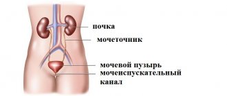
The urinary system is located entirely in the pelvic cavity. It, like the previous one, differs depending on gender. The need for the system is to remove toxic and foreign compounds, an excess of various substances through the urine. The urinary system consists of the kidneys, urethra, ureters and bladder.
Its function, logically based on its name, is to extract and deliver nutrients to cells. The location of the human abdominal organs gives a general idea of the digestive process. It consists of mechanical and chemical processing of food, absorption, breakdown and removal of waste from the body.
The respiratory system consists of the upper (nasopharynx) and lower (larynx, bronchi and trachea) sections.
The immune system is the body's defense against tumors and pathogens. It consists of the thymus, lymphoid tissue, spleen and lymph nodes.
The skin protects the body from temperature changes, drying out, damage and the penetration of pathogens and toxins into it. It consists of skin, nails, hair, sebaceous and sweat glands.
Features of blood supply
From 1500 to 1800 liters of blood passes through the kidneys per day.
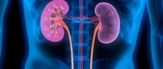
Renal circulation is completely provided by the renal arteries and veins. The artery gives rise to posterior and anterior branches. Segmental arteries branch off from the anterior one and supply the segments of the kidney. Accompanying the pyramids, then follow the interlobar arteries, followed by the arcuate arteries between both layers, then the interlobular, or radial cortical arteries, the branches of which also supply the fibrous capsule. In addition, the interlobular arteries extend further into the afferent glomerular arterioles, forming the glomerulus corpuscle. The efferent glomerular arteriole arises from the latter.
All efferent arterioles form a network of capillaries. The capillaries further unite into venules, forming interlobular or radial cortical veins. They unite with the arcuate veins, followed by the interlobar veins, which then merge into the renal vein, which leaves the renal hilum. Accordingly, blood enters the kidneys through arteries and leaves them through veins. Due to the fact that the vascular system of the kidneys is designed in this way, they carry out their main functions.
Return to contents
Hydronephrosis
The kidney is enlarged in the case of a pathological condition when its cavities are stretched due to impaired outflow of urine. This deviation is called hydronephrosis. As this disease progresses, the kidney parenchyma atrophies and, as a result, its functional capacity decreases. Often the pathology is observed in women 25-35 years of age.
Hydronephrosis is divided into two types. Primary is a consequence of congenital anomalies of the urinary system, secondary occurs due to complications of any of the diseases in it.
Innervation of the kidneys and its features
The nerves of the kidney accompany the renal artery and its branches.
The nerve innervation of the kidney structures occurs through the nerve plexus, which is formed by three types of fibers: sensory, parasympathetic, and sympathetic. The latter give rise to the superior mesenteric and abdominal nodes, the parasympathetic ones originate from the vagus nerve, and the sensory ones - from the vagus nerve and the upper lumbar and lower thoracic spinal nerve nodes. Sympathetic fibers are responsible for vasoconstriction and increased filtration in the glomeruli, parasympathetic fibers stimulate the synthesis of renin and expansion of the caliber of the glomerular tubules.
Return to contents
Uncertain symptom
When the kidneys hurt, what symptoms of ailments can manifest themselves this way? Anyone who has experienced this state at least once wants to know the answer to this question and one more - how to deal with it? In this case, you should find out whether the pain is a sign of kidney pathology. After all, pain in the lumbar region of the back often indicates other pathologies. Abnormalities in the functioning of the kidneys can be taken as disturbances in the functioning of the following systems: reproductive, nervous, musculoskeletal system, and other organs located in the abdominal cavity. Therefore, if you experience any pain in the lumbar region, you should not self-medicate. Kidneys are organs, the improper treatment of diseases of which can lead to unpredictable consequences. Some of their pathologies require urgent diagnosis and assistance from qualified physicians.
Work process
The structure and are deeply interconnected, and the rotary-countercurrent, or countercurrent-multiplying system of tubules is responsible for the process of kidney function and urine excretion. The renal corpuscle, due to the increased capillary pressure of the glomerulus, cleanses the blood plasma - this is the start of urine formation. The result of purification is up to 120 liters of primary urine per day. Further, the complex of tubules, by secreting various substances and reabsorption, or reabsorption of water from primary urine, forms secondary urine. Then it enters the papillary calyx through the collecting duct, after which through the papillary openings it ends up in the small renal calyces, then in the large calyces, then in the renal pelvis, and then in the ureter. In just one day, human kidneys produce and excrete approximately 1.5-2 liters of secondary urine per day.
This difference in quantity between secondary and primary urine is possible due to the concentration function of the kidneys.
Return to contents
How does it hurt?
The nature of lower back pain helps in the first stages of diagnosing diseases:
- It's a dull pain. Occurs during pregnancy, when consuming more fluid than the daily norm.
- Nagging pain. It is most likely that a recent injury occurred in the lumbar region. Pain sensations decrease if a person is in a calm state and does not make sudden movements. A warm compress or ointment with Diclofenac will help in this situation.
- Sharp pain is a dangerous symptom, characteristic not only of kidney disease, but also of other abdominal organs. The most common causes are ectopic pregnancy, appendicitis (inflammation of the appendix) or pancreatitis (inflammation of the pancreas). If sharp pain appears in the abdomen and lower back, call an ambulance, and take the most immobilized position.
- Severe pain occurs with renal colic, increased water consumption before bed, or due to stress. Accompanied by nausea, vomiting, dizziness. The kidney's excretion of urine is impaired.
- Acute pain accompanied by cramping sensations in the lumbar region is renal colic. It occurs suddenly and can stop just as suddenly. It is not possible to find an advantageous position in which pain discomfort will disappear.
- Dull pain appears as a result of spinal injuries with damage to the nerve roots or signals the occurrence of pelvic diseases. The prolapse of the kidney is accompanied by a dull pain in the lumbar region.
- Throbbing pain is characteristic of an exacerbation of a chronic disease such as pyelonephritis. The pain is localized in the lumbar region on the side of the affected organ.
Reference! Insular renal colic can be treated with a pleasant folk method: a glass of cognac and a hot bath. The effectiveness of this method of self-medication is recognized by world nephrologists.
There are situations when a patient comes to the doctor with complaints of general malaise, high fever and aches throughout the body, especially in the lower back.
From the anamnesis it turns out that the patient has been taking drugs aimed at treating ARVI for a week, and they do not bring improvement in the condition.
The definition of Pasternatsky's symptom comes to the rescue:
- The principle is based on the fact that the doctor taps the costovertebral angle with his fist.
- In the presence of kidney disease, pain occurs - a positive Pasternatsky symptom.
- If the kidneys are healthy, this initial diagnostic procedure will not harm the patient.
What to do in case of lower back pain is described in the video:
From the article you learned where the kidneys are located in a healthy adult, how they are located relative to other internal organs. And most importantly, we learned to differentiate (distinguish) pain that may arise in the lumbar region.
Important! You should not self-medicate (except for the issue of renal colic, treatment with a bath and cognac is recommended by urologists), it is better to consult a specialist.
If you experience a sharp, acute pain that does not go away after a few minutes, call an ambulance; there is a possibility that you will need surgical help to eliminate the disease.
Take care of yourself and your kidneys.
Often people begin to think about the location of their kidneys only when they begin to hurt. But relying solely on pain, it is quite difficult to determine where the kidneys are.
Pain syndrome in various nephrological diseases is diffuse, sometimes it can indicate dysfunction of internal organs or diseases of the musculoskeletal center.
Ignorance of the anatomical location of the kidneys, as well as the nature of the pain, complicates early diagnosis. Therefore, for timely treatment, it is necessary to know the physiology of the organ, as well as the principles of first aid in the event of kidney pain.
- Where are the kidneys located?
- Structure and functions of the organ
- Kidney pain: causes and symptoms, treatment
The kidney is an important paired bean-shaped organ. It is located on the surface of the posterior wall of the peritoneum on either side of the spinal column. The location of the kidneys corresponds to the level of the XII thoracic and three upper lumbar vertebrae.
Normally, the kidneys are reliably isolated from other organs by a connective membrane (fascia), which attaches them to the diaphragm. Their fixing apparatus is additionally represented by a fatty capsule, a bed of abdominal and back muscles, and vascular pedicles.
The right organ is projected onto the anterior wall of the peritoneum, its membrane is in contact with the duodenum and liver. Left - located in the projection of the epigastric region. The spleen hangs over the left kidney, the organ is covered from behind by the colon, and the anterior wall borders the pancreas, the middle section of the small intestine.
The location of the kidneys depends on gender and age category:
- in men they are slightly larger, the height of their projection in the lumbar region corresponds to the level of the XI thoracic and second lumbar vertebra;
- in women they are slightly lowered, but only by half a vertebra;
- in childhood, the kidneys, despite their large size, occupy a low position in the extraperitoneal space. Closer to 10 years, they rise to the third lumbar vertebra.
In cross-section, the kidney is represented by a yellow-red cortex (outer layer), located on the periphery of the organ, and a lilac-red substance (marrow). The approximate weight of the organ is 130-200 g. The average size is 10-12 cm in length, no more than 5 cm in width. The thickness can be about 4 cm.
The parenchyma (internal tissue) of the kidney is densely dotted with renal tubules covered with an epithelial layer. They interact with the capillary network of the organ, creating a functional renal unit - the nephron. It filters primary urine from the plasma. One organ can have more than 1 million such structural units.
The dimensions, location and structure of a person’s kidney may differ slightly. But exceeding the statistical average can affect the functionality of the organ, and is considered a pathology. As for kidney function, it is vital, and without kidneys the body cannot exist. In addition to performing a cleansing function, they actively maintain the acid-base balance, take part in hematopoiesis, and maintain blood pressure. The functioning of the endocrine system and the regulation of the balance of micro- and trace elements depend on their work.
Discomfort in the lower back is a common symptom of diseases of the urinary system. But only a doctor can determine exactly what is hurting, the kidneys or another organ. Therefore, it is important to know how kidney pain differs from other pathologies.
The location of the kidneys most often contributes to the appearance of pain in the back, where the organ is projected. However, the lesion is accompanied not only by pain, but also by weakness, problems with urination, and other symptoms.
Kidney pain is mainly concentrated in the lower back. But its localization may be on the inner thighs or along the ureter. Also, a painful lumbago can radiate to the perineum and navel.
The cause of lower back pain can be two diseases with similar clinical manifestations - kidney inflammation and rheumatism. At the first stage of the disease, you need to be able to distinguish kidney pain from rheumatic pain.
This is done according to three criteria:
- With an exacerbation of rheumatism, the pain is aching, shooting, aggravated during movements, which are often constrained. Kidney pain is sharp, impulsive, and does not depend on body position.
- Inflammation is accompanied by fever and pain, nausea and vomiting. With rheumatism, this picture is absent.
- When the organ is diseased, pain occurs when urinating, the color of the urine changes, and sediment, flakes, and threads of mucus appear in it.
Other symptoms of kidney damage include:
- general weakness;
- sleep disturbance (kidneys begin to hurt more at night);
- swelling of the limbs, face (mainly in the morning);
- hypertensive crisis;
- increase or decrease in daily diuresis;
- headache.
If on the eve of the onset of pain there was an injury to the kidney area or severe hypothermia, you can be sure that this is definitely kidney pain.
- The provocateurs of kidney pain in humans can be infections and injuries, as well as congenital pathologies and oncology. Common causes of organ damage are:
- Inflammation of tissues, for example, pyelonephritis, exacerbation as a result of the formation of a kidney stone, glomerulonephritis, inflammation of the urethra.
- Congenital anomalies that lead to inadequate outflow of urine (enlarged pelvis).
- Neoplasms of various etiologies (cysts, oncological tumors, polyps).
- Dysfunction of internal organs causing insufficient kidney function.
- Trauma (blood appears in the urine, colic, blood pressure drops).
The sudden appearance of pain in the kidneys is a reason to immediately seek medical help. And if the outflow of urine is disrupted, there is a high temperature, or renal colic, especially if the child is sick, it is necessary to call an ambulance.
Before visiting the doctor, you can take any drug with an analgesic and antispasmodic effect: Spazmalgon, No-Shpa, Maxigan, Drotaverine.
In some cases, soaking in a hot bath can help relieve pain. But until the diagnosis is clarified, you should not resort to this method.
To determine the cause of pain, a general analysis of biological fluids, ultrasound, urography, and MRI of the organ are performed. Once the diagnosis is made, appropriate treatment is prescribed.
Inflammatory diseases are treated with antibiotics. The class of antibacterial drug, dose and duration of treatment are selected individually based on the results of urine culture. The most commonly prescribed drugs are Norfloxacin, Cephalexim, Verapamil, Cefazolin, Ciprofloxacin or Amoxicillin.
Additionally, the patient is prescribed a salt-free diet and drinking plenty of fluids. Cranberry-lingonberry juice has a good antibacterial effect, which sanitizes the urinary tract well. Also recommended is the use of kidney herbal preparations based on bearberry, lingonberry leaf and other medicinal herbs.
Treatment of nephrological diseases accompanied by an attack of hypertension involves taking diuretics. A good diuretic and vasodilating effect is shown by: Urolesan, Canephron, Nephroleptin. To relieve swelling and destroy pathogenic flora, Furagin with Veroshpiron is prescribed.
For kidney stone pathologies, the emphasis is on taking drugs that dissolve urinary stones - Urodan, Allopurinol. A strict diet is also indicated, excluding spicy, fatty foods, and caffeine-containing drinks.
Regardless of the cause of kidney pain, ignoring the disease leads to chronic failure of this organ. Therefore, doctor’s consultations and further treatment are the key to the patient’s health.
For medical students, an introduction to the urinary system is usually preceded by the phrase: remember, a person has two kidneys, this is a paired organ.
And only then comes the answer to the question: where are the kidneys?
It includes two concepts: skeletopy and syntopy, that is, the orientation of the kidneys in relation to the bones of the skeleton and their location relative to other organs.
Developmental anomalies
The most common factor is the genetic factor in the development of renal abnormalities.
As a rule, anomalies occur when there is a violation of the formation and development of organs in the prenatal period. They are quite rare and their appearance is usually facilitated by many factors and causes, among which are genetic diseases, the effects of adverse factors on the fetus’s body: infectious diseases of the mother, taking certain medications, smoking, alcohol, drugs, radiation. Examples of renal anomalies include aplasia (missing one kidney), third kidney, dystopia (improper placement of the kidneys), fusion of the kidneys, congenital cysts, vascular abnormalities (eg, duplication of the renal artery, stenosis, aneurysm). Abnormalities of the ureters, such as a ureteral valve, are also common. These valves usually lead to the development of the disease hydronephrosis.
Return to contents
Urolithiasis disease
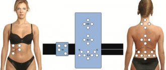
The representation is located in the area of the condyle of the elbow joint. 22. Parenchyma of the right kidney. Located in the upper part of the iliac crest on the right side of the body. It manifests itself as painful sensations when touching this area and palpation. The representative zone is located in the area of the large gluteal line on the gluteus maximus muscle, towards the superior iliac spine. Manifested by pain on palpation. The representation is located above the region of the greater trochanter of the femur, the region of the gluteus minimus and gluteus medius muscles. The pathology is manifested by pain in the joint and muscle representation.
The representation is located in the area of the large gluteal line, below the area of the sacral articulation. 13. Right kidney with bladder. Back pain is a fairly common symptom of various diseases.
The kidneys are a bigeminal (paired) organ. It is a bean-shaped formation, the concave side is located towards the spinal column.
On average, the weight of one kidney of a sexually mature person is 155-200 grams. The length reaches 12.5 centimeters, and the diameter is a maximum of 6, while the thickness of a healthy organ does not exceed 4-5 centimeters.