Brief anatomical and physiological characteristics of the kidneys
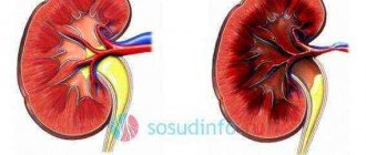
The kidneys are paired organs. Outwardly, they resemble the shape of a legume fruit. Located on both sides of the spine, in the area of 11-12 thoracic and 1-2 lumbar vertebrae. The average size of the organ does not exceed 12 cm in length and 3–5 cm in thickness. Their concave surfaces face the spinal column, and their convex surfaces face away from it.
Each kidney consists of several layers of dense tissue.
- Connective tissue capsule. It covers almost the entire organ. In the sinus region (located almost in the center of the side facing the spine), it passes into the membrane of the ureter. There, two large vessels enter the kidney. These are the renal vein and artery.
- The cortex is located directly under the capsule. Immediately below it lies the brain matter. It contains nephrons. Processes extend from the medulla towards the sinus. Blood and lymphatic vessels are located here. On the section, the color of the cortex, medulla and processes is the same. Therefore, they are considered a single anatomical structure.
- Almost in the center of the organ, between the processes lie the so-called renal pyramids. So named for their trapezoidal shape. The base is directed towards the cortex, and the apex is directed towards the sinus.
- Kidney calyces. The cavities into which the pyramids pass. They open into the pelvis, from which, in turn, emerges the ureter.
The basic functioning of the kidneys can be represented as follows. The renal artery, after “entering” the organ, begins to branch into several branches. They pass into the tissue of the processes and penetrate the medulla. Here they are further divided into smaller vessels up to the size of arterioles, which go into the nephron. There, due to even finer branching, they form a vascular tangle. Due to its dense surrounding with special cells (podocytes), primary filtration of blood occurs here. Then only blood cells, part of the plasma and some proteins go into the venules.
Everything else, and these are waste products, electrolytes, carbohydrates, lipids and some proteins, are retained by podocyte filters and, together with part of the plasma, enter the nephron tubules. This is called primary urine. It accumulates in the Bowman-Shumlyansky cup, which surrounds the vascular glomerulus in a semicircle. Its base is the beginning of the renal tubule. Through it, primary urine enters the tubule, which is long enough that it goes from the medulla to the pyramids. Here it makes a loop and “rises” into the medulla.
All along the way it is densely shrouded in venous vessels coming from the efferent vessel. This is necessary for the reabsorption (reabsorption) of proteins, lipids, carbohydrates, some electrolytes and water. As a result, only waste products, electrolytes and part of the water enter the renal calyx from the tubules. This is called secondary urine. It goes into the pelvis, from where it is excreted through the ureter.
It is important! A person normally produces up to 180 liters of primary urine per day. Of this amount, about 90−95% is reabsorbed. Thus, the daily volume of secondary urine does not exceed 2 liters. One can only imagine how many organic molecules pass through the kidneys. This circumstance provides a clear explanation why metabolic disorders often affect the kidneys.
HEREDITARY CARBOHYDRATE DYSTROPHY (GLYCOGENOSIS)
Glycogenoses
- a typical form of pathology of carbohydrate metabolism of hereditary origin, characterized by the accumulation of glycogen in cells, which causes disruption of the body’s vital functions.
main reason
- an inherited or congenital anomaly of genes encoding the synthesis of enzymes for the breakdown (less often - formation) of glycogen. Inherited in an autosomal recessive manner. There are more than 10 types of glycogenosis. Among them, Gierke's disease is the most common. Pompe, fetal cystic fibrosis, as well as Forbes-Cori, Andersen, McArdle diseases.
Gierke's disease
occurs in the absence of the enzyme glucose-6-phosphatase, which leads to the accumulation of glycogen in the cells of the liver and kidneys, but to the absence of carbohydrates in the blood. This is accompanied by secondary pituitary obesity. Most children die from acidotic coma.
Pompe disease
is associated with the absence of acid alpha-1,4-glucosidase in lysosomes, which leads to the accumulation of glycogen in the heart, striated and smooth muscles, including intercostal, diaphragmatic, tongue, esophagus, stomach, etc. Children die at an early age from heart or respiratory failure.
For the rest, cystic fibrosis is a disease associated with genotypic fermentopathy. leading to disruption of the exchange of mucoids that are part of the secretion of many glands. As a result, the secretion of the glands becomes viscous and thick, is difficult to remove, which leads to stretching of the glands, turning them into cysts, especially in the pancreas, mucous membranes of the gastrointestinal tract and respiratory tract, salivary, sweat, lacrimal glands, etc. In this case atelectasis often develops in the lungs with the development of pneumonia and bronchiectasis. Death most often occurs from pulmonary heart failure.
The term element “dystrophy” is a derivative of two terms and in translation means metabolic disorder. It affects a large number of organs. This is especially true for those that play an important role in metabolism. It is not for nothing that kidney dystrophy can serve as a marker for many similar diseases.
Mechanisms and causes of dystrophy
Common to all types of dystrophies is the initial stage of development. It is as follows:
- Violation of the exchange of a group of molecules.
- Excessive accumulation of these molecules in the cells of the organ.
The further mechanism varies depending on the specific type of dystrophy. But this will be discussed below. As for the causes of dystrophy, they are divided into two large groups.
- Congenital dystrophies. Caused by genetic and/or congenital defects at any stage of the metabolism of a substance: from synthesis to breakdown. For the kidneys, among the congenital dystrophies, the most characteristic is a violation of protein and fat metabolism.
- Acquired dystrophies arise as a result of disruption of any stage of metabolism of a substance under the influence of external causes. The most likely causes are viral infections, chronic bacterial infections, radiation, chronic intoxication, and some diseases. These include diabetes mellitus, some neoplasms, and injuries.
Fatty liver, or fatty hepatosis
Rice. 4. Fatty degeneration of the myocardium (“tiger heart”). Yellow-white stripes are visible under the endocardium, corresponding to areas of lipid incorporation in cardiomyocytes.
At the same time, with many intoxications and infections, a mechanism of decomposition of the membranes of intracellular structures with the disintegration of their fat-protein complexes is possible. Finally, fatty hepatosis develops as a result of the transformation of proteins and carbohydrates into lipids, which is observed, for example, with chronic alcohol intoxication. In any case, in the cytoplasm of hepatocytes, mainly in the periphery of the hepatic lobules, dust-like obesity first develops, which transforms into small-droplet obesity, and then into large-droplet obesity. In this case, the nucleus and intracellular structures are pushed to the periphery of the cells, which often die. In these cases, the fatty inclusions of dead hepatocytes merge, forming fatty cysts. Macroscopic changes in the liver depend on the severity of dystrophy. In severe cases, for example with alcoholism, the liver is enlarged in size, flabby, and on a section it is ocher in color - “goose liver”. With less pronounced fatty degeneration, the liver is also enlarged in size and has a yellowish-gray color when cut. With fatty hepatosis, liver function is preserved for a long time, but as the underlying disease and fatty degeneration progresses, it decreases, sometimes quite significantly.
Fatty kidney degeneration
develops by infiltration of the tubular epithelium during hyperlipidemia, observed, in particular, with nephrotic syndrome.
In this situation, lipids end up in the primary urine ( hyperlipiduria
) and are intensively reabsorbed by tubular epithelial cells, but in such large quantities that these cells are not able to metabolize the lipids that have entered them - small droplet obesity of the tubular epithelium develops. Usually it is combined with their hyaline-droplet dystrophy. In this case, the kidneys are little changed in appearance, but in severe cases of the main pathological process, they acquire a grayish-yellow color, and when cut, their pyramids can take on a yellow color.
The outcome of parenchymal fatty degeneration depends on the degree of its severity - dust-like and small-droplet obesity is reversible when the cause that caused it is eliminated, large-droplet obesity can result in cell death.
Types of dystrophy
It is important! All kidney dystrophies can be divided depending on the causes of occurrence and the specific substance. In the first case, these diseases are divided into congenital and acquired dystrophies. But from the point of view of practical medicine, it is considered more correct to divide dystrophies by type of substance.
According to the above situation, dystrophies are divided into three categories:
- Dysproteinoses. These are dystrophies caused by protein metabolism disorders. They can be either congenital or acquired. The following types are characteristic of the kidneys:
- Granular dystrophy. It develops as a result of intracellular edema and the release of protein into the cytoplasm. But since protein is an insoluble substance, it forms peculiar “grains” in the cytoplasm. That's why granular kidney dystrophy got its name.
- Hyaline droplet dystrophy. It is the result of further development of the first type. Here, as a result of significant accumulation, protein molecules begin to unite into peculiar “drops” of cartilage-like consistency and color. In their structure, these protein formations resemble hyaline, the main protein “building material” of cartilage. This circumstance explains why hyaline drip renal dystrophy received such a name.
- Horny dystrophy. Develops with excessive accumulation of keratin. This white is normally characteristic only of epithelial cells. But in the case of constant exposure of a non-epithelial cell to a damaging factor, the “assembly” of keratin molecules begins from the fibrils of the cytoplasm.
- Hydropic dystrophy. A special type of protein dystrophy. It occurs when the cell's water vacuoles become excessively enlarged. Most often, a cell contains one giant vacuole containing a clear liquid. At the same time, there are practically no other organelles. Including the missing nucleus, which is destroyed by this vacuole. The main cause of dystrophy is a viral infection. For kidney structures, this type is most characteristic of the tubules. However, hydropic dystrophy of the epithelium of the kidney tubules here is caused not so much by viral infections, but by chronic inflammation in general.
- Fatty degenerations or lipidoses. Slightly less common. Moreover, they are often acquired in nature than protein ones. For which both options are equally characteristic. And one of the main factors of fatty degeneration is considered to be ischemia - lack of oxygen. This is well evidenced by the fact that fatty kidneys are often observed in chronic infections and intoxication with certain inorganic substances: arsenic, bismuth, mercury, as well as chronic alcoholism.
- Carbohydrate dystrophies are less typical for the kidneys. This is largely due to the fact that the kidneys play a small role in carbohydrate metabolism. However, in some diseases, carbohydrate kidney dystrophy can occur quite often. It is based on the deposition of carbohydrate molecules when they are in excess. The most striking example of this type of dystrophy is diabetes mellitus.
CONGENITAL PARENCYMATOUS LIPIDOSES
Congenital parenchymal lipidoses are hereditary enzymopathies, inherited in an autosomal recessive manner, and are characterized by the accumulation of lipids in cells that damage cell structures and are often accompanied by the death of the cells themselves. The most common lipid thesaurismoses are:
- Gaucher disease
is caused by a lack of the enzyme beta-glucocerebrosidase. As a result, glucocerebrosides accumulate in the liver, spleen, bone marrow, brain, endocrine glands and lymph nodes, which leads to the death of cells in these organs and to progressive dementia, increased weight of the liver, spleen and wasting (cachexia). - Niemann-Pick disease
develops in the absence of the enzyme sphingomyelinase, which breaks down sphingomyelin, which is part of many tissues, but especially nervous tissue. In sick children, it accumulates in the cells of most organs and at the same time there is an increase in the mass of the liver and spleen (hepato- and splenomegaly), a lag in mental development, neurological symptoms, hypotension, and exhaustion appear. Children die at the age of 2-3 years.
general characteristics
Dystrophic disorders in the kidneys have a clear location, that is, they are located within one organ. But despite this, the urinary system subsequently begins to suffer, and then the entire body as a whole.
To make an accurate diagnosis, it is necessary to undergo a set of diagnostic procedures, since each type of dystrophy has its own characteristics
The kidneys perform an excretory function. They are necessary for a person to rid the body of breakdown products that are formed as a result of metabolism. Metabolic processes occur in the glomeruli of the kidneys, where the interaction of blood and water solution occurs, and it is in them that all the components unnecessary for the human body then accumulate. All toxic substances are also released through the kidneys, so the paired organ always suffers from various poisonings, to the same extent as the liver.