Cystitis is one of the most common pathologies of the genitourinary area. It refers to inflammatory diseases of the bladder. It is characterized by damage to the mucous membrane by pathogenic bacteria. The disease affects people of any gender. But women especially often suffer from this disease due to their special anatomical structure, which facilitates easy penetration of pathogenic microflora (saprophytic bacillus, intestinal staphylococcus and other microorganisms) into the urinary cavity. The disease can have an acute or chronic course.
For a comprehensive diagnosis of urinary tract, not only routine methods such as general urine and blood analysis are used, but also functional diagnostic methods. The main one is ultrasound of the bladder. Ultrasound examination (ultrasound, sonography) - examination of internal organs, performed using ultrasonic waves, for cystitis allows you to obtain information about the condition of the organ itself and the tissues surrounding it.
Types of ultrasound examination
Diagnosing cystitis these days is not difficult, since urologists have various types of diagnostic searches in their “arsenal”.
First of all, this is ultrasound diagnostics. Ultrasound of the bladder in women and men is carried out using various methods, which are determined by the doctor, analyzing the clinical picture of the disease and the individual characteristics of the particular patient. The transabdominal ultrasound method is the most common type of instrumental diagnosis.
The organ is examined by moving an abdominal sensor along the anterior wall of the peritoneum. This method makes it possible to clarify the size, structure and shape of the organ, but is not effective if the patient is clearly obese or is unable to hold urine. Since a mandatory condition for the procedure is a filled bladder.
"TVUS" method (transvaginal). An ultrasound probe is placed in the vagina (vagina). It is considered the most informative diagnostic method, allowing to accurately and correctly detect various pathological processes. It is carried out with an empty urinary reservoir.
"TUUS" (transurethral method). Diagnosis is carried out by inserting a sensor into the urethral cavity, thus providing excellent visualization. It is carried out using anesthesia. This method allows you to assess the condition of the urethral wall, the severity of its damage and possible pathological processes in nearby organs. It is used in exceptional cases, since there is a high probability of damage to the walls of the urethra by the sensor and the development of complications.
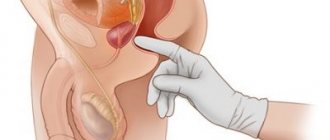
TRUS technique (transrectal method). The sensor is inserted rectally (into the rectum). It is mainly used when it is necessary to perform an ultrasound of the bladder in men. This method reveals the pathological connection between the bladder and prostate organs. It is sometimes used when examining girls for whom the transabdominal method is contraindicated, but the presence of a hymen is an obstacle to another method.
Doppler diagnostics. Allows you to identify changes in the structural tissues of the bladder walls and study the residual volume of urine in the bladder reservoir. Diagnostics consists of two stages - scanning the organ when it is completely full and when it is empty.
results
The results of an ultrasound examination of the bladder indicate several parameters that help make a final diagnosis:
- bubble shape;
- its volume;
- amount of residual urine;
- bubble structure;
- Wall thickness;
- rate of bladder emptying.
Ultrasound allows you to determine whether an inflammatory process is developing in the urinary organ.
The echo picture of a patient with acute cystitis shows accumulations of cells - epithelium, erythrocytes and leukocytes, which are described in the study results by the term “sediment”. If the patient lies down during the ultrasound, the sediment is localized near the posterior wall of the bladder. When the patient stands up, the sediment will move to the front wall.
In the chronic form of the pathology or with the progression of acute cystitis, the results of the study will show that the organ has an uneven contour and the walls are thickened. The presence of blood clots in the bladder cavity is shown on the echo picture.
- Is it possible to eat before an ultrasound of the kidneys: diet before ultrasound diagnostics
The results of an ultrasound examination must be deciphered by the urologist who referred the patient for the procedure. If necessary, the doctor selects a treatment course.
The results of an ultrasound examination must be deciphered by the urologist who referred the patient for the procedure.
Norms
The results of the bladder examination are normal:
- Form. In the transverse projection the bubble should be round, in the longitudinal projection it should be ovoid. The shape of the female organ is influenced by the number of pregnancies and births.
- Structure. Normally, it is echo-negative, but the parameter depends on the age of the person: the older you are, the higher the echogenicity should be.
- Volume. Average values for women are 250-550 ml, for men - 350-750 ml.
- Walls. The same thickness over the entire surface - 2-4 mm. If any area shows thickening or thinning, this indicates the presence of pathology in the organ.
- Residual urine. Its quantity should not be more than 50 ml. When conducting a study, it is mandatory to measure.
Preparation
Preparation for the study depends on the method of conducting it.
4 known ultrasound of the urinary tract. This:
- transvaginal;
- transurethral;
- transabdominal;
- transrectal.
Ultrasound is accompanied, if necessary, by other types of studies.
Also, to make a diagnosis of cystitis, a method is often used that helps to identify all the obstacles that urine overcomes when entering or leaving the bladder.
The effectiveness of this method lies in the study of the patient's residual urine.
For what purpose is ultrasound prescribed for cystitis? Possible contraindications
The doctor refers the patient for an ultrasound of the bladder if there is a suspicion of the presence of space-occupying formations, stones in its cavity, which cause damage to the mucous membrane and its inflammation, in case of complicated or long-term cystitis, when the disease is difficult to treat.
Ultrasound examination is indicated in the following situations:
- the appearance of pathological impurities in the urine - blood, pus, sand;
- frequent painful urination, false urge to empty the bladder with a small volume of urine;
- outflow disturbance or acute urinary retention;
- prolonged course of cystitis that is not amenable to antibiotic therapy;
- severe intoxication due to cystitis - fever, weakness, pain in joints and muscles;
- urinary tract infection during pregnancy;
- intense pain above the pubis.
You need to understand that ultrasound is an additional way to clarify the diagnosis. First of all, a general urine test is prescribed, as well as microflora culture and determination of its sensitivity to antibiotics.
Bladder functions: do not treat and you will pee
Treatment for cystitis is prescribed by the attending physician based on the results of tests and ultrasound diagnostics. Therapeutic measures depend on the form of cystitis and the causative agent of the disease. Usually prescribed:
- Antibiotics (Nolicin, Tsiprolet, Furamag) for a course of 5 to 10 days. Early withdrawal of antibacterial drugs is prohibited, even if the symptoms of the disease have disappeared during treatment. To completely destroy the pathogen, antibiotics must be taken for at least 5 days.
- Herbal medicines based on lingonberries, woolly erva, bear's ears. It is possible to use combination herbal medicines: canephron, cystone, phytolysin. They also help dissolve stones in the urinary tract.
- Antispasmodics - no-spa, papaverine - are necessary for severe pain or impaired urine outflow.
- In rare cases, doctors prescribe diuretics to improve urodynamics.
- Medicines based on methyluracil and actovegin improve the regeneration of the mucous membrane and are used in the complex treatment of chronic cystitis.
- If cystitis has developed against the background of urolithiasis, then surgical treatment is performed according to indications: crushing or extraction of stones.
- Physiotherapy is an auxiliary therapeutic technique. It should not be prescribed before an ultrasound scan, since physiotherapy simulates the growth of space-occupying formations.
- Patients with cystitis are advised to drink plenty of fluids and eat a diet that excludes spicy, salty foods and alcohol.
If complications of cystitis develop - bleeding, abscess formation, acute urinary retention - treatment is carried out in a hospital setting.
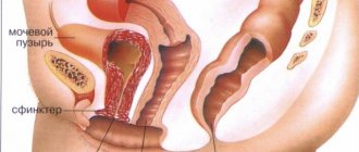
The main job of the bladder is to collect and remove urine (urine) from the body. The organ is equipped with nerve endings, so when the bladder fills, signals about filling are sent to the brain. An adult can control the urge by tensing the muscles to drain urine. When they relax, urine flows out through the urethra.
Various pathologies disrupt the normal functioning of the organ, which significantly reduces the patient’s quality of life. The result is pain when urinating, incontinence or stagnation of urine. Sometimes the organ is completely removed, bringing the urine collector out.
You can check the condition of the organ by undergoing an ultrasound examination of the bladder, which is usually performed together with a kidney examination or separately. The examination shows changes invisible to the eye that have occurred in the internal organs. The big advantage is the speed and painlessness of the method.
The study of the kidneys and bladder is a complex undertaking, so before starting the diagnosis you should do blood and urine tests and take them with you.
When is it prescribed?
The main indications for ultrasound if cystitis is suspected are:
- rare or, conversely, too frequent urination;
- the presence of pus or blood clots in the urine;
- the appearance of large white flakes in urine;
- false urge to go to the toilet, when only a couple of drops of urine containing impurities of pus or blood are released from the bladder (often this phenomenon is observed with cystitis, which was caused by a specific flora);
- change in urine color;
- decrease in the total amount of urine produced per day;
- pain or discomfort when going to the toilet “in a small way”;
- discomfort in the pubic area;
- an increase in low-grade fever to 38 degrees or more.
It is important to note that these symptoms can characterize not only cystitis, but also other pathologies of the bladder or the entire excretory system (pelvic organs). Therefore, the patient is prescribed an ultrasound, with the help of which the diagnosis will be established accurately. The question “is it necessary to do an ultrasound” in such a situation does not arise.
Reference! In advanced forms of cystitis, the procedure is performed not only to examine the condition of the urinary organ, but also to identify the dynamics of the disease. This allows doctors to monitor the patient’s condition, as well as avoid the transition of chronic cystitis to acute.
Interpretation of results and norm
Diagnosis of cystitis, performed in the acute phase, reveals the following picture: inside the bladder, tiny particles endowed with high echogenicity are clearly visible. They are usually united into foci. As a rule, these particles are an accumulation of a large number of cells - leukocyte, epithelial or erythrocyte. Crystals of salts (oxalates) can also be found in them.
Reference! If a person lies down during an ultrasound, the focus with sediment will be located on the back wall of the bladder; if the patient stands, particles will be found on the front wall of the organ.
The outflow of urine when it reaches its maximum peak should be less than 15 cm/s - otherwise we can talk about the development of cystitis or other diseases of the urinary organs.
- Is it possible to get pregnant with cystitis and what is the effect of cystitis on conception?
What the study shows and what indicators are the norm
Interpretation of the results of ultrasound of the bladder is carried out according to the following parameters:
- organ size;
- its volume;
- form;
- wall structure;
- content;
- residual urine volume (RUV).
Normally, the bladder is visualized as a formation of a regular ovoid shape, normal size, with clear, even contours and anechoic homogeneous contents. TOM should not exceed 50 ml.
With cystitis, the internal contours of the bladder become uneven, flakes and conglomerates are visible in the lumen of the organ - particles of desquamated epithelium, accumulations of microbes, sand, clots of pus or blood. The ulcerative form of cystitis is characterized by defects in the mucosa in the form of depressions - erosions. The inflamed walls are usually thickened, and the bladder itself increases in size during an acute disease, and may decrease in size during a chronic disease.
In addition to signs of cystitis, ultrasound shows the following changes:
- polyps - small outgrowths of the mucous membrane on a stalk, with clear contours;
- benign or malignant neoplasms - in this case, the contours of the bladder become lumpy and the shape is asymmetrical;
- stones in the organ cavity or at the mouth of the urethra;
- violation of the integrity of the bladder wall due to trauma.
Symptoms
• Pain in the suprapubic region;
• Irregular or improper hygiene (in girls);
Diagnostics
• Collection of medical history and complaints;
Ultrasound of the bladder for cystitis is performed after special preparation of the patient. The patient should drink 1-1.5 liters of still water or another drink (not milk) 1-1.5 hours before the scheduled procedure. With chronic cystitis, ultrasound shows thickened walls, as well as sediment at the bottom of the bladder.
Historical data
Oddly enough, the forefathers of modern ultrasound machines are the English military-industrial sonar and radar systems (RADAR and SONAR), which work on the principle of reflecting a pulse of sound waves from certain objects. And the pioneers of scanning the human body were American researchers (Hour and Holmes). They placed a “volunteer” in a tank filled with water and passed ultrasound around him.
But the era of real ultrasound diagnostics began in 1949, when the American D. Hauri first created a functioning ultrasound machine.
An important contribution to the modification of this new diagnostic method, allowing to expand its capabilities, was made by the Austrian mathematician and physicist K. Doppler. His developments in comparing and recording impulses and speed of the object of study made it possible to study blood circulation in large vascular beds.
Since 1960, ultrasound examination has been firmly established in medicine. Soon (1964), a group of Japanese researchers proposed using sensors of various modifications when examining the bladder and prostate - rectal, which allows one to obtain an image of the organ in a cross-sectional view, and intracavitary (urethral), which allows one to diagnose various pathological changes in the tissue structure of the cavity of the urinary reservoir.
Today, there are several modes of ultrasound machines - one-dimensional and echography (“M” and “A” modes).
With their help, all anatomical components of the human body are examined, visualized and measured. Mode “B” is called scanning or sonography. It allows you to obtain more effective information - a two-dimensional picture on a monitor with the ability to observe the process in motion (Doppler effect).
- Kotervin or stop cystitis which is better
What does cystitis look like on ultrasound?
The organ itself is not echogenic, meaning it appears on the screen in dark shades. On ultrasound for cystitis in women and men, light small particles are clearly visible - blood clots, pus, fungal formations, salt crystals, grouped into small lesions. All of them are designated as “sediment in the bladder”, while during an ultrasound in a vertical position (standing), sediment will accumulate on the front wall of the organ, and in a supine position - near the back.
The thickness of the walls of the bladder remains unchanged in the initial stages of the disease, the walls of the organ are smooth, symmetrical, and have the correct shape. They noticeably thicken during the transition to the acute form of the disease. With the development of pathology, one can observe a change in the contours of the organ, a violation of symmetry and a change in proportions. The same features can be seen in the photo after an ultrasound for gangrenous cystitis.
With chronic cystitis, the walls of the bladder also thicken, and specialists note the sediment as “flakes in the bladder.” In advanced disease, blood clots are clearly visible on the screen, characterized as hyper- and hypoechoic structures. In some cases, they are attached to the mucous membrane of the bladder. During the liquefaction stage, these structures are not echogenic and create an uneven outline on the image.
Sometimes a repeat ultrasound of women who have had hydrosalpinx shows cystitis. This is an indication for undergoing another examination after identifying problems with the fallopian tubes.
Ultrasound signs of breast cancer
Preparation for an ultrasound scan if cancer is suspected is similar to that indicated above. Using an ultrasound examination, the localization of the tumor, its structure, size, and blood supply characteristics are determined. At the same time, involvement in the tumor process of the ureters, prostate in men, ovaries and fallopian tubes in women is examined, and the presence of metastases in regional lymph nodes is determined.
Ultrasound signs of cancer located in the bladder are:
- iso- or hyperechoic formations on the walls of an organ with a heterogeneous structure,
- hyperechoic small formations at the bottom of the bladder (accumulations of necrotic masses and blood clots with a disintegrating tumor);
- uneven walls of the organ, uneven changes in their thickness and uniformity;
- bubble deformation.
To clarify the diagnosis, cystoscopy and histological examination (studies the structure of tissue) of biopsy specimens - pieces of tissue obtained during cystoscopy (insertion of a magnifying device into the cavity of the bladder) are also prescribed.
How is ultrasound performed?
Ultrasound examination is carried out in 4 ways:
- A common method of delivery is through the anterior abdominal wall. This technique is called transabdominal (classical method). The peculiarity of the procedure is that the study is carried out with a full bladder. This method is the least invasive for patients, but cannot be performed in cases of pronounced subcutaneous fat.
- Another method that is common among women is transvaginal examination. It is carried out when the bladder is emptied and conducts a detailed examination. The technique causes a number of inconveniences for patients.
- Transrectal examination is suitable for both males and females. Provides more information than transabdominal. During the examination, the condition of the prostate is assessed in men, because inflammation of the bladder spreads to it and vice versa.
- Transurethral ultrasound. This examination is not widespread, since it requires anesthesia and can lead to injury to the urethra. The sensor is inserted into the urinary cavity through the urethra. The value of transurethral is that it assesses the condition of the walls of the urethra.
Transabdominal examination requires preparation of the intestines and the bladder itself. To do this, for several days they follow a diet that prevents flatulence and reduces the deposition of toxins in the intestines.
Since the examination is carried out with a full bladder, drink at least 1.5 liters of water 1.5-2 hours before the examination. If the examination is carried out in the morning, then you do not need to urinate before the examination.
Transrectal ultrasound requires cleansing the rectal capsule from feces. To do this, use an enema or laxatives.
If the examination is carried out through the vagina or rectum, then it is worthwhile to thoroughly clean the genital organs to prevent infection and the development of inflammatory diseases, proctitis, vulvovaginitis, etc.
Indications for the procedure
A number of symptoms from the urinary system are indications for an ultrasound scan for cystitis. Among them:
- the appearance of bloody impurities or pus in the urine;
- frequent urge to urinate or acute urinary retention;
- small volume of urine;
- pain in the suprapubic area that appears periodically.
The indication for an ultrasound is a frequent urge to urinate.
A number of symptoms from the urinary system are indications for an ultrasound scan for cystitis. Among them is the appearance of bloody impurities in urine.
If the volume of urine is small, it is necessary to undergo an ultrasound.
Cystenal: instructions for use. The benefits of herbs for cystitis. Nephrosten: reviews from urologists. More details>>
What is the price
You can undergo an ultrasound examination in any city in Russia. When choosing a clinic or ultrasound room, you should rely both on the recommendations of the attending physician and patient reviews, and on your financial capabilities. The average cost of ultrasound in Russian cities is 1,000-1,500 rubles. and is determined by such factors as location, degree of qualification of the clinic and doctor, equipment characteristics and others.
The average cost of ultrasound in Russian cities is 1,000-1,500 rubles.
In large cities and regional centers, the price of an ultrasound of the urinary system is at least 800 rubles. Prices for ultrasound also vary depending on the volume of the procedure (the urinary system as a whole or individual organs) and the type of examination. A separate ultrasound of the urethra is cheaper (from 200 rubles).
What does it show?
Is the disease visible on the study? When performing an ultrasound, doctors can detect diverticula - these are peculiar sac-like neoplasms located on the walls of the bladder or growing into its cavity. also possible to detect sand or oxalate (salt) stones , which significantly disrupt the integrity of the mucous membrane and are also considered the main factor in the development of cystitis.
Video 1. Cystitis on ultrasound.
During certain forms of the disease, such a study will be endowed with specific manifestations.
Ulcerative and herpetic forms
For these forms of cystitis, a characteristic symptom of the development of the disease will be the appearance of erosions and small ulcers in the inner part of the bladder. At first they will develop on the mucous membrane, and then begin to spread into the deeper layers of the organ. This form is accompanied by severe pain , so the patient should be treated immediately after signs of cystitis are identified.
Candidiasis form
With the development of candidal cystitis, ultrasound will show formations that have appeared in the urinary cavity. They can have different shapes and sizes. The rate of growth of neoplasms depends on the state of the patient’s immunity and the duration of cystitis.
Acute form
Significant thickening of the walls of the bladder becomes noticeable only with the onset of an acute form of pathology. At the beginning of its development, an ultrasound will show an even contour of the organ, which will be completely free of deformation. However, as inflammation progresses, the walls of the bladder will gradually thicken , the contour will become more crooked, and the shape will be uneven - with the help of ultrasound, such negative changes in the organ can be noticed without problems.
Chronic form
With the development of this form, thickening of the walls of the organ also occurs. Ultrasound shows the presence of flakes in the bladder, which indicates advanced disease.
If the inflammation is too advanced, hypo and hyperechoic areas can be found in the inflamed organ. They may be blood clots . They also cause disruption of the contour of the urinary organ while in a liquefying phase, causing it to appear asymmetrical.
Healthy Bladder
In a normal and healthy state, the organ is smooth, symmetrical, without protruding walls or an uneven contour. The mucous membrane should be free of deformations, ulcers, spots and thickenings. A healthy organ has a wall thickness of 5 mm.
Video
Primary diagnosis of cystitis is carried out using urine and blood tests. After the doctor receives the results, he can refer the patient for an ultrasound examination of the urinary system. Ultrasound of the bladder for cystitis is a necessary measure.
If the doctor has difficulty making a diagnosis, this diagnostic method helps to obtain an accurate picture of the development of the disease, since ultrasound visually shows the structure of the bladder, in which characteristic signs of inflammation are visible when cystitis occurs.


