Where to get diagnosed
You can undergo an ultrasound of various organs, including the kidneys, at the urological center of branch No. 1 of the Federal State Institution “GVKG im. Academician N.N. Burdenko". The advantages of our multidisciplinary hospital include:
- significant experience in the field of diagnosis and treatment of diseases of a urological nature (the hospital became an independent structural unit back in 1974);
- professional staff of specialists (all diagnostic and treatment procedures are carried out by doctors of the highest category);
- a full range of services in the field of urology - from thorough diagnostics to conservative treatment and removal of kidney tumors;
- technical equipment that meets modern criteria (hospital equipment allows for highly accurate diagnostics and minimally invasive interventions that improve the quality of life of patients);
- comfortable rooms equipped with everything necessary (in which the patient can comfortably recover after treatment).
Surgery to remove a kidney with a tumor
Surgical intervention may involve excision of the pathological formation itself with small areas of nearby tissue or removal of the entire organ. The only criterion for choosing which kidney operation will be performed is the tumor and its characteristics. Radical nephrectomy (complete removal of an organ with adjacent structures) is performed if the neoplasm has one of the following properties:
- malignancy;
- very large size;
- localization near the renal vessels;
- multiple metastases;
- rapid growth.
The final decision on the type of surgery is made by the urologist. Bilateral nephrectomy is disabling, but in some situations, open surgery is the only way to prolong the patient's life and prevent extensive and irreversible damage to the cancer's target organs - the brain, spine and lungs.
Laparoscopy of a kidney tumor
The gentle operation is carried out using special microscopic equipment through small punctures in the abdominal wall. It is recommended for small benign kidney tumors, if the pathological accumulation of cells is not prone to degeneration into cancer and growth. This type of surgical manipulation ensures the preservation of the organ and the rapid return of the patient to normal life. During the operation, the kidney tumor and a thin layer of surrounding tissue are removed. This helps prevent the re-formation of benign cellular structures.
Rehabilitation after removal of a kidney with a cancerous tumor
Due to unilateral nephrectomy, the second paired organ takes on the entire load of the urinary system. The main task of a person who has undergone this procedure is to preserve the functions of a healthy kidney. Recommended:
- water hardening;
- daily walks;
- avoiding hypothermia;
- protection against any infectious diseases;
- regular examinations by a nephrologist or urologist.
The diet after removal of a kidney with a tumor should include easily digestible foods low in protein. The basis of the diet are:
- low-fat varieties of fish and meat;
- Rye bread;
- some fermented milk products;
- fruits and vegetables.
All dishes are steamed, boiled, stewed or baked. It is important to completely exclude:
- smoked meats;
- pickles;
- canned food;
- fried and fatty foods.
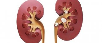
Kidney tumors and causes of their development
A kidney tumor is a pathological process characterized by the proliferation of tissue consisting of qualitatively changed cells. Depending on the nature of growth, benign and malignant neoplasms are distinguished. The latter are more common and account for about 94–95% of all detected tumors. The average age of detection of kidney tumors is 70 years. According to statistics, tumors occur 2 times more often in men than in women. In children they are detected with a frequency independent of gender. Factors contributing to the development of tumors in the kidneys include:
- hereditary causes;
- bad habits (smoking, drinking alcohol);
- exposure to radiation or carcinogenic substances;
- disruptions in the immune system;
- the presence of chronic foci of infection in the body or kidneys.
Treatment of benign kidney tumors
A single simple kidney cyst does not require additional treatment; the patient is recommended to undergo an ultrasound scan of the kidneys annually and avoid hypothermia and infectious diseases. The need for treatment arises in the event of complications of the disease: pyelonephritis or chronic renal failure.
The possibilities for conservative treatment of kidney cysts are quite limited and are reduced to compensation for emerging abnormalities (inflammation, anemia, arterial hypertension, impaired excretory function of the kidneys).
For large uncomplicated cysts that compress adjacent tissues, puncture emptying of the cyst (ignipuncture) is performed.
Surgical treatment - enucleation (enucleation) of a cyst, resection of a kidney or cyst, removal of a kidney (nephrectomy) - is used in the case of:
- compression of the urinary tract by a cyst;
- compression of the cystic tissue of the kidney;
- infection of the cyst cavity and abscess formation;
- cyst rupture;
- large cysts.
Essential drugs
There are contraindications. Specialist consultation is required.
| 1 | Enalapril (hypotensive drug, nephroprotector) |
Dosage regimen
Take 2.5-10 mg orally 2 times a day, constantly.
| 2 | Amoxicillin + Clavulanic acid (Flemoklav Solutab) - an antibiotic for infections of the kidneys and lower genitourinary tract |
Dosage regimen
Inside, intravenously. The dosage is indicated in terms of amoxicillin.
The dosage is prescribed individually, based on the severity of the course and localization of the lesion, as well as the sensitivity of the pathogen. For children under 12 years of age, the medicine should be given in liquid form, for example, drops or syrup for oral administration. A one-time dose and its dosage are calculated depending on age. Children under 3 months of age - 30 mg/kg/day. in 2 doses, at 3 months of age and older for simple infections - 25 mg/kg/day. in 2 doses or 20 mg/kg/day. in 3 doses. If the infection has become severe, then take 45 mg/kg/day. in 2 doses or 40 mg/kg/day. in 3 doses.
For adults and children aged 12 years and older, or if the patient's body weight is 40 kilograms or more, the daily dose is: 500 mg 2 times a day. or 250 mg 3 times/day. For respiratory tract infections and severe infections, the dosage is 875 mg 2 times a day. or 500 mg 3 times/day.
For adults and children over 12 years of age, the maximum dose of amoxicillin used per day is 6 g, for children under 12 years of age - 45 mg/kg body weight. The use of clavulanic acid is limited to 600 mg per day for adults and children over 12 years of age. Children under 12 years of age should take 10 mg/kg body weight. The treatment period is up to 14 days.
| 3 | Ciprofloxacin (Ciprinol) is a broad-spectrum antimicrobial drug, a second-generation monofluorinated fluoroquinolone. |
Dosage
The drug is dosed individually, it all depends on where the focus of the disease is located and the severity of the infection, as well as on body weight, body condition, and age characteristics.
For diseases of the urinary tract and kidneys without complications, the drug is prescribed at 250 mg 2 times a day, for complications - 500-750 mg 2 times a day.
Treatment must be continued for 14 days.
| 4 | Iron (III) hydroxide polymaltosate (Maltofer) - replenishes iron deficiency in the body, stimulates erythropoiesis, restores hemoglobin |
Dosage
The drug is taken after or during meals. The treatment period and dosage depend on the level of iron in the body. Maltofer can be taken either in one dose or divided into several doses throughout the day.
The tablets should be swallowed whole, either chewed after or during meals. If there is a pronounced lack of iron, you should take 1 tablet 1-3 times a day for 3-5 months until hemoglobin normalizes. After a course of treatment, to restore iron reserves, it is necessary to take 1 tablet of the drug per day for several months.
To prevent iron deficiency and treat latent iron deficiency, you need to take 1 tablet of the drug per day.
The drug in drops can be mixed with various juices (fruit and vegetable), or drops can be added to nutritional mixtures. When mixed, the activity of the drug will not decrease. 20 drops (1 ml) of the drug contain 176.5 mg of iron (III) polymaltose complex hydroxide (50 mg of elemental iron), 1 drop. equal to 2.5 mg of elemental iron.
Dosage for the treatment of clinically significant iron deficiency. Adults and children over 12 years of age are prescribed to take the drug 40-120 drops per day. The course duration is at least 60 days. With clinically pronounced iron deficiency, hemoglobin normalizes 2-3 months after treatment is started. To restore internal iron reserves, it is necessary to continue taking the drug prophylactically for several months.
Dosage for the treatment of latent iron deficiency. Adults and children over 12 years of age are prescribed 20-40 drops per day. For prevention, adults and children need to take 4-6 drops of medicine per day. 1 ml of syrup contains 10 mg of iron (III). For the treatment of clinically significant iron deficiency, adults and children over 12 years of age are prescribed 10 to 30 ml of the drug per day. For latent iron deficiency, the dosage is from 5 to 10 ml per day.
| 5 | Furosemide (loop diuretic) |
Dosage
It is recommended to take from 40 to 80 mg of the drug per day. The dosage is individual for each person and is selected depending on the diuretic response. The daily dose of the drug is divided into 2 doses, or taken at a time. The drug in tablets is taken, without chewing, on an empty stomach. You need to drink enough water. The maximum daily dose for adults is 1500 mg. The duration of treatment is determined by the doctor individually.
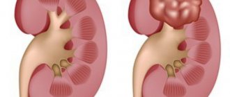
Symptoms indicating the possible presence of a malignant neoplasm
Treatment of malignant tumors in the kidneys is most effective in the initial stages. But during this period, the neoplasm usually does not manifest itself in any way. A doctor may accidentally detect a kidney tumor on a routine ultrasound even in the complete absence of clinical signs of the disease. As the tumor grows, symptoms appear that indicate a deterioration in the patient’s general condition: nausea, weakness, loss of appetite, sudden weight loss. Blood in the urine, complaints of pain in the lumbar region, swelling in the sides of the abdomen, and renal colic are more likely to indicate that the kidney tumor has reached an advanced stage.
Types of formations
Hyperechoic
The formation of increased echogenicity differs from the surrounding tissues in structure. It is very dense, so when examined with ultrasound, the waves are reflected well.
On an ultrasound monitor, hyperechoic formations in the kidney are displayed as white spots. Most often, the echogenicity of the renal parenchyma is increased if stones (calculi) are present in it.
Because of them, other objects in the organ cavity may not be visible during the examination. Kidney size may increase.
The presence of kidney stones indicates dysfunction of the kidneys, their sinuses and metabolic problems.
Hyperechenogenic
Hyperechogenic - inclusion of increased echogenicity. The pathological change differs from other tissues in its high density, which is easy to detect on ultrasound.
On the monitor, changes are presented as white spots of impressive size. With kidney stones, the echogenicity will be higher than usual.
Due to such changes, the size of the kidneys increases and other structures become invisible. Kidney stones indicate dysfunction of the organ.
A kidney cyst is a space-occupying formation that is a cavity filled with liquid contents with clear, even edges. The capsule of the cyst is formed by tissues of the parenchyma of the organ, which have become denser and formed a cavity.
This occurs as a result of the influence of unfavorable factors on the kidney parenchyma. The glomerular excretory ducts become clogged, and primary urine accumulates in one place, forming a capsule.
Over time, as fluid contents accumulate, the cyst increases in size.
When examined by ultrasound, the contents of the cyst have an anechoic structure, because the fluid located in the cyst capsule is not able to reflect the sound wave. A simple cyst has one chamber with clear contours.
A kidney cyst, which has septa and several chambers, is more often prone to degeneration into a malignant neoplasm
Single cysts less than 5 cm in size rarely bother the patient and cannot be treated. Such cystic formations should be monitored.
Multiple cysts of different sizes are called polycystic kidney disease.
A complex cyst is a cyst with a septum or a multi-chamber cyst and is subject to careful, additional examination, as it often develops into a malignant formation. The treatment for such a cyst is surgical.
There are many different classifications of kidney cancer, but we will focus on a simpler one that is accessible to everyone.
Pelvic cancer is one of the most common malignant neoplasms of the kidney. It is asymptomatic for a long time, but as soon as the tumor grows, symptoms also increase. The first symptoms are low-grade fever, then gradually other symptoms characteristic of cancer appear.
Malignant neoplasms are prone to metastasis
In addition to urination disorders and hematuria, patients complain of severe, unbearable pain, exhaustion of the body as a result of loss of appetite and increasing intoxication. In addition, against the background of weakened immunity, organs in which metastases have already appeared begin to manifest themselves pathologically.
Depending on in which tissue the “failure” occurred; cell division, adenomas, lipomas, angiomas, fibromas and other types of tumors are distinguished. For quality treatment, it is necessary to know the nature of the formation as accurately as possible.
For example, some of them are absolutely harmless and can remain in the kidney for decades without harming health. There are also those that should be removed as soon as possible due to the rapid growth of altered tissue and the high risk of malignancy (transformation into a malignant tumor).
This is the most common pathological growth of the glandular tissue of the kidney. It is characterized by a slow growth rate, as well as small size of the formations. At the moment, doctors cannot explain the reasons for its appearance in the human body.
Kidney cancer is an oncological disease that in most cases affects the epithelial tissue of the organ. Men and women aged 40–70 years are at risk. Heavy smokers increase their chances of developing a neoplasm by 2–4 times.
Depending on the tissue in which the cell division “failure” occurred, adenomas, lipomas, angiomas, fibromas and other types of tumors are distinguished. For quality treatment, it is necessary to know the nature of the formation as accurately as possible.
For example, some of them are absolutely harmless and can remain in the kidney for decades without harming health. There are also those that should be removed as soon as possible due to the rapid growth of altered tissue and the high risk of malignancy (transformation into a malignant tumor).
There are many different classifications of kidney cancer. but we will focus on something simpler and more accessible to everyone.
Hypoechoic
Hypoechoic and isoechoic formation
A mass that takes on a hypoechoic appearance may be a harmless abnormality when examined. It can also be a symptom of a disease.
Diseases that provoke formation in the left or right kidney include inflammatory processes. For example, this could be an abscess or carbuncle growth.
Bleeding that forms hematomas will be shown as hypoechogenic on ultrasound. A mass formation in the right or left kidney is observed during the formation of a cyst or tumor.
A cyst, as a rule, is a formation of the left or right kidney, which is filled with a liquid substance. If such anomalies are found, this indicates the presence of changes in the pyelocaliceal region of the internal organs.
If the doctor suspects that the formation of the right or left kidney is the beginning of the development of one of the above diseases, then it is necessary to conduct an MRI study. In addition, a biopsy procedure of an internal organ or x-ray diagnostics may be prescribed.
A urine test is required and blood is donated for testing. Only after these procedures can the patient’s diagnosis be clarified.
Indications for ultrasound
Ultrasound examination of the kidneys is recommended periodically for people of any age, even in the absence of signs of illness. It is also indicated for those who do not suffer from diseases of the urinary system. In addition, the procedure is recommended if symptoms such as:
- abnormalities in urine tests;
- palpation of a tumor in the kidney during a medical examination;
- regular aching or sharp pain in the lumbar region;
- frequent increases in blood pressure;
- renal colic;
- regular headaches (if their origin is not determined during the examination);
- severe swelling of the lower extremities or face;
- injuries or bruises in the kidney area;
- the presence of chronic and acute inflammatory diseases of nonspecific and specific types.
Symptoms
Due to the absence of pain receptors in the kidney parenchyma, the disease is characterized by a predominantly asymptomatic course. There are known cases of arterial hypertension of renal origin in patients with small tumors, but it can be caused by other mechanisms. Obvious symptoms appear at the moment when the tumor begins to put pressure on the kidney capsule, which is equipped with nerve endings. This is manifested by prolonged nagging pain in the lower back, initially without any specific irradiation in any direction. As renal angiolipoma progresses, pain is concentrated on the side of the affected organ. Simultaneously with the pain syndrome, hematuria occurs, which is first determined only by laboratory examination of urine, and then becomes visible to the naked eye. Prolonged course of the disease leads to the development of anemia and hypoalbuminemia. Sometimes there is difficulty in the outflow of urine due to blockage by the site of angiomyolipoma with the development of renal colic. A sharp increase in pain, its spread to the entire abdominal area, pallor and tachycardia are signs of rupture of the tumor and retroperitoneal bleeding. With such symptoms, the patient must be urgently taken to the hospital for surgical care.
What tumors can be detected on ultrasound
Ultrasound examination of the kidneys allows you to detect secondary cancer (metastases) of malignant tumors of the lungs, intestines, uterus, etc. Also, using this procedure, you can detect a number of primary tumors, which include the following.
- tumors . Benign neoplasms in this category include fibroids, adenoma, hemangioma, fibroma, lipoma, dermoid, myxoma, etc., and malignant neoplasms include myoangiosarcoma, lipoangiosarcoma, fibroangiosarcoma and renal cell carcinoma. In addition to them, experts include Williams’ tumor in this category. This disease is most often recorded in preschool children, but can also be detected in adults.
- Tumors of the pelvis. This group includes benign neoplasms, such as angioma, papilloma and leiomyoma, and malignant ones: squamous cell carcinoma, sarcoma of the pelvis, mucoglandular and transitional cell carcinoma.
What diagnostic options are optimal?
You should start with simple and minimally invasive techniques.
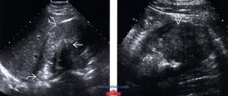
Ultrasonography
Echography allows you to quickly, painlessly, without trauma to tissues and with high reliability detect any of the tumor-like neoplasms. Main criteria for ultrasound diagnostics:
If an ultrasound scan reveals an isoechoic structure of the tumor, then it may be a benign neoplasm, which is almost no different from normal renal tissue. If the doctor identifies a hypoechoic formation, then the presence of a cyst can be assumed. Hyperechogenicity is characteristic of solid tumors without fluid inclusions. In addition to the hypoechoic option, cysts and tumors with cavities may have an anechoic appearance on ultrasound scanning.
A significant disadvantage of ultrasound is the likelihood of not noticing small neoplasms and difficulties during examination against the background of obesity or intestines distended by gases. In addition, you need to take into account the accuracy of ultrasound diagnostics, which is no more than 70%.
X-ray examination
The most informative are x-ray techniques, in which a special contrast agent is injected into the urinary tract and subsequent images are taken. These include:
If a hypoechoic renal formation is detected on ultrasound, the diagnosis must be continued: a safe and non-traumatic method of echography reveals a tumor, but does not allow making a correct diagnosis. X-ray techniques help confirm the assumption, clarify the location of the large tumor in the right or left kidney and make a decision on treatment tactics.
Source: https://pochkimed.ru/kista-pochki/obemnye-obrazovaniya-na-pochkax.html
Differentiation of neoplasms by ultrasound examination
Benign kidney tumors. Such neoplasms look like round echogenic space-occupying formations, usually small in size.
Malignant kidney tumors. A common sign indicating their presence is the detection of space-occupying formations with high echogenicity. Unlike cysts, they do not have a capsule, and their edges are always uneven. Sometimes it is possible to visualize a hypoechoic pseudocapsule formed by renal parenchyma compressed around the tumor. In some cases (10–20% of the total), calcifications are detected in neoplasms (in the form of extremely echogenic central or peripheral areas). Sometimes the tumor has necrotic areas or hemorrhages. They are visualized as hypoechoic areas on a hyperechoic background. As the tumor grows, its contours on ultrasound extend beyond the contour of the kidney.
Metastases. These malignant neoplasms are usually found in the cortex. Metastases can be found in one or both kidneys. Such tumors have the appearance of hypoechoic space-occupying formations of various sizes and shapes.
Diagnosis and treatment
Hypoechoic formations
If a congenital pathology was hidden under a hypoechoic formation, the doctor decides to carefully monitor the patient. When the problem threatens the normal functioning of the organ, surgical elimination of the anomaly is performed using kidney plastic surgery.
If the patient has suffered an injury and a hematoma has formed in the kidney cavity, it does not require therapy, since it soon goes away on its own. Serious measures are taken when blood accumulation negatively affects the kidneys.
Medicines are used to treat suppuration in the kidneys. Surgery is required only in extreme cases.
And cysts do not require radical treatment. Only a doctor can make a decision on how to treat a patient, based on the results of tests and studies performed.
Hypoechoic
In case of congenital pathology, the patient is under close medical supervision from the first days of life. If the problem begins to interfere with the functioning of the organ, then a decision is made to surgically remove it.
Dark spots also indicate hematomas that could be caused by injury; they do not require intervention, since they resolve on their own after a while. The operation is also performed in case of purulent accumulations and the risk of infection spreading to neighboring tissues and organs.
Drug therapy is relevant for cysts and benign tumors; such treatment will help prevent their further growth.
In some cases, cysts cause complications in the form of a secondary infection; with multiple cysts, atrophy of the paired organ is possible.
The key point in choosing treatment tactics is the size of the tumor. In the absence of symptoms and the small size of the tumor, you can limit yourself to dynamic observation. If the tumor does not grow and does not cause negative consequences, then it is usually left alone.
When a tumor that impairs kidney function is actively growing, the main treatment method is surgery. In this case, it is also important what size the tumor is, but it is also necessary to take into account its nature.
For a benign tumor, as a rule, kidney resection is performed, removing only the tumor itself. In cases with malignant tumors, this is not enough, and they resort to surgery to remove the affected kidney.
A malignant kidney tumor, the consequences of uncontrolled development of which are extremely sad, is successfully treated. With timely diagnosis, the patient can count on a positive outcome in 90% of cases. Treatment methods depend on the specific situation. Most often, surgery is the main cure.
In this case, resection or complete removal of the diseased organ can be performed. When deciding on the nature of surgical treatment, the doctor is guided by the principle “do no harm.” After removal of a malignant kidney tumor, the struggle for health does not end. For complete cure, drug and radiation therapy are used.
Like any disease, benign or malignant formations directly depend on the individual characteristics of a person’s health. Different individuals will experience kidney tumors, symptoms, and treatment differently.
How long do patients with this diagnosis live? Again, it all comes down to specific data. Benign tumors usually do not interfere with living a full and long life.
Malignant tumors without metastases are cured in 90% of cases.
Echogenic focal formations
Echogenic tumors are treated surgically.
The choice of treatment method directly depends on the type of formation and its size. When the patient has no symptoms and the formations are small, constant medical supervision is necessary.
If there is no growth and nothing interferes with a person’s life, therapy is not applied. Changes that lead to kidney dysfunction and active growth of the formation are considered signals for surgical intervention.
Echo-dense benign lesions are removed surgically, and doctors are usually able to save the kidney. Malignant tumors require radical therapy, so complete removal of the damaged kidney is performed.
In addition to surgical treatment, chemotherapy and radiation are used. They are necessary in the presence of cancerous tumors. Most often they act as an auxiliary therapy to suppress the development of malignant tumors. Used before and after surgery to remove a tumor. They can be the basis of therapy if it is impossible to excise the tumor.
Echogenic
After the examination, the doctor decides which method will be used to treat the echogenic focal formation. If there are no symptoms or dysfunction, then regular monitoring is sufficient.
Echodense benign tumors are excised in a minimally invasive manner, while maintaining the integrity of the organ. It is much more difficult to treat a malignant mass formation of the right and left kidney; in most cases, a radical method of organ removal is used.
In addition, treatment includes a course of chemotherapy and radiation, which will prevent the formation of metastases.
Objectives of ultrasound examination
The main purposes of an ultrasound examination for suspected kidney tumors are:
- identification of formation and clarification of its localization;
- determining the size of the tumor and the level of its penetration into the kidney tissue;
- monitoring tumor regression in case of conservative treatment;
- postoperative monitoring to exclude relapses in case of surgical resection of a tumor in the kidney;
- visual control during puncture biopsy of a tumor;
- detection of kidney metastases in other types of malignant tumors (melanoma, lung cancer, breast cancer, etc.), as well as determination of their prevalence and quantity.
Types of tumor neoplasms
Simple kidney cysts are benign tumors that exhibit typical features at the time of diagnosis. Simple cysts cannot transform into a malignant tumor and, as a rule, do not require further treatment.
Complex cysts are not always benign and may contain cancer cells. When diagnosing complex cysts, an operation is required to organize a histological examination.
Another type of tumor neoplasm is a solid tumor or, in other words, a parenchymal tumor that does not contain fluid. Such neoplasms can be benign, but are more often malignant.
Preparing for an ultrasound
Ultrasound examination of the kidneys is a procedure that requires minimal preparation. The main recommendations for patients are as follows:
- For three days before the kidney examination, you should follow a diet: food should be well digestible and light. Foods that cause bloating or increased gas formation (brown bread, sauerkraut, carbonated drinks, fruit juices, raw fruits, whole milk, etc.) must be excluded;
- the last meal should be no later than 8 hours before the kidney examination;
- The procedure requires a full bladder, so it is recommended to drink several glasses of still water an hour before the ultrasound;
- Before an ultrasound examination of the kidneys, you should not take diuretics;
- 4 hours before the procedure, you must refrain from smoking: changes in the vascular system caused by nicotine can blur the picture and interfere with tumor detection and accurate diagnosis;
- If there are constant problems with the intestines that are not corrected by diet, the patient is recommended to take medications that reduce gas formation before the examination.