Warehouse relocation to Europe. We sell hepatitis C drugs in Russia at the purchase price - warehouse liquidation Go to website
Disease of the two tubular organs that help urine leave the kidneys and travel to the bladder is called dilatation of the ureter. Due to problems with urine transport, a person experiences dangerous disturbances in the functioning of the urinary system. This is a fairly serious illness.
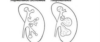
What is the dilation of the ureter called? Megaureter is an acquired or congenital lesion that provokes problems with the functioning of the kidneys, and with bilateral inflammation in a person leads to kidney failure. When the tubular ureters dilate, the outflow of urine does not occur, which can lead to an inflammatory process in the kidneys and problems with the circulatory system.
Expansion of the tubular process
The walls of the ureter have a three-layer structure, which helps urine move slowly to the bladder. The outer muscular layer includes nerve and collagen fibers that help move urine up to five contractions per minute. As the size of the ureter increases, contractile force begins to decrease, movement of urine becomes difficult, and the patient's intrarenal pressure increases. Prolonged stagnation of urine provokes the onset of infection, which only worsens the person’s condition. If treatment of the lesion is not started in a timely manner, then problems with the functioning of the kidneys will soon arise.
Often, infectious processes in the ureter only increase the expansion of the organ. Enlargement of the ureter and renal pelvis is diagnosed using ultrasound examination of the fetus.
If after birth the child does not have a megaureter, then expansion of the tubular organs will not occur in the future. In a normal state, the diameter of the ureter does not exceed 5 mm; if an expansion was detected during the examination, the doctor prescribes a more extensive examination of other internal organs.
In adolescents with this form of damage, the following symptoms most often present: the presence of blood in the urine, incontinence, complaints of persistent pain in the abdomen and lower back, and the formation of stones in the urinary organs.
Causes of ureteral tumors
The urothelium in the ureter is highly sensitive to the chemical composition of urine. Among the factors that increase the risk of developing a ureteral tumor, as well as other neoplasms, smoking is the leading one. Statistics indicate that 70% of men and 40% of women diagnosed with ureteral tumors are long-term smokers.
Urothelial cancer is also more likely to appear in people taking large amounts of analgesics or diuretics. Thus, when treating arterial hypertension, the risk of developing ureteral tumors increases due to the use of diuretic drugs. People whose professional activities take place in oil refining, plastics and plastics production enterprises are also at increased risk.
Doctors say that chronic pyelonephritis, stones in the urinary tract and ureteral injuries slightly increase the risk of neoplasms. Genetic predisposition, Lynch syndrome and malignant tumors of the pelvic organs (uterus, ovaries, intestines, prostate, etc.) also have an impact.
Main types of damage
Experts identify the following forms of damage:
- The primary type is a congenital disease. It occurs when there is a lack of coordination between the muscle and connective tissues of the ureter. In this case, the organ lacks the strength to move urine normally through the tubes. Megaureter can appear in a child during its development in the womb. Most often, the congenital form of the disease appears in boys.
- Secondary type - occurs with high pressure in the bladder. Most often, this condition is caused by regular nervous disorders, emotional outbursts, or chronic cystitis. Most diseases, after a comprehensive diagnosis and effective treatment, resolve in the first years of a newborn’s life.
Diagnostics
Congenital pathology of ureteral dilatation is detected long before the baby is born.
Three weeks after birth, the child is sent for a comprehensive urological examination to establish the exact causes and determine the stage of development of the pathology, and then prescribe effective treatment.
Urography
In other cases, such a pathology is diagnosed when a patient presents with certain complaints.
In most cases, an infection of the urinary system is suspected, since many of the symptoms are the same.
Initially, the patient is sent for an ultrasound examination, which allows one to determine the presence of stones in the ureter and inflammatory diseases.
During an ultrasound examination, the doctor determines the location of the internal organs, thus detecting possible prolapse of the kidney and the subsequent bending of the ureter.
Ureteral examination
Options for promising tools for conducting urological studies also include urography, which involves the use of a contrast agent.
During urography, images are taken that show the movement of this radiopaque component along the urinary tract.
Such a study makes it possible to detect in what place there is an obstruction, where the stones are located, and what their sizes are. The doctor can evaluate the size and pathological changes of the diverticulum.
Possible kinks of the ureter and prolapse of the kidney are also visible on urographic images.
Urethrography also allows you to observe changes in the body.
This type of diagnostic study also involves the introduction of a contrast agent, but it is only administered against the flow of urine. A radiopaque contrast agent is also required when performing cystography.
Laboratory diagnostics
Absolutely accurate information about the corresponding disorders can be obtained during cystoscopy, when a small sensor with a video camera installed in it is inserted into the bladder, allowing the doctor to observe all changes from the inside.
In the same way, you can observe changes in the urethra by performing urethroscopy.
Laboratory research methods are useful, since the results of blood and urine tests can indicate an inflammatory process, as well as urolithiasis.
With conservative treatment, diagnostic studies every six months are considered mandatory in order to be able to monitor the effectiveness of treatment and, if necessary, make the necessary adjustments.
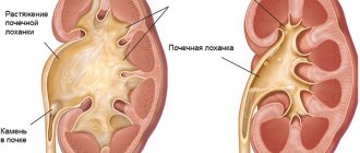
Reasons for the appearance of the extension
There are several reasons for dilation of the ureter. The main ones include high ureteral pressure and problems with urine outflow. There were situations when, after normalization of pressure, the ureter continued to remain dilated.
Often the patient is diagnosed with congenital insufficiency of the muscles of the tubular organ. In this case, the ureter is greatly weakened and loses its contractile ability to move urine to the bladder. Another reason for this condition is the narrowing of the tubes at the site of their attachment to the bladder.
The main reasons for an enlarged ureter:
- increased pressure inside the tubular organ, which provokes expansion of the ureter and kidney, as well as problems with the outflow of urine;
- weakness of the membranes in which the muscles are located;
- problems with the formation and development of nerve endings;
- urine backs up into the pelvis due to narrowing of the ureter.
Organ laying in intrauterine development
The development of the urinary tube occurs during pregnancy. Moreover, these organs are able to stretch due to the fact that they have longitudinal folds in the mucosa. Under the mucous membrane, glands are located similar in structure to the prostate. A newborn's ureters may develop long after birth. The ureter develops throughout pregnancy.
To see where the ureter originates and how it can be located, you need to buy a purchased anatomy textbook, which has visual drawings.
Characteristic symptoms of damage
There are a large number of reasons for dilation of the ureter in a child. In the absence of a primary lesion, megaureter occurs in a latent form. In this case, the person does not have pronounced symptoms of the disease, he does not suspect anything about his condition. In another case, a person may feel unpleasant pain in the abdomen and lower back, and tumor-like formations can be easily felt or blood may be noticed in the urine coming out. When the acute form of the lesion develops, a person is diagnosed with a high number of leukocytes in urine, nausea, vomiting and an increase in body temperature.
The most unpleasant symptoms of this disease appear at stages 2 and 3 of its development, it is at this time that a person develops such a dangerous complication as chronic kidney failure or pyelonephritis.
When the processes expand or are double affected, the child often experiences double urination. This condition occurs because after the first emptying of the bladder, it again fills with urine from the dilated organs and the need to urinate reappears.
The second time, urine comes out in large quantities, with an unpleasant odor and a cloudy sediment. Due to the fact that the weakened body of a newborn child is very susceptible to various infections, problems with physical development or skeletal abnormalities may begin. Most often, when the ureter dilates, newborns lose their appetite, their skin turns pale, thirst and urinary incontinence appear.
Prevention and consequences
It is impossible to prevent the occurrence of congenital pathology, but there is a chance to cure it in a timely manner. If the ureterocele is not treated, the prognosis for life is extremely unfavorable; the pathology can lead to uremia and, accordingly, death.
The secondary type of ureterocele, which appears against the background of urolithiasis, can be prevented by regularly conducting urine tests and getting rid of kidney stones in a timely manner. In the video about the causes, symptoms, diagnosis and treatment of ureterocele:
Degree of development of the problem
After carrying out diagnostic measures, the attending specialist assesses the condition of the kidneys and prescribes effective treatment. Doctors distinguish three main stages of the development of the disease:
- Easy stage. There is a moderate dilation of the lower ureter. This condition often goes away on its own without outside influence.
- Average degree of damage. The diameter of the ureter is greatly expanded. With timely and high-quality treatment, you can easily get rid of the problem.
- Severe degree. Megaureter can cause kidney problems. In this case, after the examination, the doctor will definitely prescribe surgical intervention to the patient.
Therapeutic measures
This pathology can be treated with the help of medications, and it is also possible to use folk recipes. It is possible to use surgical intervention, but only in cases where hydrocalycosis is combined with urolithiasis or abnormalities of the genitourinary system.
Use of medications
Medicines for the treatment of enlarged renal calyces are used to eliminate the symptoms of the pathology. In the presence of a pronounced inflammatory process, non-steroidal anti-inflammatory drugs are used. Among them are: Dicloberl, Diclak, Voltaren, Indomethacin.
Folk recipe
Traditional methods are most often used for preventive purposes, very rarely for treatment. If there is an expansion in the pelvicalyceal area, infusions and decoctions from medicinal plant materials are used to restore it, namely:
- A decoction of lingonberry leaves: to prepare it you will need 2 tbsp. l. leaves and 200 ml of fresh boiling water.
- Decoction of bearberry herb: 1 tbsp. l. raw materials must be mixed with 200 ml of boiling water.
On sale you can find medicines that contain medicinal plants: Phytolysin, Trinephron, Canephron, Uronephron, Cyston.
Before using traditional recipes, you should consult your doctor.
Surgical intervention
Surgical treatment is used if there are problems associated with the outflow of urine. It is necessary to restore the patency of the urethra; for this, shock wave lithotripsy can be used in the presence of small stones. To avoid such health problems, you should listen carefully to your body, undergo the necessary examinations in a timely manner, and also lead a healthy lifestyle. By following these simple rules, kidney problems can be avoided.
How does it proceed in a small child?
With the advent of modern equipment in clinics, diagnostics make it possible to determine the presence of megaureter and anomalies of the genitourinary system even at the stage of intrauterine development. Early diagnosis and identification of megauretera may lead to unnecessary surgery. This can be explained by the fact that in most cases the process of expansion of the child’s ureter stops, and the size of the ureter is restored within several months of the baby’s life.
At this age, the doctor should regularly monitor the baby’s condition and prescribe a urine test and ultrasound examination. Timely detection of the lesion will help to avoid complications and exacerbation of the disease, as well as prevent unnecessary surgery for the child. The baby's organs continue to actively develop for some time; for this reason, in the first few months of life, the doctor cannot always accurately determine the state of the urinary system and kidney function.
When carrying out diagnostic measures, the attending physician must be especially attentive and careful, since the risk of error in this case is very high. Elimination of the lesion is possible only with timely identification and administration of effective and correct treatment. It often happens that the dilation of the ureter in a newborn goes away on its own. Very often no outside intervention is required. In an adult, in the acute stage of dilatation of the left ureter, a mandatory operation is performed.
Diffuse changes
If the ultrasound report says that you have diffuse changes in the kidney parenchyma, you should not take this as a final diagnosis. The term diffuse in medicine means numerous and widespread tissue changes in adults and children. Diffuse changes in the parenchyma indicate that a person needs additional examination in order to determine the exact causes of physiological abnormalities. Most often, diffuse changes in the parenchyma are observed if the size of the kidney changes. In acute diffuse type disorders, the size of the kidneys of children and adults increases. In chronic diffuse pathology, the parenchyma is thinned.
If diffuse disorders are moderate, this may indicate:
- about congenital renal anomalies in children;
- about age-related changes that kidney tissue has undergone. In this case, diffuse changes may be normal;
- about previous infections;
- about chronic renal pathologies.
That is, any changes that are unusual for the physiological norm of renal tissue are considered diffuse. These are increased echogenicity, thickening or thinning of the kidney tissue, the presence of fluid, etc. The most striking examples of diffuse parenchymal disorders are a cyst of parenchymal tissue or its thinning.
Parenchyma cyst
It can form in both the left and right kidney. It can be congenital or acquired. If a congenital cyst of parenchymal tissue is detected in children, then the formation of an acquired cyst is typical for people over 50 years of age.
A parenchymal tissue cyst is a more serious disease than a cyst located in another area of the right or left kidney. Representing a limited cavity filled with fluid or serous secretion, the cyst compresses the tissue, disrupting the process of formation and excretion of urine. If the cyst in the left or right kidney is solitary, does not grow and does not affect the functioning of the organ in any way, it is enough to monitor it. There is no treatment for such a cyst.
If multiple cysts form in the parenchymal tissue, doctors decide on surgical removal. There is no fundamental difference in the location of the cyst. It requires the same treatment tactics in both the left and right kidneys.
Thinning of the parenchyma
Diffuse changes indicating thinning of the parenchyma indicate not only the patient’s advanced age. If an elderly person is being examined, the doctor will most likely associate the thinning with age-related changes. The symptom also occurs in young people. Here, the main reason for thinning tissue is due to past diseases that the person did not treat or treated incorrectly.
The thinned kidney parenchyma is unable to perform its usual functions in full, therefore, if a person does nothing and continues to be treated, a chronic disease occurs. And he joins the ranks of patients of nephrologists and urologists.
Ultrasound examination (ultrasound, sonography) is an effective method for diagnosing various diseases and pathologies of the urinary system. This study guarantees an accurate and reliable diagnosis, and also allows you to clearly visualize the area under study. is aimed at assessing the organization of the structure, identifying pathologies and disorders in volume, shape and contour. Sonography reveals abnormal position of the organ, as well as disorders in the urinary system.
Interpretation of an ultrasound examination provides the most complete picture of the condition of these organs, and can also confirm or refute the doctor’s preliminary diagnosis. In order to correctly decipher ultrasound results, it is important to know the criteria and norms by which the general state of the environment is determined. The main criteria when interpreting sonography results are the shape and size of the kidneys, as well as the size of the parenchyma (the upper layer of kidney tissue, which performs the task of balancing the internal environment).
Relative readings
A relative indication is a disease that is at stage 1 of development and does not pose a particular danger to a person’s life, but significantly affects his condition. For example, it brings fatigue, headaches, reduces performance, and provokes nausea.
The ureter in this case is minimally dilated. The patient has time during which he can take a course of effective medications, which will help prevent even further expansion of the ureter. This will help prepare the patient's body for surgery.
Location
The right kidney is located lower than the left. Thanks to ligaments and muscles, internal organs are able to move slightly during breathing and movement, otherwise any sudden tilt or turn could cause injury.
But the physiological displacement of the kidneys should not exceed two to three centimeters! If the organ is constantly located lower than necessary, or moves strongly inside the body, the ultrasound report indicates its atypical location - dystopia or right- (left-sided) nephroptosis -.
Carrying out electronic urography
An effective diagnostic method is urography, which does not cause discomfort in the patient and helps to obtain accurate information about the condition and functioning of organs, the location of the lesion, the anatomical structure of the ureters, as well as dilated areas.
Contraindications to the procedure are severe kidney disease, nervous breakdowns, problems with concentration abilities and other processes in which, due to the accumulation of large amounts of urine in the blood, it is impossible to accurately determine the clinical picture of the disease.
Women carrying a child are prohibited from performing the procedure or only for special indications. For example, if there is a suspicion of malignant or benign formations in the ureter.
Layers of the walls of the ureter
- Adventitium. This is fibrous connective tissue with admixtures of elastic fiber. Nerve plexuses, veins of the ureter and arteries pass through its thickness. The renal fascia descends and surrounds all parts of the ureteric tube, but it is poorly developed.
- The muscular layer has three layers at its base:
- Internal longitudinal,
- Medium circular,
- External longitudinal.
The last layer has separate bundles, their increase is observed at the bottom of the organ.
- The mucous membrane contains longitudinal folds; from the inside, the organ resembles a stellate structure. In the depths lie the tubular-alveolar glands.
The topography of the ureters differs significantly on the right and left sides. The position of the ureter on the right at the beginning is located behind the intestine. The distal part of the urinary tube crosses the base of the suspensory apparatus of the ileum of the small intestine. When passing into the intramural section of the ureter, the iliac arteries appear in front.
On the left side, the urinary tube may be located behind the bend of the intestine; in the small pelvis, a crossover occurs between the vessels. The ureter in men along its length crosses with the testicular artery, and in women with the ovarian artery.
Inside the pelvis, the topography is the same on both sides, but differs depending on gender.
In men, before entering the bladder, the vas deferens is attached, which runs along the inside.
In women, the urinary tube penetrates the periuterine tissue.
The anatomy and structure of the ureters are the same in both sexes.
Vaccine cystourethrography
Another method for diagnosing ureteral enlargement is cystourethrography, which helps to examine the presence of enlargement and reflux (reflux of fluid from the bladder) on X-ray images.
In children who cannot empty their bladder on their own, the procedure is performed under anesthesia. Urine is removed from the bladder by pressing on it with your hands.
It is prohibited to conduct such an examination in the following cases: acute form of cystitis, urethritis, as well as increased sensitivity to contrast agents used during the procedure.
Treatment
The only way to treat ureterocele is a surgical solution to the problem. If the size of the formation is small and the functioning of the kidneys is sufficient to purify the blood, the operation is performed by endoscopic dissection, that is, without external incisions, but with internal access through the urethra.
The surgeon removes the area affected by the cystic formation and creates a new opening, allowing filtered urine to freely leave the body. It is important to form a new valve (anti-reflux), which will prevent urine from being thrown up the filtration system.
If the ureterocele is large and the walls of the bladder are affected, then there are indications for open access surgery.
At an advanced stage, the question may arise not only of reconstruction of the urinary tract, but also of nephrectomy, complete or fragmentary.
Basic information about ureterocele
Therapy
The most common and effective treatment for ureteral enlargement is reimplantation. In this procedure, a new anastomosis is created between the ureter and bladder.
Surgeries can be minimally invasive and open. The first operation lasts for 125 minutes and requires hospitalization of the patient for up to a week. The second type is an operation that lasts the same amount of time but requires hospitalization for 14 to 16 days.
After the operation, the child may experience the following complications: acute pyelonephritis, colic in the kidneys, wound bleeding and stent migration in the lumen of the upper bladder.
Structure
Genitourinary system
The ureter belongs to the category of paired organs. It is the link between the renal pelvis and the bladder.
Its functions include ensuring the normal (natural) outflow of urine from the kidneys. Externally, it is very similar to cylindrical tubes, which are slightly flattened in diameter.
Their length depends directly on the height of the kidney organs; most often it ranges from 24 to 35 cm.
In newborns, the length of the ureter is only 7 cm; as the child grows, the length of the organ increases.
At the age of two it already reaches 14 cm, at the age of three – 21 cm, and by the age of eighteen the growth of the ureter stops and the length is finally fixed.
The ureter consists of three parts:
- abdominal;
- pelvic;
- intramural.
Organ functions
In three places, narrowings are observed, predetermined by its physiological structure. They are located in the area of the connection with the kidneys, bladder and in the area of \u200b\u200bthe intersection of the common glomerular vessels.
The mucosa, adventitia and muscular layers make up the three-layer walls of the ureter. The mucous membrane differs from the rest in that it forms folds throughout the ureter.
The muscular one is also noted to have a three-layer structure; muscle bundles are located on each section of the ureter in completely different ways.
In the upper part there is a longitudinal and circular arrangement, in the abdominal part there is a spiral shape, and in the pelvic part there are twisted shapes. The outer layer is equipped only with horizontal muscle fibers.
The functioning of the ureter depends to a large extent on the functioning of the kidneys, renal pelvis, and bladder.
The coordinated work of the entire urinary system ensures a normal urination process. Urine moves freely through the ureter due to peristaltic contractions.
Recovery after surgery
The patient's recovery continues for quite a long time. Evaluation of the results of the operation will be long-term. Doctors will judge the quality and effectiveness of the surgical intervention only after several years have passed after the operation itself.
It is important to remember that there is nothing scary or dangerous about the operation. You should not be afraid of it and put it off for some time. According to statistics, success with surgical intervention is observed in 90% of patients. The sooner treatment for the disease is started, the higher the chance of getting a positive result.
When treating ureteral enlargement, special attention should be paid to its severity. The severity will be determined after receiving the results of clinical diagnostics and a multifactorial assessment by a physician. In normal cases of ureteral dilatation, the patient's condition will recover within a few weeks after the operation. In more complex cases, the patient will need 10-15 weeks for rehabilitation.
To avoid dilation of the ureter, it is important to monitor the condition of the body and promptly treat diseases of the genitourinary system. It is also important to stop taking large amounts of fluid if the urinary system does not have time to remove it from the body in time.
Forms
When studying etiological factors, three main forms of ureteral dilatation are determined: obstructive, refluxing, and bladder-dependent.
Detection of pathology
Obstructive expansion is characterized by all kinds of anatomical obstacles. Most often, obstructions are located in the lower part of the ureter, where it connects to the bladder.
Due to the fact that urine cannot flow freely into the urinary organ, it acts on the walls of the ureter, exerting extreme pressure. This is what provokes its further expansion.
If such a deviation is not diagnosed and, accordingly, the correct treatment is not prescribed, this will lead to serious kidney damage.
Unfortunately, in most cases, obstruction cannot be treated conservatively, so doctors resort to surgical intervention.
Refluxing dilation is caused by vesicoureteral reflux, when urine from the bladder flows through the ureter back to the kidney.
If no pathological changes are observed in the urinary system, then the urine does not tend to move backward.
Pathologies of the genitourinary system
Vesicoureteral reflux can provoke not only dilation of the ureter itself, but also an enlargement of the bladder itself, due to the fact that emptying does not occur, and urine circulates between these two organs through the vesicoureteral junction.
This deviation is classified as severe. More often diagnosed in newborn boys. During the first year of life, in some cases positive changes are noted.
If such encouraging prospects are still not observed, doctors have to perform surgical operations, during which actions are aimed at narrowing the diameter of the ureter or its reimplantation.
If neither obstruction nor reflux are responsible for the fact that the ureter suddenly turns out to be dilated, then non-refluxing non-obstructive dilation is diagnosed, which quite often can resolve completely without medical intervention.
But even this form of disorder must be under constant medical supervision in order to exclude other anomalies.
Refluxing and obstructive dilatation is the most dangerous form, since the ureter increases excessively in size, which subsequently leads to complete obstruction.
Stages
The initial stage is practically unnoticeable, the disorders are not severe and are discovered accidentally during ultrasound examinations.
The second stage is accompanied by an increase in the renal collecting system and difficulty in excreting urine.
Third stage: transition of the disease to the chronic stage, deterioration of health.
In the final stages, fluid accumulating in the body provokes swelling, changes in the color of urine, manifesting itself in difficult-to-bear high-intensity pain in the lower back and abdomen.
Symptoms and signs of ureteral abnormalities
It should be noted that there are no specific clinical manifestations associated with ureteral anomalies.
In most cases, the anomaly is detected incidentally during routine prenatal ultrasound examination. The pathology must be eliminated before any symptoms or infection occurs. Some patients have frequently recurring inflammation of the urinary tract, abdominal pain and blood in the urine.
In children, the symptoms are similar, so in case of recurrent abdominal pain and changes in the general urine test, a urological examination should be performed.
Anomalies in the development of the ureters in adults can be detected during diagnostic workup for hypertension, proteinuria, or even renal failure, which is often caused by bilateral lesions. About 50% of women with ectopia suffer from persistent urinary incontinence or complain of watery vaginal discharge.
Experts recommend examining boys with recurrent epididymitis before puberty for abnormalities in the structure of the genitourinary system. In men with frequent complaints typical of chronic prostatitis and pain during ejaculation, in some cases, ectopic ureters are detected, but urinary incontinence is atypical.