Doctors often observe an anomaly in which the orifice of the ureter is located atypically. The pathology is called ectopia of the ureteral orifices. Intravesical or extravesical location is observed. In the first case, the development of pyelonephritis is possible, and with an extravesical location, a person leaks urine after urinating. Ectopia requires timely diagnosis using ultrasound and other procedures. Ectopia has several types, each of which requires a special treatment.
Structure, location and functions of the bladder
The bladder is an organ for the accumulation and temporary storage of urine, which is excreted at regular intervals through the urethra. Its main role is to store and release urine into the urethra.
Its volume ranges from 500-800 ml and varies depending on the individual characteristics of the person.
The shape and position in relation to other organs and tissues depend on the degree of its fullness with urine and the gender of the patient.
- Location in women. When the bladder is empty, in women it lies in the pelvic cavity. It is separated from the rectum by the vagina and uterus. When filled with urine, it changes shape and, in cases of severe stretching, reaches the level of the navel. The wide part (bottom) is directed towards the vagina, and the narrow part goes into the urethra. The anterior surface of the uterus is adjacent to the anterior surface in women.
- Location in men. An empty bladder is found in the pelvic cavity; in men, it is separated from the rectum by the seminal vesicles and a section of the vas deferens. The bottom or wide part of the bladder in men faces the rectum. The lower part is fused with the prostate gland, and the upper part remains mobile. In men, intestinal loops are adjacent to the upper surface.
- Location in newborns. The ureter is different in newborns. In newborn children, the bladder is located much higher than in an adult. Immediately after birth, it begins to gradually descend downwards, and by six months of the child’s life it is determined at the level of the upper edge of the fusion of the pubic bones.
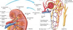
Internal structure and blood supply
The bladder consists of three layers: muscular, mucous and serous.
The muscle layer includes three types of fibers that stretch and contract as needed. Where the bladder passes into the urethra, the muscle layer forms a sphincter (sphincter), which contracts involuntarily (regardless of the patient’s wishes).
The mucous membrane is pink and covered with folds. It contains small mucous glands and lymphatic follicles.
This organ is supplied with blood from the superior and inferior cystic arteries, which belong to the basin of the large iliac arteries. Lymph flows to nearby inguinal lymph nodes.
Every few minutes, the openings of the ureters involuntarily open into the cavity of the bladder and throw out a small amount of urine. Having reached a certain position and size, it performs its functions and expels urine into the urethra, from where it is discharged.
Possible anomalies
Common developmental anomalies include:
- diverticulum It is a bag-like protrusion of the wall. There are single and multiple options. Urine stagnates in such diverticula, which contributes to the development of cystitis;
- non-closure and fistulas of the duct, which connects the bladder through the umbilical cord with amniotic fluid during intrauterine development;
- its absence or underdevelopment is rare. A vice incompatible with life;
- doubling. In the cavity of the bladder there is a septum that divides it into two parts; it prevents the bladder from fully performing its functions. Treatment is surgical.
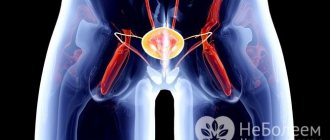
Difference between ectopic ureteric orifices in men and women
Doctors have noticed that this problem is diagnosed 3-4 times more often in the fairer sex. If a man encounters a deviation, usually such an anomaly is independent (primary). When the female body is affected, ectopia and a double ureter are diagnosed simultaneously. Leakage of urine is typical for women, but this symptom is not observed in men, so they do not always seek help. This fact is explained by the different structure of the urinary system. In the female body, the opening of the internal organ is located below the urethral sphincter, so urine flows out. In most men, the mouth of the internal organ flows into the prostate gland, and ectopia with outlet into the ejaculatory duct is extremely rare. In representatives of the fairer sex, the pathological process occurs in the urethra, and in a smaller part, the mouth of the ureter is located in the uterus.
Structure, location and tasks of the ureter
The ureter originates from the renal pelvis. It is a hollow tube with a diameter of about 4-6 mm and a length of about 300 mm. Its task is to deliver urine from the kidney to the bladder and prevent the reverse flow of urine.
From the renal pelvis it goes down behind the peritoneum to the bottom of the wall of the bladder, through the wall of which it penetrates in an oblique direction.
The diameter of the ureter depends on the site, since there are anatomical narrowings:
- immediately after the transition of the pelvis to the ureter;
- on the border between its transition into the pelvic cavity;
- throughout the pelvic cavity;
- at the point of entry into the bladder (narrows by 2-3 mm).
The ureter in women is 20-25 mm shorter than in men. It runs along the free edge of the ovary and passes at the base of the broad ligament of the uterus. Penetrates into the space between the vagina and bladder in an oblique direction. At the junction it forms a muscular sphincter.
The wall of the ureter consists of three layers: connective tissue, muscle and mucous.
At the exit of the pelvis there is a sphincter that prevents urine from flowing back into the kidney. And the sphincter in the bladder area helps expel urine.
On x-ray, the ureter appears as a long, narrow shadow that extends from the kidney to the bladder. Its size and length differ among patients. The contours are smooth and clear. A number of physiological curvatures and narrowings are noted on x-rays.
Postoperative period and complications
After surgery, the patient is advised to undergo repeated radiography, ultrasound diagnostics and voiding cystourethrography. These studies are important to rule out the presence of vesicoureteral reflux and ureteral obstruction. If the patient had hydronephrosis, then after surgery it can persist for several years. It is recommended to control arterial hypertension and proteinuria in patients who have had renal dysplasia.
If the deviation is not detected in a timely manner, then various complications are possible that are difficult or impossible to cure. After surgery, infection of the urinary tract is possible. Hematuria often persists, which is not normal. At the late postoperative stage, complications such as recurrence of vesicoureteral reflux or ureteral obstruction are possible. In rare cases, a patient may experience devascularization of the lower part of the kidney after partial nephrectomy.
Questions
Question: How much urine does the bladder hold and hold?
Answer: According to some reports, up to 1-1.5 liters, but the capacity of the bladder is an individual indicator.
Question: How many ureters are normal?
Answer: A healthy person has two ureters - one from each kidney. With developmental anomalies, there are double or triple ureters or the absence of a ureter on one side.
Question: What is the diameter of the ureter in mm and how long is it?
Answer: The average diameter of the ureter in a healthy person is within 5-6 mm, the length depends on gender and individual structure, on average it is 200-300 mm.
The ureters are paired organs that connect the renal pelvis to the bladder. The diameter of the ureter depends on the location of the latter. This organ serves as a kind of connecting link of the urinary system: urine flows through it from the kidneys to the bladder, from where it will be released from the body to the outside.
Functions in the human body
The urinary ducts perform the function of transporting urine from the renal collecting system to the urinary tract. The paired organ has motor function. Its middle layer, the smooth muscle layer, stimulates the wave-like contraction of the canal. Peristalsis is provided by a pacemaker (pacemaker), which is located in the pelvis zone of the duct.
The rhythm frequency and cyclicity of contractions depend on several factors:
- volume of urine excreted by the kidneys,
- human physical activity,
- the presence of pathologies,
- body position,
- correct functioning of the nervous system.
The urge to contract the walls occurs when a sufficient amount of calcium accumulates in the muscle layer.
Anatomy and topography
The length of the ureter generally varies between 30-35 cm, which is typical for a mature person. This organ is hollow and looks most like an elastic tube. It starts from the narrowest part of the kidney, in the area of its gate, which also includes blood vessels. The ureters are located in the retroperitoneal space, that is, they are not included in the number of abdominal organs.
The body has departments, called areas, in which its progress is monitored. The first part, the initial one, is called the abdominal part. It is located on the outer surface of the psoas major muscle, which is one of the walls of the retroperitoneal space. Anterior to the right ureter is the downward part of the duodenum (the initial part of the intestine). On the left there is a bend that corresponds to the place where the same intestine flows into the next section of the intestine - the jejunum. In front there are also blood vessels of the ovaries (in men - testicles), the peritoneum, which serves as a barrier between the abdominal cavity and the retroperitoneal space (it is called the parietal).
At the beginning of the ureter there is the first bend - it corresponds to the place of exit from the kidney. This part is its first anatomical narrowing. The second is located at the point of transition of the abdominal section to the next one, which runs mainly in the small pelvis (pelvic). At this point, the passage of the left ureter through the root of the mesentery of one of the sections of the large intestine - the sigmoid colon, is monitored, and the right - through a similar small intestinal structure. The mesentery is a piece of the parietal peritoneum, which is needed to attach any organ to the wall of the abdominal cavity.
The pelvic region on the right and left is surrounded by large blood vessels. In this part, the ureter descends into the pelvis. The diameter of the pelvic part is smaller than the abdominal part by several millimeters. Its course in the small pelvis differs between men and women. In the female sex, the organ lies behind the ovary, then it comes into contact with the uterine cervix and passes between the vagina - the initial part of the woman's internal reproductive system - and the bladder, into which it empties. In men, the ureter runs next to the vas deferens, then crosses it and goes into the bladder. The parietal part of the organ has the smallest extent (1.5-2 cm) and is located directly in the wall of the bladder next to the opening through which the ureter flows into it. At the site of entry into the bladder, a third anatomical narrowing is observed.
What is reflux or urine backflow into the kidneys in children?
Reflux is a fairly common pathology; it occurs more often in boys than in girls. If treatment is not timely, severe complications may occur in the form of hydroureteronephrosis or chronic and acute pyelonephritis. In this article we will tell you why urine backflows into the kidneys, and we will analyze the symptoms and methods of treating the disease. Reflux is a pathological condition that is accompanied by the return movement of urine through the bladder channels to the kidneys. This pathology is classified into two types:
- vesicoureteral, characterized by reflux of urine into the ureter;
- pelvic-renal, characterized by the flow of fluid from the pelvis to all parts of the kidneys.
Reflux classification
Have you been trying to cure your KIDNEYS for many years?
Head of the Institute of Nephrology: “You will be amazed at how easy it is to heal your kidneys just by taking it every day...
Read more "
Reflux is a pathological condition that is accompanied by the return movement of urine through the channels of the bladder to the kidneys.
In medicine, there are several types of reflux, which can affect not only the kidney organs, but even the reproductive function in women. Each type of pathology has its own causes of formation and symptoms. Main types of reflux:
- Gastric form - accompanied by a violation of the physiologically natural pattern of movement and digestion of products, most often the failure occurs at the last stage. This form of pathology affects the esophagus, duodenum and stomach;
- Ureteral form - accompanied by disruption of the ureter, i.e. the body receives severe intoxication. Poisoning occurs due to the fact that the liquid that should come out naturally is thrown back into the ureteral channels;
- The esophagitic form is rare and is accompanied by heartburn and unpleasant belching. This form is dangerous because it can proceed for a long time without any special complications and as a result lead to esophageal cancer;
For information! Reflux can affect the upper gastrointestinal tract, gallbladder ducts, liver, pancreas and reproductive organs. Do not ignore any symptoms and do not self-medicate. Remember the complication of reflux is oncology.
Experts also divide pathology into primary and secondary forms. The primary form is formed as a result of congenital damage to the walls of the bladder or the beginning of the ureteral canals; the disease develops in the prenatal state. The secondary form is formed as a result of improper treatment of diseases of the urinary system, most often appearing after surgery or untreated cystitis of any form.
Causes and symptoms of pathology
There are several causes of provocateurs that can cause reflux of urine into the kidneys, as well as disrupt the functioning of the ureter and bladder channels
There are several causes of provocateurs that can cause urine to reflux into the kidneys, as well as disrupt the functioning of the ureter and bladder channels:
- immature state of the channel;
- dysfunction of the bladder;
- damage to the orifice of the ureter;
- chronic form of cystitis;
- the presence of dystopia of the mouth;
- presence of closing valve dysplasia;
- blockage of the urinary canals, leading to disruption of urine flow through the urinary canals.
The symptoms of reflux are quite pronounced, so if you feel at least one of them, you should consult a doctor. Main symptoms of casting:
- attacks of severe headaches;
- constant feeling of a full stomach;
- thirst;
- swelling of the limbs;
- chills and sudden increase in body temperature;
- urine takes on a dark, red color and also becomes foamy;
- increased blood pressure;
- pain when urinating and in the lumbar region.
For information! There are passive and active reflux. With passive, urine enters the kidney regardless of the process of bowel movement; with active, reverse reflux occurs exclusively during the period of urination. The symptoms of the passive and active types of the disease are the same as those of the primary and secondary ones.
Characteristics of vesicoureteral reflux
This type of reflux has five stages that can impair the functioning of the kidneys and urinary system
This type of reflux has five stages that can impair the functioning of the kidneys and urinary system. Stages of pathology:
- the first stage is manifested in the reverse flow of urine into the pelvic part of the ureter;
- the second stage is manifested in the reverse flow of urine throughout the ureter and the renal collecting system;
- the third stage is manifested in the reverse flow of urine into the pyelocaliceal part of the kidney, which has an increased size in diameter;
- the fourth stage is formed due to the reverse flow of urine from the bladder, the pyelocaliceal system and the ureter are increased in size;
- The fifth stage occurs with a reduction in the functionality of the kidney, due to the reverse flow of urine into the kidney and from the bladder, while the kidney is damaged and depleted.
For information! During the period of reflux, the functionality of the renal system is assessed by degree and reflects a decrease in performance:
- first degree - by 30%;
- second degree - by 60%;
- third degree - more than 60%.
The period of the disease can be permanent or transient:
- A persistent disease is always present in the patient;
- A transient disease occurs against the background of exacerbation of pathologies of the urinary system (cystitis, acute prostatitis).
Urine reflux into the kidneys in children
Vesicoureteral reflux in children or VUR occurs as retrograde movement of urine from the bladder into the ureter
Vesicoureteral reflux in children, or VUR, occurs as retrograde movement of urine from the bladder into the ureter. The pathology is manifested by the fact that the reverse movement of urine towards the kidneys is disrupted and causes PMR. If the disease is not treated in a timely manner, severe chronic inflammation of the bladder may occur.
For information! According to statistics, PMR occurs in 1% of newborns. If chronic inflammation of the bladder is not eliminated in a timely manner, the child may become disabled.
Reasons why urine may be thrown into the kidneys:
- pathological impairment of the renal system;
- metabolic disorders and increased internal urine pressure;
- cystitis of any form;
- organ shrinkage;
- neurogenic dysfunction;
- damage to the orifice of the ureter.
There are several types of VUR in children:
- low pressure reflux - manifests itself at the initial stage of filling the urinary system;
- high blood pressure caused by congenital insufficiency;
- the primary form is formed as a result of an infection of the genitourinary organs;
- chronic form, manifested by a high frequency of VUR.
Symptoms of childhood PMR
Children's PMR manifests itself in a pronounced symptom complex, which manifests itself in a high temperature that is difficult to bring down
Children's PMR manifests itself in a pronounced symptom complex, which manifests itself in:
- slow physical development;
- underweight in newborns;
- high temperature, which is difficult to bring down;
- attacks of pain, colic, cutting;
- presence of bloody discharge during urination.
During the period of inflammation, the child’s temperature remains at 39C for a long time; clinical analysis shows the presence of a high level of leukocytes in the urine; the child, as a rule, has a painful appearance and pain in the lumbar region and side.
For information! In children of the first year of life, reflux is detected by ultrasound of the genitourinary system.
Treatment of PMR
Modern medicine offers two types of health therapy - conservative treatment or surgical treatment. The conservative method is based on maintaining urine purity and reducing the level of reflux nephropathy. Drug treatment involves taking antibiotics, trimethoprim and sulfatrim. Also prescribed is therapeutic and preventive massage of the lumbar region, adherence to diet therapy, with the mandatory inclusion of food to normalize metabolism and reduce urine production.
Recommendations for parents regarding the treatment and prevention of childhood MTCT can be found in the video
Surgical treatment is used if conservative therapy has failed. Operations are carried out in two ways:
- laparotomy - performed under general anesthesia, a small incision is made in the abdominal cavity and the anatomical defect of the valve is removed;
- endoscopic method - performed by inserting an implant, the main task of which is aimed at blocking the reverse flow of urine into the kidneys.
For information!
To determine the stage of the disease, every six months the patient must undergo kidney ultrasonography. The folk way to cleanse the kidneys! Our grandmothers were treated using this recipe...Cleaning your kidneys is easy! You need to add it during meals...
Timely diagnosis and treatment of the disease gives a fairly good prognosis. Thanks to a large number of medications, adherence to special diet therapy and modern methods of treating PMR completely goes away and does not manifest itself in any way in the future.
Wall structure, blood supply and innervation
The organ wall consists of three layers. The first, most internal, is the mucous membrane. It has a folded structure. The middle layer is muscle. In the upper part of the ureter, it consists of an internal circular (circular) and external longitudinal muscle layers. In the middle part, the circular layer is located between two longitudinal ones. On the outside, the organ is covered with adventitia, a connective tissue membrane.
The ureter has an abundant blood supply from various communications of the vascular system. Blood comes to it from the renal, ovarian (testicular) arteries. Its middle part is supplied with blood by the ureteral branches of the abdominal aorta, the common and internal iliac arteries. From below, the rectal and vesical arteries participate in the blood supply. Venous drainage goes to the iliac veins. The ureter is innervated by the vagus and pelvic nerves.
What is special about the lumen of the ureter? On average its diameter is 8 mm. However, in places of anatomical narrowing of the ureter, it decreases to 3-4 mm. The wall is quite extensible and elastic, therefore it is able to stretch, allowing large portions of urine to pass through, in which case the diameter increases to 1.2 cm. The organ also has the property of peristalsis - it is capable of making forward contractile movements that promote the outflow of urine into the bladder. At the same time, some of its sections narrow, while others, on the contrary, expand, which leads to movement of the fluid flow. This progression is especially clearly visible with contrast-enhanced fluoroscopy in a living person.
Ureter
The ureter, ureter, is a paired organ located in the retroperitoneal space and subperitoneal tissue of the small pelvis. Accordingly, it is divided into the abdominal region (pars abdominalis) and the pelvic region (pars pelvina). The length of the ureter in men is 30-32 cm, in women - 27-29 cm.
In the same subject, the right ureter is shorter than the left by about 1 cm. About 2 cm of the length of the ureter is in the intravesical part, and the ratio of the length of the intramural and submucosal segments is 1:2. Throughout the rest of the length, the ureter is divided almost equally between the abdominal and pelvic sections.
There are three narrowings in the ureter, the location of which is important when a stone passes through the ureter: at the junction of the pelvis with the ureter in the ureteropelvic segment (UPS), at the intersection with the iliac vessels at the entrance to the pelvis and near the bladder. The lumen of the ureter in narrowed areas has a diameter of 2-3 mm, in expanded areas - 5-10 mm.
The projection of the ureter on the anterior abdominal wall corresponds to the outer edge of the rectus abdominis muscle, on the lumbar region - the line connecting the ends of the transverse processes of the vertebrae. The ureter is surrounded by fiber and layers of retroperitoneal fascia; through the fascia it is quite closely connected to the parietal peritoneum by connective tissue bridges.
In the retroperitoneal space, the ureter lies on the psoas major muscle with its fascia; above the middle of this muscle, the ureter crosses the testicular vessels in men and the ovarian vessels in women, located posterior to them. At the terminal line of the pelvis, the right ureter crosses the external iliac artery, the left ureter crosses the common iliac artery, located anterior to them.
Above this intersection, the ureters with their posterior surface come into contact with the genital femoral nerve, which innervates the skin of the groin and perineum, where pain in renal colic can radiate. Inward from the right ureter is the inferior vena cava, outward - the inner edges of the ascending colon and cecum, in front and above - the descending part of the duodenum, in front and below - the root of the mesentery of the small intestine.
Medial to the left ureter is the abdominal aorta, lateral to the inner edge of the descending colon, in front and above is the small intestine, in front and below is the root of the mesentery of the sigmoid colon and the intersigmoid recess of the peritoneum. In the pelvic region, the ureter, adjacent to the side wall of the male pelvis, crosses the iliac vessels, then the obturator vessels and the nerve and is 2.5 cm from the side wall of the rectum.
Approaching the bladder, it bends anteriorly and inwardly, passes between the posterior wall of the bladder and the anterolateral wall of the rectum outward from the vas deferens, crossing the latter at a right angle, then goes between the bladder and seminal vesicles and pierces the wall in the bottom area bladder from top to bottom and from outside to inside.
Located on the lateral surface of the female pelvis, the ureter goes anterior to the internal iliac artery and the uterine artery extending from it, then at the base of the broad ligament of the uterus at a distance of about 1.5-2.5 cm from the cervix it once again crosses the uterine artery, passing behind the uterine artery. On average, the distance between the ureter and the cervix is 2.3±0.8 cm (from 0.1 cm to 5.3 cm), if it is less than 0.5 cm, which is observed in 12% of women during surgical interventions on the uterus with ligation of the uterine arteries increases the incidence of ureteral damage.
The ureter then goes to the anterior wall of the vagina and flows into the bladder at an acute angle. The upper wall of the ureter at the confluence is a fold lined on both sides with mucous membrane, which, due to the presence of muscle fibers in its thickness, is capable of contracting, closing the lumen of the ureter and playing the role of a valve.
Character traits
The ureters in both sexes begin in the renal pelvis. Then they penetrate through the peritoneum into the bladder in an oblique vector.
The walls of the paired organ consist of the following layers:
The diameter of the paired organ is not constant. This value may vary in different areas. In a healthy body, the tubes have several natural narrowings in certain places:
- transition of the pelvis to the ureter;
- exit of the ureter into the pelvic cavity;
- several areas in the small pelvis, or throughout the entire size in the same area;
- before the ureter and bladder merge.
The length of the urethral tubes differs in each sex. Their parameters depend on the body, age, and individual anatomical properties of the person.
Female ureters
In women, the paired tubes are 2-2.5 cm shorter than in men. Due to the anatomical features in the pelvic area, the ureters are bent due to the presence of genital organs in that place.
The upper sections of the ducts pass along the ovaries, then next to the broad uterine ligament. Then the tubes enter obliquely into the bladder close to the vagina. At the transition point, the muscular sphincter is formed.
Male paired duct
In men, in the pelvic cavity in front of the ureter, the ureter bends forward and inward, passes between the walls of the rectum and bladder and approaches the vas deferens at a right angle. Then it moves past the seminal vesicles and rests against the wall of the urinary tract.
The size and diameter of the tubes also differ for people of different sexes. Women have shorter ducts. Their length normally does not exceed 20-35 cm, the average diameter along the entire length is 5-6 mm. The male urinary organ is 2-2.5 cm longer, and the width is almost the same.
Because the tube is narrowed in certain places, slight pressure may be felt along the entire length of the organ. If the ureter is healthy, the movement of urine is not difficult.
Bladder
The bladder, vesica urinaria, has the shape of an ovoid with a physiological capacity of
200-250 ml in men, 300-350 ml in women. The capacity of the bladder can reach 500-600 ml, in pathological conditions - 1 liter or more.
The urge to urinate occurs when the bladder volume is 150-350 ml. The bladder consists of an apex, body, bottom and neck, which passes into the urethra.
In the bottom area, the vesical triangle (Lieto) is distinguished, which is a smooth section of the mucosa, devoid of a submucosal layer, the apex of which is the internal opening of the urethra, and the base is formed by the interureteric fold - a transverse ridge connecting the orifices of the ureters. The orifices are located at a certain elevation and have a variety of shapes (point, funnel-shaped, triangular, crescent-shaped, oval, comma-shaped, slit-shaped), differing both in different individuals and from different sides in one person.
Their diameter is approximately 1 mm. At the moment of opening, the mouth looks like a regular round hole or a fish mouth. Using native anatomical preparations of the bladder with ureters, we measured the diameter of the orifices at their maximum opening by introducing a conical probe. On the right it averaged 3.20±0.10 mm, on the left - 3.20±0.05 mm.
On both sides of the vesical triangle to the internal opening of the urethra there are muscles (Bella), capable of displacing the orifices of the ureters downwards and medially, the function of which is of anti-reflux significance. They lengthen the intramural portion of the ureter. The latter shortens when the bladder is stretched, which entails a decrease in its hydrodynamic resistance.
The bladder is located at the level of the pubis. After 40-45 years, together with the urogenital diaphragm, it goes down somewhat. The peritoneum covers the upper and partly the posterior and lateral surfaces of the bladder. When filled, the bladder rises above the pubic symphysis, and the parietal peritoneum, passing onto it from the anterolateral wall of the abdomen, extends upward. In old people, the unfilled bladder is located below the symphysis.
The anterior wall of the bladder is separated from the pubic fusion and the horizontal branches of the pubic bones by the prevesical cellular space. The prostate gland is adjacent to the bottom of the bladder in men, surrounding the neck of the bladder and the beginning of the urethra. The posterior wall of the bladder borders the ampullae of the vas deferens, seminal vesicles, ureters and rectal ampulla. From above and from the sides, the bladder is in contact with the loops of the small intestine, the sigmoid, and sometimes the cecum. In women, the bottom of the bladder is located on the urogenital diaphragm. The uterus is adjacent to the bladder and the vagina is located in the subperitoneal space.
The ureter is a paired urinary organ that serves to drain urine into the bladder.
Diagnostics
With ectopia of the ureteral orifices, a comprehensive diagnosis is necessary, consisting of instrumental and laboratory studies. First of all, the doctor is interested in the present symptoms that are observed in the patient. Then the patient is examined, after which diagnostic measures are prescribed. First of all, you should undergo ultrasound diagnostics of the kidneys and organs of the urinary system.
Effective diagnostic methods are urethroscopy and cystoscopy. With cystoscopy, it is possible to determine the pathology in which the openings of the accessory ureter are not found in the urinary triangle. Urethroscopy allows you to determine the ectopia of the orifice and its degree. In most cases, the patient undergoes excretory urography, which reveals a double kidney or ureter. Using ascending urethrography, the location of the ureter is determined.
To make a diagnosis, a color test is used using indigo carmine, which is injected into the bladder through a catheter. If urine leaks through the catheter, this indicates ectopia. Computed tomography is used to determine the location of the dysfunctional kidney. Using voiding cystourethrography, you can find out where the ureter joins the bladder. The method determines the presence of vesicoureteral reflux.
Laboratory diagnostic methods are also important, especially when pyelonephritis is suspected. In this case, a complete blood count will indicate a significant presence of leukocytes and bacteria. Bacteriological culture is prescribed to determine the causative agent of the infection. Another method for studying infectious injury is a smear from the urethra.
Structure of the ureter
The ureter begins from a narrowed section of the renal pelvis, where urine formed in the kidney flows. Its outlet end ends in the wall of the bladder. In this place, the mucous membrane forms a fold that prevents the reverse flow of urine. The fold works like a valve, since thanks to the muscle fibers it contains it can actively close.
Externally, the ureter has the appearance of a thin tube, which has an outer shell of connective tissue, a middle muscular layer, the fibers of which are intertwined in different directions, and an internal mucous membrane that forms longitudinal folds along the entire length of the ureter.
Part of the ureter is located in the abdominal cavity, and part is located in the pelvic cavity. Along its entire length, segments of narrowing alternate with expansions. On average, the diameter of this organ in the abdominal cavity is from 8 to 15 mm, in the pelvis – up to 6 mm. Significant elasticity allows the ureter to expand when there is difficulty in the outflow of urine up to 8 cm, for example, if there are stones in the ureter. The narrowest point is the exit from the renal pelvis, and this is biologically expedient.
Peristalsis
The ureters work in the same way as the intestines. The movement of fluid inside the ducts is ensured by peristalsis. The pacemaker causes the muscles to contract when a sufficient amount of urine accumulates in the renal pelvis. All parts of the organ have autonomy and contract regardless of the peristaltic activity of other parts of the duct.
Mechanism of rhythmic contractions:
- the accumulation of urine in the pelvis is accompanied by stretching of the ureteropelvic region, which becomes an impulse for spastic contraction of the wall,
- the nerve impulse reaches all parts of the urinary canal, which ensures its wave-like contraction,
- circulatory muscles of the ducts drive fluid to the bladder,
- the moment urine enters the bladder, the wave-like contractions stop.
Ureteral examination
The examination begins with the collection of complaints. Most often, patients with ureteral diseases complain of pain. The pain can be stabbing, aching, paroxysmal, and radiate down the abdomen. Damage to the pelvic region can cause a disturbance in the rhythm of urination - dysuria.
When palpating the abdomen, there may be tension in its anterior wall and pain along the ureter. The lower segment of this organ can be palpated during examination through the vagina in women or the rectum in men.
In urine tests for pathology of the ureters, leukocytes and red blood cells can be detected. Most often this is evidence of inflammatory changes or stones in the ureters.
Cystoscopy allows you to examine the orifices of the ureters in the bladder - their shape, size, location, the presence of blood or purulent discharge in them. Chromocystoscopy allows you to determine the blockage of urine flow due to a stone in the ureter or damage. The level of damage can be more accurately determined by catheterization, and it can also provide a means of treating the ureter if urinary diversion is necessary.
During survey urography, the ureters are not visible, but radiopaque stones can be seen in them. Their course is visible during a study with contrast - excretory urography. In these cases, asymptomatic duplication of the ureters can also be detected. When contrast is administered from the bladder cavity, the study is called retrograde ureterography.
Contractile capabilities are studied using X-ray cinematography and electroureterography. These types of examinations make it possible to identify such dysfunctions of the ureter, hypo- or hyperkinesia, hyper- or atony.
Symptoms of diseases and pathologies
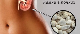
One of the most frequently diagnosed pathologies, which leads to the development of complications and a significant deterioration of the patient’s condition, is the formation of sand or stones in the ureteral ducts. According to practicing specialists, such a disease can be a consequence of poor nutrition, bad habits, or leading a predominantly unhealthy lifestyle.
In order to promptly identify an existing disease, it is necessary to know what clinical signs may be its manifestation. These include, for example:
- One of the most common symptoms is an acute and sudden attack of severe pain. Most often, discomfort occurs during fast walking, running or other active activities.
- Urine incontinence. As a rule, such a symptom is one-time in nature and occurs against the background of blockage of the duct with its subsequent release.
- Frequent and very strong urge to urinate.
- If the stones have blocked the area of the mouth, that is, the area where the ureter flows into the bladder, there is a high probability of disruption of the outflow of urine, the appearance of symptoms of general intoxication of the body, which include: pallor of the skin, lethargy, weakness, increased general body temperature, severe nausea.
Disturbance in the activity of the ureters due to the negative influence of any factors, among other things, can cause poisoning of the body with decay products, toxins and wastes. To prevent possible complications, you should consult a doctor immediately after the appearance of disturbing symptoms.