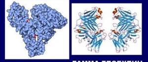
The protein, which was discovered by the English scientist Henry Bence-Jones, became the first tumor marker in 1847. If Bence-Jones protein is detected in the urine, then in most cases the patient is diagnosed with myeloma, a cancer that occurs in the bone marrow.
The presence of protein in the urine is called proteinuria. If, as a result of the study, traces of this protein are detected in the urine, then the analysis must be retaken. Bence-Jones protein should normally be absent.
A urine test for Bence-Jones protein is performed by a urologist, hematologist or oncologist in a situation where a pathological process in the bone marrow is suspected. This analysis is an addition to confirming the diagnosis.
Urine testing for Bence-Jones protein can be prescribed for a number of cancer diseases, and this method confirms the cancer process and determines the doctor’s actions, as well as treatment tactics.
If the result is positive
The urine of a healthy person does not contain proteins, including Bence Jones proteins. Its appearance in the studied samples indicates a high degree of proliferation of plasma cells, which occurs in a number of malignant processes.
- Myeloma (myeloma disease) is a pathology of the circulatory system in which uncontrolled proliferation of red bone marrow cells occurs.
- Plasmacytoma is a tumor consisting of plasma cells that mutate into myeloma.
- Osteosarcoma is a malignant tumor originating from bone tissue.
- Lymphogranulomatosis (Hodgkin's lymphoma, malignant granuloma) is an oncological pathology that is characterized by the proliferation of lymphoid tissues.
- Chronic lymphocytic leukemia is a disease in which abnormal B lymphocytes accumulate in the bone marrow, blood, liver, or spleen.
- Primary amyloidosis is a pathology in which the accumulation of amyloid, a complex protein-polysaccharide complex, occurs. Primary amyloidosis may be caused by multiple myeloma, in which case the amyloid consists of light chains of immunoglobulins.
- Macroglobulinemia is a pathological condition in which mononuclear macroglobulins are detected, synthesized by B lymphocytes.
Bence-Jones protein is a tumor marker; when it is detected, a full diagnosis of organ systems is carried out to identify the type and localization of the malignant process. Its determination in the blood is not informative, since due to its low molecular weight it is quickly filtered by the kidneys and excreted in the urine. The presence of Bence Jones protein can also indicate kidney pathologies, since its high concentrations destroy tissue and impair organ function. Proteinuria causes renal dystrophy, Fanconi syndrome or renal amyloidosis. When exposed to other factors (dehydration, increased calcium levels in the blood, exposure to x-rays, taking certain medications), there is a risk of developing chronic renal failure.
Bence Jones protein is the light chain of immunoglobulins produced in urine. This protein appears in the urine during a group of diseases classified as monoclonal gammopathy, most notably multiple myeloma.
These diseases result from the cancerous proliferation of a single clone of cells called plasma cells. These cells produce, in excess quantities, one type of immunoglobulin, the so-called M protein, the light chains of which are filtered by the kidneys into the urine and are detected in studies as Bence-Jones protein
.
Treatment
And Bence-Jones myeloma is performed only in a hospital setting. After confirmation of the diagnosis, radiation therapy and cytotoxic drugs are prescribed. Cyclophosphamide and sarcolysine have a pronounced effect in chemotherapy. Sarcolysin is prescribed intravenously at 300 mg per day. Most often in combination with prednisolone, which increases the effectiveness of the drug by 70%.
Maintenance therapy is required, as well as medications to relieve associated symptoms (vomiting, diarrhea, increased nervous excitement, etc.). To improve the functioning of the nervous system, cerucal, tizercin or haloperidol must be prescribed. The course of treatment is more than a month and all this time the patient must remain in a hospital setting. Between chemotherapy and radiation therapy blocks, outpatient maintenance treatment is carried out according to the scheme described above. Corticosteroids are also effective and can be prescribed in high doses for hypercalcemia and autoimmune complications.
An important stage of treatment is constant monitoring of the presence of a specific protein in the urine. The analysis is carried out weekly and treatment is considered effective if the amount of Bence Jones protein constantly decreases.
Radiation therapy can prevent multiple bone fractures, with total doses of up to 4000 rads. Plasmophoresis is also popular. This operation involves removing blood from the patient’s body (up to 1 liter) and returning it with an increased content of red blood cells. This procedure is especially important for severe anemia and azotemia.
Since during treatment there is a high risk of contracting an infection, antibacterial drugs are prescribed that have a strong antibacterial effect. Among such drugs, donor gamma globulin is required to be administered in 6-10 doses intramuscularly. Other complications are treated symptomatically, and since with myeloma of this type they almost always occur, treatment should be comprehensive and prevent possible consequences.
Thus, the blood disease myeloma is a serious disease belonging to the group of oncological diseases. If you suspect the presence of myeloma, you must urgently contact a hematologist, who will conduct the necessary diagnostic tests and draw up a competent and effective treatment regimen. It is worth remembering that timely therapy is the key to recovery and a high quality of life in the future.
High levels of protein in the urine indicate the presence of dangerous diseases occurring in the human body, for example, oncology. To exclude the latter, the patient is prescribed a urine test for Bence-Jones protein. This organic compound is one of the tumor markers and makes it possible to diagnose a malignant tumor of cells of the immune system.
A disease in which protein appears in the urine is called proteinuria. In most cases, such urine contains protein bodies - albumin. However, in some cases, laboratory tests reveal another type of organic formation - Bence Jones protein. These antibodies consist of light chains, therefore they are not able to linger in the blood for a long time and are filtered by the kidneys, subsequently excreted in the urine. Bence Jones proteins have an extremely low molecular weight, and the protein itself is produced by plasma cells during the formation of a malignant tumor.
Method for determining Bence-Jones protein
The content of this protein is checked on a urine sample. During its collection, the same rules are followed as when collecting urine for a general study. Before collecting a urine sample, wash your intimate area with soap and water and dry thoroughly. The first few drops should be flushed down the toilet and then filled into a portion of a sterile container and taken to the laboratory as quickly as possible.
Sometimes the study is carried out by collecting urine over several days, then a portion is placed in a special container on the first day, then a portion the next day, and so on, and delivered to the laboratory.
In healthy kidneys, plasma proteins pass into the urine in very small quantities due to their large size and negative charge. Bence-Jones protein, however, is so small that it easily passes through the filter membrane into the urine.
A general urine test can detect the presence of proteinuria, but more detailed tests are needed to accurately determine the type of protein being secreted. Previously, the thermal precipitation phenomenon was used to detect Bence-Jones protein in urine. In this study, heating urine to 60°C caused the formation of clots from the light chains of monoclonal immunoglobulins.
Currently, the method of protein electrophoresis
on agarose gel of urine, which allows you to accurately determine the type of proteinuria, including the detection of Bence-Jones protein.
How to test urine for tumor markers
In order for the analysis results to be reliable, you need to prepare and correctly collect the biomaterial. Preparation begins approximately a week before the scheduled test. From this moment on, you need to remove liver and meat from your diet. The day before the analysis, foods that can change the color of urine (carrots, beets, blackberries, etc.) are excluded from the menu. It is better not to drink carbonated and alcoholic drinks.
Urine is collected in a sterile container, which is sold in pharmacies. If you don’t have time or don’t want to go to the pharmacy, you can use a small glass container with a lid. The urine container must be washed, doused with boiling water, and dried. For analysis, as a rule, the middle part of urine is taken. It is collected in the morning on an empty stomach - the genitals are washed with clean water without soap, dried with a clean towel, then they begin to urinate into the toilet, continue into the container for analysis and finish into the toilet. For laboratory testing, 50 ml of urine is sufficient.
In women, urine collection is more difficult, since vaginal mucus and menstruation may be included in the analysis. Therefore, the collection of material should be carried out in the middle of the menstrual cycle; to be completely sure, before washing, you can place a tampon in the vagina.
The analysis must be taken to the laboratory within 2 hours after collection.
Interpretation of Bence-Jones protein results
In a healthy person, Bence-Jones protein is not detected in the urine. This test is performed when monoclonal antibody gammopathy is suspected, for example multiple myeloma
or
Waldenström's macroglobulinemia
. This study is one of the most important diagnostic criteria for these diseases.
Bence-Jones protein can also be detected completely incidentally when proteinuria is diagnosed during a general urine test, and in more detailed studies it turns out to be Bence-Jones proteinuria
.
In this case, it is necessary to immediately begin further diagnostics in the direction of monoclonal antibody gammopathy. The very presence of this protein in the urine often causes kidney failure, because the accumulation of light chains of proteins has a destructive effect on the kidneys.
Oncological diseases are characterized by chaotic proliferation of pathogenic cells. Symptoms indicating the presence of a dangerous pathology are the reason for diagnosis.
The patient must attend an ultrasound, computed tomography, biopsy, and take a urine test. The development of cancer contributes to changes in urine, the formation of Bence-Jones protein in it. This protein is a light chain of immunoglobulins, which is formed under the influence of cells of the immune system.
Types of secreted protein
In clinical practice, the secretion of immunoglobulin is divided into two types: the secretion of Bence-Jones protein and other immunoglobulins.
In the first case, we are talking about diseases in which free light chains of immunoglobulins are formed, but serum Pig is absent (about 20-22% of clinical cases of myeloma).
In the second case, we talk about glomerulopathies of various natures, which are based on inflammation of the glomerular apparatus of the kidneys with a different combination of characteristic symptoms.
Norm
The urine of a healthy person contains a small amount of protein - 0.033 g/l. A slight abnormality, called transient protein, is present in the body of newborns. Organic matter levels return to normal within a few days.
The excess protein indicator in children aged 12-13 can reach 0.01 g/l. Changes in the composition of children's urine are provoked by the child's activity and intense physical activity. In medical practice, the phenomenon is called orthostatic proteinuria.
The urine of a healthy person contains a small amount of protein
The presence of Bence-Jones protein is not considered a pathology if its formation is explained by organ transplantation or treatment with cytotoxic drugs. In all cases, protein sediment in the urine is a symptom of diseases - chronic leukemia, Waldenström's macroglobulinemia, myeloma. These pathologies are characterized by an increase in the amount of the substance to 0.02 g/l.
Causes
Excessive amounts of Bence Jones protein are recruited by cells that are responsible for producing antibodies. Once in the bloodstream, the substance moves throughout the body, settles in the kidneys, and then comes out through urination.
Protein sediment in the urine occurs in diseases:
- Myeloma is a malignant tumor that develops in the bone marrow and causes problems with the circulatory system.
- Plasmacytoma is a neoplastic disease that provokes the destruction of bone tissue.
- Primary Amyloidosis is a pathology that is characterized by improper protein metabolism in the body.
- Chronic leukemia is a cancer that damages the lymphatic system, liver, and bone marrow.
- Osteosarcoma is a malignant tumor that affects bone tissue. A feature of the disease is the formation of metastases in the early stages.
Late diagnosis of pathologies and lack of treatment can lead to serious consequences.
Secretion
Based on the type of immunoglobin produced in the body, they are divided into:
- Pathological changes in light chains - production of Bence-Jones uroprotein.
- Glomerulopathy, that is, the production of other types of immunoglobulins.
Other combinations of pathological changes in the kidneys cannot be excluded. Developing nephropathies are a consequence of diseases such as myeloma, Waldenström's disease, and lymphocytic leukemia with a chronic course.
Bence-Jones proteins have molecules with a mass of no more than 40 kDa, due to this they easily pass through the kidney filters, after which they are broken down into amino acids and oligopeptides with the help of lysosomes.
An excess of light chains passing through the kidneys provokes the development of catabolic dysfunction, which can cause necrotic changes in the tissues of the renal tubules.
Protein bodies interfere with reabsorption, and if light chains combine with proteins called Tamma Horsfall, protein casts begin to form in the distal renal tubules.
Diagnostic methods
To identify the Bence-Jones protein, the following methods are used:
- The immunosification method is considered accurate and reliable. An agarose gel is applied to the plate and electrophoretic separation is performed. Half of the organic matter is stained and antiserum is applied to the other half. Protein bodies interact with whey and form a precipitate. After the sediment has hardened, the second half is colored, and the doctor makes a comparative analysis. Normally, immunoglobulins form wide stripes; in the presence of deviations, thin stripes.
- The thermoprecipitation method is used when the protein concentration is high. Urine and acetate buffer are mixed in a 4:1 ratio, after which the liquid is boiled at temperature for 15 minutes. During the heating process, protein bodies sink to the bottom, creating sediment. If the liquid begins to boil, the organic matter disappears. The result of the study may be affected by high acidity and low density of urine.
- The precipitation reaction is a method aimed at identifying a small amount of protein. Filtered urine is added to a vessel with acid and examined using a special apparatus. If the composition of the biological fluid contains protein, the color of the test tube will change.
Several methods are used to detect the Bence-Jones protein.
It is impossible to diagnose the presence of Bence Jones protein using indicator paper.
About the Bence Jones tumor marker
Any protein is a high-molecular organic protein substance containing alpha amino acids united by a peptide bond into a single chain. Due to its small molecular weight, Bence Jones protein does not linger in the systemic circulation, but penetrates through the blood, directly into the organs of the urinary system, and is excreted from the body in the urine.
Therefore, the presence of a tumor marker, otherwise Bence-Jones proteinuria, is determined in urine, and not in the blood, like most tumor indicators. The severity of the malignant process is assessed by protein concentration. Low molecular weight protein increases the permeability of the glomerular filter of the renal apparatus and damages the tubular epithelium. The process of reabsorption (reabsorption) is disrupted.
While alpha amino acids are in the urinary system, its molecules harm the walls of the bladder, urethra, and damage the tissue of the kidney calyces and pelvis. Possessing high toxicity, the substance poisons and destroys the kidneys, causing organ dysfunction, threatening the development of kidney failure.
In addition to the fact that Bence-Jones protein is not absorbed by the kidneys, it is able to enter into a correlation with other proteins, which leads to the formation of protein casts from the destroyed epithelium (the so-called cylinders in the renal tubules), the formation of calculi (stones) in the bladder.
Any proteinuria (the presence of excess protein in the urine) is a sign of glomerular inflammation of the kidneys. Detection of Bence Jones protein is a clinical sign of a malignant process. Upon further diagnosis, it is confirmed in 75% of cases. The protein received its name after the British physician Henry Bence-Jones, who first identified the tumor marker in 1847.
Bence Jones proteinuria
The definition of Bence-Jones proteinuria, first of all, indicates multiple myeloma. This tumor is malignant in nature, but is not cancer, since it does not originate from tissues, but from plasma cells. The disease has three forms:
- diffuse, damaging the bone marrow;
- diffuse-focal, affecting the bone marrow and some other organs (in particular, the kidneys);
- multiple myeloma, which affects the entire body.
Myeloma is characterized by excessive bone fragility, frequent fractures, and bone pain. The disease spreads to the renal apparatus, the patient quickly develops myeloma nephropathy, which leads to renal decompensation.
In addition to myeloma, detection of Bence Jones protein in urine can be a sign of the following pathologies:
- Fanconi syndrome and renal amyloidosis are diseases characterized by impaired protein metabolism;
- malignant disease of lymphatic tissue, spleen, liver, lymph nodes (lymphocytic leukemia);
- tumor of the hematopoietic and lymphatic system (lymphosarcoma);
- new bone formation, with early manifestation of metastases throughout the body (steosarcoma);
- damage to plasma cells (plasmocytoma);
- malignant degeneration of the endothelium of blood vessels (endotheliosis);
- systemic pathological process associated with impaired mineralization of bone tissue (osteomalacia);
- oncology of lymphoid tissue (lymphogranulomato);
- malignant tumor of the hematopoietic organs (Waldenström macroglobulinemia).
To accurately confirm each of the possible diagnoses, a single urine test for Bence Jones protein is not enough. The patient needs a comprehensive examination.
Myeloma
The cause of this pathology is not known. Plasma cells in the bone marrow begin to produce proteins of irregular structure in increased quantities. Nonspecific symptoms, as a rule, do not alarm patients. The disease is characterized by late seeking medical help, when pain appears in the bones of the limbs, pelvis, ribs, and spine. Often the diagnosis is made when the patient has fractures of the vertebral bodies and Bence-Jones protein appears. At this stage, radical help is no longer possible.
Spinal fractures observed in multiple myeloma are called pathological, i.e. when exposed to minimal traumatic force, the vertebral bodies break, which is accompanied by increased pain. It is enough to lift a heavy bag and drive along a bumpy road for the affected vertebra to fracture. When X-ray examination of the flat bones of the skeleton and spine, the bones have a specific appearance. One gets the impression that “the bones are moth-eaten.” They have round, clear shadows - this is an unambiguous sign of myeloma.
Depending on the predominant clinical picture of the disease, the following types of malignant lesions are distinguished:
- Solitary plasmacytoma is a single focal bone lesion, accompanied by the appearance of Bence Jones protein in the urine, M-gradient in the blood, impaired renal function, and increased calcium in the blood. the prognosis is more favorable than with multiple myeloma. Laboratory indicators may not change, then no changes will be detected in the urine. In this situation, the diagnosis is often made at the stage of renal failure, infectious, autoimmune complications, neurological disorders, amyloidosis of the kidneys and other parenchymal organs.
- Smoldering myeloma is an asymptomatic disease. The general condition of the patients does not suffer. There is only an isolated increase in the M-gradient in the blood. The level of calcium in the blood and Bence Jones protein in the urine are not detected. The patient's treatment tactics are mainly expectant; dynamic observation is required before chemotherapy is prescribed.
- Acute plasmablastic leukemia - a large number of plasmacytic cell precursors appear in the blood - young forms, the function of which is not full, the effect on the body is toxic. It develops as a primary disease or as a secondary complication of multiple myeloma.
- Five types of myeloma affecting mainly bone tissue include forms classified on the basis of immunohistochemical tests. Therefore, to make a correct diagnosis, an in-depth examination, urine and blood tests are required.
In addition to the skeletal system, the pathological process of myeloma involves the kidneys; myeloma develops, which is manifested by one or more signs:
- Frequent exacerbations of pyelonephritis and other inflammations caused by reduced immunity, defects in immunoglubullins and protein bodies in the body;
- rapidly progressing symptoms of renal failure;
- Bence Jones protein, glucose in urine;
- increased calcium levels, appearance of an M-gradient in the blood;
- arterial hypertension, amyloidosis, nephrocalcinates.
Indications for the study
A urine test for Bence Jones protein is a specific test that is not carried out in every laboratory. A referral to donate urine is issued by an oncologist or urologist if the patient has symptomatic complaints that correspond to signs of one of the possible oncological pathologies.
The study is prescribed for a diagnosed disorder of protein-carbohydrate metabolism with extracellular deposition in the renal tissue of a complex protein-polysaccharide compound (amyloid) - amyloidosis disease of the renal apparatus.
On a regular basis, analysis is taken from patients with malignant lesions of the bone and lymphatic system to monitor the dynamics of therapy. In addition, the study is recommended for people with an unfavorable family history (oncological diseases in the listed body systems).
Rules for preparing for analysis
Objective results of a urine test for the detection of Bence Jones protein are guaranteed only by compliance with the preliminary preparation conditions. The patient needs to purchase a special sterile container for urine from the pharmacy. It is convenient, inexpensive, and reliable. It is recommended to change your diet for seven days before donating urine. It is necessary to eliminate protein products (meat and offal, fish, seafood, smoked meats).
For two days, foods that tend to stain urine (beets, asparagus, carrots, rhubarb, blackberries) are excluded from the menu. Alcohol-containing drinks, carbonated water, and lemonade are prohibited. For 2–3 days, you must stop using medications (including multivitamins that change the color and smell of urine).
For women, the ban applies to vaginal contraceptives, medicinal suppositories, and douching with antiseptic solutions. On the eve of the test, you should interrupt sports training, exclude physical activity, and avoid hypothermia. It is necessary to collect urine in the morning, after waking up. The most accurate research data is provided by the average portion of urine.
To do this, you should first urinate into the toilet, then into the prepared first aid container, and then back into the toilet. For microscopy, 50 ml of urine is sufficient. Before collecting urine, you should perform a hygiene procedure for the external genitalia. The use of soap or gel is not recommended. For women, the optimal time to get tested is the middle of the menstrual cycle.
Before collecting urine, the vagina should be covered with a cotton swab or tampon to prevent secretions from the vaginal glands from entering the container. The container with urine must be tightly closed and delivered for examination within two hours. If necessary, the doctor may prescribe a study of the daily urine rate. In this case, all the urine excreted per day is collected in a container with a capacity of three liters.
Important! Failure to comply with the preparation conditions may distort the results of the study, as a result of which the wrong treatment will be prescribed.
How to prepare for submitting biomaterial?
Examination for Bence Jones protein requires mandatory preliminary preparation of the patient:
- Alcohol is completely avoided the day before the biomaterial is submitted.
- Within two days, the course of diuretic medications is stopped.
- For seven days, a diet is followed, heavy physical activity and hypothermia are excluded.
Attention! Diagnostic screening involves collecting a daily volume of urine or a morning urine sample.
You should not collect biomaterial in household containers that have been previously emptied of their contents. Only using a sterile plastic container purchased from a pharmacy or taken from a laboratory will give an accurate result. The collected urine is immediately delivered to the laboratory (pre-laboratory storage is allowed at above-zero temperatures of 2-8 C).
Laboratory method for protein determination
After filtration and reabsorption in the kidneys, no more than 30–150 mg/day of proteins are determined in the urine, most of which is albumin. This is called microalbuminuria and is not detected by routine urine tests. When the norm is exceeded, proteinuria develops, indicating kidney disease.
At the same time, only Bence-Jones protein has an oncological nature. In benign paraproteinemias, this substance is not detected in the urine. Electrophoresis is used to identify paraproteins in urine. Using the electrophoretic method of analysis, the molecules of protein fractions are grouped by category and size.
Protein molecules, like any electrically charged particles, move in an electric field, therefore, when a constant voltage is applied to a urine sample, they begin to move: positively charged - to the cathode, negatively charged - to the anode. After the molecules are separated, serum is added, which reacts with it to color the paraproteins. Bence Jones proteinuria is manifested by intense coloration.
Another method for detecting Bence Jones protein is to isolate the substance using oxidation and heating. A buffer solution containing sodium acetate and acetic acid (in a ratio of 4:1) is added to the urine sample, then heated to 60 °C and cooled. As a result, the protein is transformed into a precipitate, which is washed in alcohol and ether. The final balance is weighed.
How to get tested?
The quality of a urine test for Bence-Jones uroproteins depends on how patients follow the doctor’s recommendations regarding the collection and donation of biological fluid.
Basically, laboratory technicians advise adhering to several rules:
- The results will be reliable if you stop eating excess liver and meat for about a week. The day before the test, you do not need to drink carbonated drinks, alcohol, and you need to exclude foods that affect the change in urine color, such as beets, blackberries, carrots, blueberries. The doctor may prohibit the use of certain medications, so you must always inform about the treatment being carried out.
- It is necessary to collect urine in a clean container - this can be a special pharmaceutical container or a glass jar with a lid. The container must first be doused with boiling water.
- Protein is detected in morning urine, in its middle portion. First you need to wash yourself without using gels or soap. For the study, 50 ml of liquid is enough.
- The test must be delivered to the research center no later than two hours from the moment of its collection.
We suggest you read: Transitional epithelium urine analysis
One of the most common ways to find Bence Jones uroproteins in urine is to mix the collected urine filtrate in a 4:1 ratio with acetate buffer. After this, the mixture is heated in a water bath to 60 degrees, this leads to the fact that the protein quickly settles and it becomes possible to calculate its concentration.
The concentration of low molecular weight protein can also be determined using immunoelectrophoretic studies. In this case, specific sera are used against the protein chains of immunoglobulin fractions.
At the lowest concentrations, Bence Jones uroprotein can be determined using a precipitation reaction performed with sulfosalicylic acid.
Determination of Bence protein in urine is carried out in laboratory conditions. At the same time, in order for the examination to give the correct result, you should carefully prepare for it.
- Eliminate meat and meat by-products from your diet. This must be done a week before going to the hospital for testing.
- The day before sample collection, it is necessary to remove from the menu foods that can change the color of urine. Doctors include carrots, beets, blackberries, and so on here.
- It is mandatory to give up alcohol a week before visiting the hospital. It is also undesirable to drink carbonated drinks.
- Some medications may also interfere with accurate research. Therefore, the doctor should be informed about what medications the patient is taking. In some cases, he may prohibit their use until the analysis is completed.
It is very important to follow the urine collection procedure itself. A clean container is used for this. It is advisable to first pour boiling water over it or sterilize it in another way.
Only morning urine is required for trace protein analysis. In this case, an average dose should be collected. Initially, urination is carried out into the toilet, then 30-50 milliliters into the container, then again into the toilet. The sample itself is sent to the laboratory no later than two hours after collection.
The exact method used to determine the presence of proteins in urine depends on the equipment of the medical institution.
Most often this is done as follows. The collected urine filtrate is mixed with acetate buffer. The ratio should be four parts urine to one part “supplement”. Next, the resulting mixture is heated to sixty degrees. In this case, a water bath is used. As a result of this treatment, the protein settles, which makes it possible to calculate its concentration.
Further diagnostics
If Bence-Jones paraprotein is detected, an extensive diagnosis of multiple myeloma is prescribed, including hardware and laboratory testing. Hardware procedures include x-rays of skeletal bones, MRI (magnetic resonance imaging), spiral CT (computed tomography).
Laboratory diagnostics include:
- General blood analysis. In pathology, a decrease in hemoglobin, the number of platelets, leukocytes, erythrocytes, as well as the presence of plasma cells is recorded.
- Biochemistry of blood. There is an increased level of concentration of uric acid, urea and creatinine, hypercalcemia (increased calcium levels).
- Coagulogram. It is carried out to assess blood clotting.
- Cytogenetic study of bone marrow cells (plasmacytes). Necessary for determining chromosomal pathologies.
The presence of paraprotein in urine means a positive test result, its absence means a negative result.
Separately, a collection of bone marrow cells (biopsy) is prescribed to compile a myelogram - assessing the qualitative and quantitative content. Myeloma, in most cases, is diagnosed in men who have crossed the age of sixty. The cause of mortality is not only the development of oncological processes, but also concomitant progressive renal failure.
The disease is asymptomatic up to a certain point, which is the main reason for timely (late) diagnosis. A urine test for Bence Jones protein makes it possible to detect cancer pathology at an early stage of development, which significantly increases the chances of effective treatment.
If the test result is positive, radiation therapy and chemotherapy are used (to eliminate malignant cells). As a special therapy, the patient is prescribed medications developed in the laboratory, which are based on monoclonal antibodies that can inhibit tumor growth.
What to do if a protein is detected
As already mentioned, Bence Jones protein is a tumor marker for myeloma. However, it is impossible to make an accurate diagnosis from just one urine test. Therefore, the doctor prescribes additional tests:
- Clinical studies of blood and urine. Patients with multiple myeloma usually have elevated ESR and decreased white blood cells. Pathological casts and red blood cells are detected in urine.
- Urine examination using the Zimnitsky method. Allows you to identify signs of renal failure, which often develops with pathologies of the hematopoietic system.
- Biochemical blood test. In patients with bone marrow diseases, the metabolism of minerals and proteins is impaired, which is reflected in the results of the study.
- Bone marrow puncture. This study makes it possible to detect malignant changes in cells with great accuracy.
- X-ray of the skull, spine and ribs. With myeloma, the image shows a significant decrease in bone density, and sometimes signs of fractures.
Bone marrow diseases require persistent and long-term treatment. Patients are prescribed a course of antitumor chemotherapy drugs, radiation therapy and blood transfusions. In severe cases, a bone marrow transplant is performed.
At the same time, therapy is carried out for developing renal failure. Patients are advised to follow a diet with limited protein in food.
It is important to remember that the earlier treatment for myeloma is started, the more favorable the prognosis of the disease. At an early stage, it is still possible to avoid the proliferation of malignant cells. In advanced cases, the survival rate of patients decreases sharply. Testing urine for Bence-Jones protein allows one to identify dangerous diseases at an early stage, begin treatment on time, and thereby save the patient’s life.


