The Apex Clinic provides MSCT of the kidneys in Novosibirsk using modern and high-precision equipment at an affordable cost. MSCT of the kidneys is prescribed to clarify the diagnosis, check the effectiveness of the prescribed treatment for the disease, as well as in a number of other cases. Multislice computed tomography allows for examination of the adrenal glands, kidneys, and abdominal space. Through the use of modern equipment, it is possible to obtain a large number of “thin” sections of the area under study, which allows for a detailed assessment of the structure of organs and to identify the smallest pathological changes and formations.
General information
MSCT of the kidneys is the name given to multi-slice computed tomography. This technique allows you to see the image of the organ layer by layer, which gives maximum information content. The monitor shows the organ in detail; the thickness of one layer is 0.5-1 mm (the diagnostician can examine the condition of the sinus, as well as the parenchyma). The study can be done with contrast to get an accurate picture of the condition of the kidneys. If a pharmaceutical drug is used, then photographs are taken at various phases of drug administration:
- arterial;
- excretory;
- parenchymatous.
Sometimes MSCT of the urinary system is performed in a delayed phase.
Operating principle of the device:
- The X-ray beams move in a spiral around the patient's body.
- The image is obtained using sensors; the picture can be seen in 2D or 3D format.
- If the doctor uses a contrast agent for a detailed study of blood vessels and lymph nodes, then the patient will first be prescribed allergy tests: in some cases, allergies may occur with the presence of undesirable reactions. The contrast is excreted by the kidneys and does not cause harm to the body.
MSCT of the kidneys - what it is and how it is performed, we will look at below. It is noteworthy that many diagnosticians prefer to first do an examination without administering a contrast agent, and only after that they recommend the patient an examination with the drug - this way the diagnostician can compare both results obtained.
CT scan of the bladder and prostate
In differentiating diseases of the male genitourinary system, multislice computed tomography is of great importance. During the examination, the patient is placed on a mobile table, which passes through the annular part of the tomograph, where the X-ray tube and special sensors for reading information are located. The emitters inside the device make circular movements, which, together with the movement of the table, provides spiral scanning of the tissues in question.
The tomograph transmits the layer-by-layer images obtained during a CT scan of the bladder to a computer screen, where, if necessary, a complex computer program recreates a 3D image of the area being studied.
Such a detailed study makes it possible to detect developmental anomalies and pathological processes occurring in the tissues of the bladder and prostate, visualize the condition of the mucous membrane of the urethra and parenchyma of the gland, determine the boundaries, size of organs, changes in shape and structure.
CT scan of the bladder and prostate is prescribed if symptoms are present:
- disturbances in the urination process;
- the presence of impurities in the urine (blood, salts, protein) of unknown etiology;
- pain in the lower abdomen, radiating to the groin;
- erectile dysfunction;
- infertility;
- injuries in the pelvic area.
The study is carried out before surgery, in order to determine the location and scope of upcoming actions, as well as in the postoperative period to assess tissue restoration.
Using multislice CT, pathological processes occurring in the tissues of the genitourinary system are identified and differentiated, and traumatic lesions in this area are localized.
Diseases diagnosed with CT scan of the bladder and prostate:
- inflammatory phenomena (acute and chronic prostatitis, cystitis);
- neoplasms (cysts, polyps, adenoma, cancer) of the bladder and prostate;
- metastases to these organs;
- lymphadenopathy of regional nodes;
- traumatic lesions of the prostate gland and bladder, perforation of hollow organs;
- the presence of stones and foreign bodies in the bladder cavity;
- vascular pathologies in the area under consideration.
Early diagnosis of pathological processes contributes to the effectiveness of treatment and guarantees a favorable prognosis in most cases.
Using Contrast
MSCT of the kidneys using a pharmaceutical drug is prescribed to patients if there is a suspicion of the presence of malignant tumors, or problems with the integrity of blood vessels or their blockage. It is important to conduct research if you need to identify the presence of kidney stones and find out what size they have reached.
Iodine-containing medications, or gadolinium, are used as a contrast. It is administered intravenously or given orally. After a day, the components are completely eliminated from the body, mainly by the kidneys. The solution does not pose any threat to the general well-being of the patient, provided that the person does not have allergic reactions to it.
The procedure involves a preliminary examination without contrast, only after this the doctor can introduce a solution. This method is the most informative; it is necessary to compare images and obtain detailed information about the condition of the organ.
Most doctors use a special injection syringe that works automatically. If the patient can drink the substance, and there is time to wait until the drug is absorbed, then you can simply drink a medicine that will highlight the vessels - this is what contrast is called.
Experts do not undertake to claim that the use of contrast does not cause any harm to the patient, but the information content of the procedure with the use of such drugs increases significantly. If a diagnostic procedure is necessary for individual intolerance to contrast, MSCT of the kidney without contrast is prescribed.
This point can be justified by the following factors:
- you can determine the exact size of the tumor, the nature of the tumor - malignant or benign;
- MSCT of the kidneys with contrast determines the presence of urolithiasis;
- prescribed to patients after severe injuries;
- before surgery.
If there is a suspicion of severe kidney pathologies, then a procedure using contrast is not prescribed. This is due to the fact that the elimination of the drug is carried out by this organ, the elimination of the drug will be slowed down, which will cause harm to the body.
What can be revealed with this research?
Tomography serves as an alternative diagnostic method when the patient or his relatives refuse cystoscopy.
Computed tomography of the bladder is of great importance for diagnosing the following diseases of the excretory system:
- benign and malignant volumetric processes in the bladder;
- metastases of tumors from other organs of the patient;
- nonspecific inflammation (cystitis);
- traumatic injury to the patient’s bladder with its rupture, hematoma and bleeding;
- renal ring with urinary obstruction;
- congenital malformations of the excretory system;
- venereal pathological processes;
- degenerative processes with stretching of the bladder walls.
Benefits of the study
The capabilities obtained with MSCT of the kidneys combine all the advantages of ultrasound, MRI and CT. Let's look at them in detail:
- You can see in detail the position of the kidneys relative to the skeleton of the patient and other organs.
- The most accurate imaging method when it comes to detecting the presence of stones.
- A relatively safe technique if the patient does not have individual intolerance to the contrast agent.
- Minimum contraindications.
- The procedure is carried out quickly, and after the examination, the diagnostic results are given to the patient.
How is it different from MRI?
MSCT and MRI of the kidneys differ in the principle of their effect. The first method involves the use of X-rays, the second - a powerful magnet. The diagnostician receives a three-dimensional image of the organ and can clearly visualize all existing abnormalities.
During MSCT, X-ray radiation is used in conjunction with modern computer technologies, which produces a layer-by-layer picture; the kidneys can be seen in a large number of sections - this does not give the pathology a chance to go unnoticed. Both examination methods are considered informative, but MRI is slightly more expensive than MSCT diagnostics.
What is the difference from SKT
During MSCT, gantry is used, this is the name of special devices that are the first models of computed tomographs. The principle of their operation is that the design rotates around the patient, showing the organs being examined in different planes. Data processing is carried out by a computer.
Gradually, technology improved, and the spiral tomography method became available, which does not separate images into separate layers. Informative and high-quality images are obtained through spiral cutting, and data can be obtained within a few seconds.
MSCT is a technique that was invented recently; its difference from SCT is that the device involves the arrangement of several rows of detectors. The rotation of the X-ray tube in this device is doubled. The examination can be completed 32 times faster due to the 16-spiral installation. The method determines the presence of complications even at very early stages, determines the degree of malignancy of the tumor, as well as the presence of metastases.
What can be found in the photographs
By conducting a unique method of scanning the urinary system, a specialist has the opportunity to detect many diseases, including very dangerous ones, at the initial stage. A CT scan of the urinary system can detect the following pathologies:
- cystitis;
- venereal diseases;
- cancer, tumor diseases;
- infectious diseases;
- nephritis, glomerulonephritis;
- traumatic ruptures, sprains of the organ being examined;
- urolithiasis disease.
In addition, the doctor may order a CT scan before the scheduled operation or to monitor the therapy.
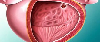
In the images obtained after a CT scan, you can see the structure of the bladder, urinary tract, urethra, and vessels of this area. Diagnostics also gives specialists the opportunity to detect a tumor of any stage, shape, or origin. The images will show neoplasms in different parts of the organ:
- inside the cavity;
- inside the bladder wall;
- in surrounding tissues and organs;
- metastasis into the lymph nodes.
When is the study carried out?
To perform diagnostics with contrast, the patient requires a referral from a specialist. The technique itself involves the administration of a special component intravenously or orally in order to obtain a clear image of the condition of the blood vessels. The contrast details the condition of soft tissues, arteries, and veins. When diagnosing oncology, a specialist can study in detail the state of the lymphatic system. The method is shown for studying the following diseases:
- congenital anomalies;
- inflammation in acute or chronic form: MSCT with contrast determines the stage at which the disease is, the presence of complications, etc.;
- bruises or other injuries to paired organs;
- determination of hemorrhage into the parenchyma;
- occlusion of organ vessels;
- the presence of an abscess or the presence of purulent formations;
- vascular occlusion;
- to determine the causes of renal colic.
During pregnancy, doctors prohibit MSCT of the kidneys with contrast, since there is radiation exposure, and the contrast agent can be passed on to the baby, causing disruptions in the development of the fetus.
Indications and contraindications
MSCT is prescribed by a doctor in case of a history of renal colic, as well as persistent pain in the lumbar region. If pathological impurities are detected in the urine (protein, blood, uric acid crystals, increased number of leukocytes). If the cause of renal failure is clarified.
Contraindications to the service are intolerance to radiopaque (iodine-containing) drugs, severe renal failure. In children and pregnant women, the study is carried out according to strict indications, since the procedure is accompanied by ionizing radiation.
Who is the procedure contraindicated for?
Despite the fact that the procedure is practically safe and does not cause harm to health, not everyone can undergo it. There are absolute (when there is more harm than benefit, the procedure cannot be performed) and relative (the doctor himself decides the feasibility of diagnostics) contraindications. Let's look at them in detail.
Absolute contraindications
During pregnancy, MSCT of the kidneys is prohibited due to the fact that there is a radiation dose that can negatively affect the development of the fetus, up to the occurrence of abnormalities. In such situations, women are prescribed an MRI or ultrasound.
The procedure with contrast is also contraindicated if a person has an allergic reaction or individual intolerance to the active substance included in the contrast. In this case, after the procedure, undesirable effects may occur - skin rash, vomiting, severe dizziness. To make sure that everything goes correctly, the doctor first sends the patient for allergy tests.
Another absolute contraindication is severe renal failure. When contrast is administered, the main active substance is excreted by the kidneys, and if they work incorrectly, the process becomes more complicated, and the contrast will remain in the body for a long time, which can aggravate the patient’s condition.
Relative contraindications
As for relative contraindications, in such cases the doctor himself determines whether it is worth conducting an examination, or whether it is better to replace it with ultrasound, CT without contrast or MRI:
- The weight is too heavy - more than 120 kg, the devices are not designed for such overweight people, they can break, and clear results cannot be achieved.
- In case of diabetes mellitus, the doctor must make sure that MSCT with contrast will be a safe procedure for the patient.
- If a woman is breastfeeding, then she needs to abandon this process three days before the CT scan, and not feed the baby after 3 days. The contrast agent tends to be excreted in mother's milk, and for a fragile body this can be dangerous. During this period of time, the child is transferred to artificial food.
The patient's serious condition may also cause the diagnostician to refuse to perform the procedure. This point is due to the fact that it will be difficult for the patient to stay in the tomograph, especially if he experiences pain. When administering painkillers and sedatives, the procedure can only be carried out with the permission of the doctor.
Pregnancy and lactation
As mentioned above, even if it is necessary to find tumor metastases, this type of diagnosis is not prescribed to women carrying a baby. At the beginning of the period, it is recommended to undergo an ultrasound, and then an MRI. The latter method is also informative, but the effect of a magnetic field on the fetus will not cause any negative reaction, it is safe. During lactation, you need to stop breastfeeding for a while and transfer the baby to artificial feeding.
Conducting a CT scan of the urinary system using contrast
It is recommended to carry out the procedure with contrast if the patient has signs of developing bladder cancer. Contrast agents help expand the diagnostic capabilities of tomography. The tissues being examined become clearer due to contrast.
Sometimes a complex diagnosis is carried out not only of the bladder, but also of the kidneys. To visualize the kidneys, urografin is used, it must be administered into the body in 2 doses:
- the first part is taken before bedtime;
- The patient drinks the second part before the procedure.
Preparation and execution
No special preparation is required; 3 days before the examination, the patient must exclude foods that cause bloating from the diet; 8 hours before the examination, do not eat at all.
The results of the MSCT procedure are delivered to your hand, the process itself lasts for one hour: all this time the patient needs to remain motionless. The method is not painful, will not cause discomfort, and allows you to obtain a clear image of the organ. It will take a specialist about three days to establish an accurate diagnosis, after which the correct treatment will be selected.
Algorithm for the procedure
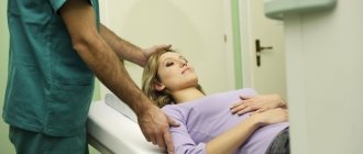
MSCT of the kidneys without contrast takes approximately 10 minutes, with contrast – about half an hour. Before the diagnosis, the patient must remove all clothing that contains metal elements and jewelry. After this, the person lies down on a special table, raises his arms, and must remain in this position until the doctor notifies the end of the study. There is a connection with the doctor; specialists are in the next room during MSCT.
Getting Results
The conclusion is drawn up by a diagnostician, which takes approximately 2 hours. The results are transmitted to the patient on electronic media, as well as in printed form. Next, you need to go to a specialist, he will make a diagnosis, and only after that will prescribe treatment. Additional diagnostic methods are used extremely rarely, since MSCT of the kidneys is informative.
Possible risks
MSCT examination cannot be called a safe technique, and we have talked about this more than once in the publication. During the procedure, X-rays are used, which carry a certain degree of radiation. Therefore, frequent (repeated) examinations are not recommended. Very rarely, MSCT of the kidneys can provoke the growth of malignant tumors.
When using contrast, there is a possibility of side effects - coffee rash, swelling of the mucous membranes. Sometimes the patient's blood pressure drops sharply, but this happens rarely.
Side effects after the procedure
Among the side effects, allergic reactions associated with the administration of contrast during MSCT can be identified:
- nausea;
- rash accompanied by itching;
- redness, inflammation of the skin;
- nausea;
- presence of a metallic taste in the mouth.
Preparing for the examination
Before the examination, the patient must undergo general clinical laboratory tests and visit the attending physician. He will give you a referral for testing and instructions on how to prepare for a CT scan.
Preparatory measures for the examination include limiting food consumption the day before the examination.
The day before the examination, you should limit the intake of foods that increase gas formation: dairy, flour, legumes, some vegetables (asparagus, cabbage, potatoes, onions), fruits, chewing gum and carbonated drinks.
For main meals, it is better to prepare porridges and light soups; for drinks, opt for tea.
In the morning before the tomography, you should avoid food altogether, limiting yourself to colorless, clear liquids. During a contrast study, the last meal should be eaten at least 6 hours before.
If a CT scan of the bladder with contrast is performed, it may be necessary to drink an ampoule of the solution 5 hours before the examination, or it will be administered intravenously immediately before the procedure, or it will be administered in the middle of the procedure with a special device after performing the diagnosis without it.
Alternative research methods
In some cases, patients are offered to replace MSCT of the kidneys with other methods:
- MRI is a safe and informative method;
- CT is in second place after MSCT, but also allows you to determine kidney diseases: tumors, polycystic diseases and other abnormal conditions;
- Ultrasound is a less informative method, but it is absolutely safe and is prescribed even to pregnant women.
Tissue irradiation with X-rays is insignificant with the MSCT method, but in some cases patients are prescribed other examination methods for safety reasons, especially if multislice tomography has been recently completed.
Which is better - CT or MRI of the bladder?
CT scan of the bladder is highly informative in identifying neoplasms and inflammatory phenomena and can reduce examination time. The price of the procedure is significantly lower than the cost of an MRI of a similar area. But the use of X-ray radiation has a number of contraindications and, in some cases, magnetic resonance imaging is advisable.
MRI is safe for the body: the magnetic field does not have a pathological effect on the cells of the human body. This diagnostic method makes it possible to evaluate the structure and structure of soft tissues, reflects the slightest changes in them and is more informative than CT of the bladder. The disadvantages include the significant duration of the procedure and high cost (more information about prices for instrumental examination methods can be found on the websites of medical diagnostic centers).
The doctor decides which method is more appropriate to use in each specific case based on the indications and clinical picture of the disease.
