Diseases of the genitourinary system are quite common. Many women know what chronic pyelonephritis is, since this pathology complicates pregnancy. Kidney diseases can lead to dangerous consequences that require constant cleansing of toxic substances from the blood (hemodialysis). The disease has several causes (for example, hereditary predisposition, hypothermia, infection from the genital organs, etc.), and is often asymptomatic, that is, it does not bother the person in any way, so everyone needs to know how to check the kidneys. This will help you seek medical help in time and avoid serious complications.
Kidney structure
View gallery
The kidneys are paired organs located in the lumbar region. Their main function is the formation of urine. The kidneys maintain the oncotic pressure of the blood and produce erythropoietin. The main structural unit - the nephron - consists of the vascular part (glomeruli) and tubules. The former are responsible for filtering the blood and forming primary urine. The second ones are involved in the reabsorption of substances necessary for the body. Ultimately, what remains is a processed waste product – secondary urine. If at any stage an obstruction occurs, then kidney function weakens. This is expressed in a change in the qualitative or quantitative composition of urine. To understand how to check the kidneys yourself, you need to know about diuresis disorders, which almost all patients have. These may include a decrease or increase in the urge to urinate, a change in the color of urine, or visiting the toilet more often or less often.
View gallery
Kidney function (screening)
A comprehensive laboratory test of blood and urine, which allows you to evaluate the excretory and filtration abilities of the kidneys and diagnose disorders of their function.
The research results are provided with a free doctor’s commentary.
English synonyms
Renal Function Tests.
Units
μmol/L (micromoles per liter), mmol/L (millimoles per liter), mg/dL (milligrams per deciliter), unit/dL (unit per deciliter).
What biomaterial can be used for research?
An average portion of morning urine, venous blood.
How to properly prepare for research?
- Children under 1 year of age should not eat for 30-40 minutes before the test.
- Children aged 1 to 5 years should not eat for 2-3 hours before the test.
- Do not eat for 12 hours before the test; you can drink clean still water.
- Avoid (in consultation with your doctor) taking diuretics for 48 hours before collecting urine.
- For women, examination (urine collection) is recommended to be carried out before menstruation or 2-3 days after its end.
- Avoid physical and emotional stress for 30 minutes before the test.
- Do not smoke for 30 minutes before the test.
General information about the study
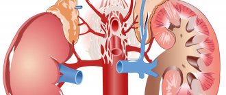
The kidneys are paired organs located in the retroperitoneum on both sides of the lumbar spine. They ensure blood filtration from excess and toxic metabolic products, removing them in the urine, and, thanks to the processes of filtration, secretion and reabsorption, regulate the level of fluid, electrolytes and trace elements, acid-base balance in the body. They synthesize biologically active substances: renin, which plays an important role in the regulation of blood pressure, and erythropoietin, which stimulates the proliferation of red blood cells in the bone marrow.
Kidney functions are regulated by hormones of the hypothalamic-pituitary system and adrenal glands, so changes in the level of antidiuretic hormone, aldosterone, and cortisol greatly affect the content of electrolytes and fluid in the blood, their ability to reabsorb and filter. In addition, under the influence of parathyroid hormone and vitamin D, the kidneys participate in the regulation of phosphorus and calcium metabolism.
With kidney pathology, the release of toxins and fluids from the body is disrupted, and the balance of osmotically active substances in the blood changes. Severe kidney disease without adequate treatment, regular hemodialysis or, if necessary, donor organ transplantation leads to death.
There are more than 100 diseases and pathological conditions that can cause progressive impairment of kidney function. The most common are diabetes mellitus, hypertension, infections, autoimmune pathology (systemic lupus erythematosus, vasculitis), neoplasms, developmental abnormalities of the urinary tract, and pathologies of mineral metabolism.
Renal dysfunction usually occurs unnoticed. Kidney disease can be suspected when puffiness and swelling appear around the eyes, in the ankles and legs, chest and abdomen, when there are changes in the color, transparency and amount of urine, when urination increases, especially at night, when there is pain in the lower back, in the area where the kidneys are projected. The progression of kidney pathology is often accompanied by itching, loss of appetite, nausea, vomiting, swelling and numbness of the arms and legs, fatigue, decreased attention, darkening of the skin, and convulsions. However, changes in laboratory parameters appear much earlier than the first clinical symptoms.
Thus, an increased number of leukocytes in the urine indicates an inflammatory process and a possible urinary tract infection. Protein and red blood cells in the urine may indicate damage to the glomerulus, and increased concentrations of urea (azotemia) and creatinine in the blood are signs of kidney failure.
Detection of acute or chronic renal failure requires urgent treatment measures to avoid progression of the disease.
Due to the variety of causes of kidney disease, a comprehensive examination of the patient is necessary, taking into account clinical data, the results of laboratory and instrumental research methods, and in some cases, a kidney biopsy.
What is the research used for?
- To assess the functional state of the kidneys.
- To evaluate renal damage in certain systemic diseases (eg, diabetes mellitus, hypertension, systemic lupus erythematosus, amyloidosis, vasculitis, myeloma) and trauma.
- For differential diagnosis of urinary tract diseases.
- For monitoring patients with chronic kidney disease, renal failure.
- For the examination of patients on dialysis.
- For preventive examination before prescribing drugs with possible nephrotoxic effects.
- To monitor the treatment of kidney pathology.
When is the study scheduled?
- For symptoms of kidney pathology (changes in color, smell, clarity and amount of urine, frequency of urination, pain in the lumbar region, swelling in the face and other parts of the body, high blood pressure).
- If the results of a general urine test are questionable or there are deviations in individual biochemical parameters.
- For kidney pathology according to instrumental diagnostic methods (ultrasound, CT).
- For systemic diseases with a high risk of damage to kidney function.
- During preventive examinations.
- Before prescribing drugs that affect kidney function and while taking them.
- When monitoring the treatment of kidney disease.
What do the results mean?
Reference values
- BUN/creatinine ratio: 0 - 100.
- Serum creatinine
| Floor | Age | Reference values |
| Male | Less than 5 days | 27 - 88 µmol/l |
| 5 days – 1 month | 27 - 88 µmol/l | |
| 1 month – 1 year | 18 – 35 µmol/l | |
| 1-2 years | 27 - 62 µmol/l | |
| 2-12 years | 27 - 62 µmol/l | |
| 12-17 years old | 44 - 88 µmol/l | |
| 17-51 years | 74 - 110 µmol/l | |
| Over 51 years old | 72 - 127 µmol/l | |
| Female | Less than 5 days | 27 - 88 µmol/l |
| 5 days – 1 month | 27 - 88 µmol/l | |
| 1 month – 1 year | 18 – 35 µmol/l | |
| 1-2 years | 27 - 62 µmol/l | |
| 2-12 years | 27 - 62 µmol/l | |
| 12-17 years old | 44 - 88 µmol/l | |
| More than 17 years | 58 – 96 µmol/l |
- GFR (glomerular filtration rate): 60 and above.
- Urea in serum
| Age | Reference values |
| Less than 1 day | 1.4 - 4.3 mmol/l |
| 1 day – 14 years | 1.8 - 6.4 mmol/l |
| 14-16 years old | 2.8 - 7.2 mmol/l |
| More than 16 years | 2.8 - 7.2 mmol/l |
- Urine sediment examination:
| Index | Reference values |
| Reaction | 5.0 — 7.5 |
| Bilirubin | None or small amount |
| Specific gravity | 1.010 — 1.025 |
| Protein | Missing or traces |
| Glucose | Up to 30 mg/dl |
| Urobilinoids | 0.2 - 2.0 units/dl |
| Ketones | Up to 15 mg/dl |
| Nitrites | Not detected |
| Reaction to blood | Negative or traces |
| Leukocytes | Not found or traces |
| Color | Pale yellow to deep yellow |
| Smell | Unsharp specific |
| Transparency | Transparent |
| Epithelium: flat | For men: 0 - 3 in sight For women: 0 - 5 in the field of view |
| Epithelium: flat (in view) | For men: 0 - 3 in sight For women: 0 - 5 in the field of view |
| Leukocytes | For men: 0 - 2 in sight For women: 0 - 4 in the field of view |
| Red blood cells: unchanged | 0 - 3 in view |
| Red blood cells: changed | 0 - 3 in view |
| Cylinders: hyaline | 0 - 1 in view |
| Cylinders: others | 0 - 1 in view |
Serum creatinine
Causes of increased serum creatinine levels:
- renal failure;
- chronic kidney disease;
- chronic nephritis;
- disturbance of urine outflow;
- pathology of muscle tissue (gigantism, myasthenia gravis, muscular dystrophy, poliomyelitis);
- rhabdomyolysis;
- dehydration;
- congestive heart failure;
- shock.
Reasons for decreased serum creatinine levels:
- short stature, insufficient muscle mass;
- severe liver pathology;
- lack of protein in the diet;
- pregnancy.
1) Density (allows you to evaluate the ability of the kidneys to concentrate urine)
Causes of increased density (hypersthenuria):
- acute nephritis;
- diabetes;
- renal failure;
- excessive fluid loss (with vomiting, diarrhea, fever, dehydration);
- stress, surgery, excessive release of antidiuretic hormone;
- gestosis during pregnancy.
Causes of decreased density (hyposthenuria):
- acute glomerulonephritis;
- acute and chronic tubulointerstitial nephritis;
- diabetes insipidus (antidiuretic hormone deficiency);
- Conn's syndrome;
- hyperparathyroidism;
- drinking large amounts of liquid;
- variant of the norm in children of the first year of life.
2) pH (acidity)
Reasons for increased pH (alkalinization of urine):
- urinary tract infection caused by bacteria of the genera Proteus or Pseudomonas;
- chronic renal failure;
- metabolic acidosis (with vomiting);
- respiratory alkalosis (with hyperventilation);
- hypokalemia.
Reasons for decreased pH (urine oxidation):
- metabolic acidosis, diabetic ketoacidosis, diarrhea, fasting, uremia;
- urinary tract infection caused by the bacterium Echerichia coli;
- respiratory acidosis;
- kidney tuberculosis;
- fever.
3) Protein
Causes of protein excretion in urine (proteinuria):
- violation of the filtration barrier - loss of albumin (glomerulonephritis, nephrotic syndrome, amyloidosis, malignant hypertension, lupus nephritis, diabetes mellitus, polycystic kidney disease);
- decreased reabsorption - loss of globulins (acute interstitial nephritis, acute renal necrosis, Fanconi syndrome);
- increased production of proteins capable of filtering (multiple myeloma, myoglobinuria);
- isolated proteinuria without renal impairment (due to fever, exercise, prolonged standing, congestive heart failure, or idiopathic causes).
4) Nitrites
Reasons for increased nitrite levels:
- presence of bacteria in urine.
5) Glucose
Reasons for increased glucose levels:
- diabetes mellitus, gestational diabetes;
- other endocrine disorders (thyrotoxicosis, Cushing's syndrome, acromegaly);
- impairment of tubular reabsorption in the kidneys (Fanconi syndrome).
6) Epithelial cells of the renal tubules
Reasons for increased levels of renal tubular epithelial cells:
- kidney infarction;
- acute glomerulonephritis;
- pyelonephritis;
- viral infection (for example, cytomegalovirus);
- rejection of a transplanted kidney;
- poisoning with heavy metals, toxins;
- overdose of salicylates.
7) Red blood cells
Causes of hematuria (blood in urine):
- glomerulonephritis;
- systemic lupus erythematosus, lupus nephritis;
- urolithiasis or crystalluria;
- neoplasms (kidney cancer, prostate cancer, bladder cancer);
- infection (cystitis, urethritis, prostatitis);
- polycystic kidney disease;
- kidney infarction;
- vasculitis;
- renal vein thrombosis;
- trauma, damage to the urethra by a urinary catheter;
- kidney tuberculosis;
- benign familial hematuria, benign recurrent hematuria;
- congestive heart failure;
- subacute infective endocarditis.
 Leukocytes
Causes of increased white blood cell levels (leukocyturia):
- urinary tract infection;
- interstitial nephritis, pyelonephritis;
- glomerulonephritis;
- kidney tuberculosis;
- fever.
9) Cylinders (indicate dysfunction of the glomerulus and tubules)
Reasons for the appearance of cylinders:
- kidney infarction;
- glomerulonephritis;
- nephrotic syndrome and proteinuria;
- tubulointerstitial nephritis, pyelonephritis;
- chronic renal failure;
- congestive heart failure;
- diabetic nephropathy;
- malignant hypertension;
- fever with dehydration, overheating;
- intense physical activity, emotional stress;
- heavy metal poisoning;
- kidney amyloidosis;
- kidney tuberculosis;
- kidney transplant rejection;
- lipoid nephrosis;
- paraproteinuria in myeloma.
10) Crystals appear in cases of mineral metabolism disorders and indicate the presence of stones or an increased risk of developing urolithiasis, nephrolithiasis.
11) Bacteria indicate a bacterial urinary tract infection.
Urea in serum
Causes of increased serum urea levels (azotemia):
- insufficient renal function in congestive heart failure, water and electrolyte deficiency, shock, stress, acute myocardial infarction;
- chronic kidney disease (chronic pyelonephritis, glomerulonephritis);
- disturbance of urine outflow;
- gastrointestinal bleeding;
- diabetes mellitus with ketoacidosis;
- excessive protein intake or increased protein catabolism (for example, with burns, neoplasms);
- taking anabolic steroids.
Reasons for low serum urea levels:
- liver failure;
- nephrotic syndrome;
- malabsorption, malnutrition, reduced protein intake;
- acromegaly;
- antidiuretic hormone deficiency syndrome.
BUN/creatinine ratio
Reasons for increasing the coefficient:
- renal failure;
- disturbance of urine outflow;
- dehydration, deficiency of electrolytes in the blood;
- heart failure;
- catabolic state with tissue damage;
- gastrointestinal bleeding;
- combination of azotemia and kidney pathology.
Reasons for lowering the coefficient:
- acute tubular necrosis;
- rhabdomyolysis;
- severe liver pathology, fasting;
- antidiuretic hormone deficiency;
- pregnancy;
- taking phenacemide.
What can influence the result?
- Excessive consumption of liquid, mineral waters, salt, alcohol, coffee, green tea before taking the test, violation of dietary recommendations.
- Parenteral administration of saline solutions, glucose solutions, contrast agents shortly before delivery of the material.
- Intense physical activity, emotional stress, pregnancy.
- Trauma to the urethra with a urinary catheter.
Important Notes
If changes in laboratory parameters are detected, it is necessary to conduct additional laboratory and instrumental (ultrasound of the kidneys, CT scan of the kidneys) studies to clarify the pathological process in the kidneys. A comprehensive assessment of the patient’s examination results, diagnosis and treatment should be carried out only by the attending physician.
Also recommended
- General urine analysis with sediment microscopy
- Leukocyte formula
- Erythrocyte sedimentation rate (ESR)
- Laboratory examination for pyelonephritis
- Diabetes monitoring
- Laboratory examination for arterial hypertension
- Urinalysis according to Nechiporenko
- Rehberg test (endogenous creatinine clearance)
- Potassium, sodium, chlorine in serum
- Total protein in whey
- Serum calcium
- Serum phosphorus
- Total protein in urine
- Albumin in urine (microalbuminuria)
- Urea in daily urine
- Creatinine in daily urine
- Erythropoietin
- Renin
- Bence Jones protein, quantitative (urine immunofixation)
- Diagnosis of autoimmune kidney damage
- Antibodies to glomerular basement membrane
- Circulating immune complexes (CIC)
Who orders the study?
Nephrologist, urologist, therapist, general practitioner.
Literature
- Fischbach FT, Dunning MB A Manual of Laboratory and Diagnostic Tests, 8th Ed. Lippincott Williams & Wilkins, 2008: 1344 p.
- Wilson D. McGraw-Hill Manual of Laboratory and Diagnostic Tests 1st Ed. Normal, Illinois, 2007: 666 p.
- Sabatine. M. Pocket Medicine. 3d ed. USA, Philadelphia: LWW; 2008.
Kidney research methods
There are many ways to identify pathology of the urinary system. In medical institutions, kidneys are checked using special tests, for example, tests according to Zimnitsky, Nechiporenko, Amburge. All these methods have been used for a long time, so their effectiveness is proven. Each sample is needed to assess a specific function, for example, analysis according to Zimnitsky allows us to identify a violation of filtering capacity, according to Nechiporenko - the presence of an inflammatory reaction and hematuria. To make a correct diagnosis, instrumental examination of the kidneys is used. These methods include excretory urography and biopsy. The gold standard is kidney ultrasound. Each of these methods, if necessary, is prescribed by a doctor and allows you to detect certain pathologies.
Radionuclide survey
Radionuclide examination has become firmly established in the medical practice of urology and nephrology. Such diagnostics helps to identify kidney dysfunctions in the early stages of their development, which is difficult to diagnose using other methods.
This method is relatively simple to implement and makes it possible to re-examine during treatment without risks to the patient’s health. Also, radionuclide compounds can be used for patients with high sensitivity to various radiocontrast agents. Based on the objectives of the upcoming diagnosis, the doctor selects a suitable radionuclide tracer.
The advantages of this diagnostic method lie in the possibility of simultaneous assessment of the total functioning of the kidneys and the functioning of each kidney separately. The method is characterized by low radiation exposure to the body and does not cause discomfort to the patient; it can be implemented many times, and this is important for monitoring throughout the course of treatment.
The disadvantage of such diagnostics is the impossibility of identifying the cause of detected violations, and errors may also occur due to incorrect installation of sensors.
How do you check if your kidneys are healthy?
In order to understand whether there is kidney disease, it is necessary to pay attention to the presence of patient complaints, especially if symptoms such as increased frequency and changes in urination, blood in the urine, and increased nocturnal diuresis are observed. An important sign is pain in the lumbar region, lower abdomen on the right or left. In addition to the main symptoms, a sharp increase in body temperature and general weakness may be observed.
View gallery
Kidney diseases are often preceded by a sore throat, acute respiratory viral infection, and hypothermia. Signs may include swelling and increased blood pressure. These symptoms appear with glomerulonephritis - an inflammatory process in the glomeruli, which has various forms. In this regard, only part of the characteristics or even one of them may predominate. How are the kidneys checked if there is edema? First of all, it is necessary to find out what is causing the symptom. If swelling predominates in the morning, and it feels soft and warm to the touch, then you need to consult a nephrologist.
Modern methods for diagnosing kidney diseases
If you are concerned about the following 17 symptoms:
- Frequent urination, especially at night, more than 2 times a night.
- Do you experience pain when you urinate?
- Blood in urine
- Different color of urine
- There are flocculent impurities
- Unpleasant smell of urine
- Blood pressure at 140/90
- Dehydration
- The kidney can be palpated (normally it shouldn’t be at all)
- Edema, puffiness appeared
- General weakness and malaise
- The temperature rose to 37.5 degrees
- An insatiable desire to drink
- Decreased amount of urine
- Decreased appetite
- Weight decreased
- Anemia, without previous blood loss
It is also recommended to take control if you:
- You eat salty, spicy, meat products, etc. (be sure to control the amount of protein consumed; per day it should not exceed 0.7 grams per 1 kg of person’s weight)
- If you use alcoholic beverages, drugs
- If you have diabetes or lupus
- When taking medications that inhibit kidney function (such as Acyclovir, Biseptol, Aspirin-diuretics)
If any of the above symptoms bother you, you need to urgently consult a doctor who will prescribe a competent examination for you.
The list of modern diagnostic methods is extensive; we’ll try to tell you everything about how to check your kidneys. Doctors classify all options as follows:
- Physical diagnostics;
- Laboratory methods for testing kidneys;
- Instrumental methods of examination.
Physical
This group includes an initial examination of the patient, collection of anamnesis, complaints and possible causes of the disease
Visual inspection is the second stage, which involves palpation of the lumbar region. Sometimes palpation is enough for an experienced professional to already know a preliminary diagnosis and write a referral for tests to confirm or refute his assumptions.
Laboratory
These diagnostic methods involve examining liquids or substances under a microscope, identifying pathological changes in the composition of elements.
These diagnostic methods involve examining liquids or substances under a microscope, identifying pathological changes in the composition of elements. As a rule, this includes blood tests for kidney and urine diseases. Urine tests may have different purposes, but the first is a general analysis. Here are the tests you need to take if there is pathology:
- Zemnitsky's samples;
- Analysis according to Nechiporenko;
- Amburger samples;
- 24-hour urine analysis;
- Ben-Jones protein test;
- Distasis fences and other elements.
Blood samples are also taken for kidney disease. Blood biochemistry is mandatory, determining the level of components and deviations from the norm, and a general analysis.
Instrumental
Perhaps this is the most extensive list of options, which helps to examine the kidneys literally “from all sides.”
Perhaps this is the most extensive list of options, which helps to examine the kidneys literally “from all sides.” Instrumental examinations are classified as follows:
- Imaging methods, which include ultrasound examination. The method is ideal for identifying pathologies in the early stages.
- X-ray methods:
- plain radiography of the abdominal area;
- infusion urography;
- excretory urography;
- retrograde pyelography;
- angiographic examination of arteries;
- CT or computed tomography.
- MRI or magnetic resonance imaging.
- Radioisotope methods:
- scanning;
- scintigraphy;
- radiography.
- Endoscopy:
- cytoscopy;
- Chromocytoscopy.
- Morphological methods or biopsy.
A huge list does not mean that you need to take all the tests to identify pathologies of kidney function. Tests, as a rule, are paid for; starting self-medication and self-diagnosis will be incorrect and costly, so you should first get a referral from a doctor; only a specialist will be able to determine which tests will have to be taken to clarify the diagnosis.
Ultrasound is one of the most frequently prescribed studies of renal pathologies.
Now let’s tell you a little more about some instrumental studies:
- Ultrasound is one of the most frequently prescribed studies of renal pathologies. According to its characteristics, the procedure is the most convenient for the patient, fast in speed and accurate in the final clinical picture. In particular, ultrasound will show the boundaries of the kidneys, interaction with other organs, the presence of developmental anomalies, mobility of the paired organ, localization of the source of infection and will give a picture of the dynamics.
- Urography is carried out with the introduction of a contrast agent, on the basis of which the presence of stones, infections of the kidneys and the entire genitourinary system, foci of inflammation, etc. are revealed. It is especially necessary to perform urography if there is a suspicion that the kidney is burdened with a tumor.
- X-ray is an instrumental study that clarifies all the information about the condition of the kidneys and bladder. X-rays are used to determine the volume of the tumor, its location and interaction with the vessels and parenchyma. The technique reduces the likelihood of errors in diagnosing urological diseases.
- Scintigraphy is always performed if the diagnosis of urinary system disorders is complicated by additional factors. The study clarifies the shape, size of the organ, its position, degree of damage, evaluates the functionality of the organs and possible disorders. In addition, only scintigraphy provides a complete clinical picture of the condition of the renal tissue, and this is extremely important in the presence of formations or enlargement of the pelvicaliceal system.
- MRI is a study that is necessary to detect organ diseases at the earliest stage. MRI is also indicated when ultrasound, CT, and X-rays are ineffective - for some types of pathologies, the listed options are not available or will not provide a complete clinical picture of the disease. Moreover, when prescribing therapy, the doctor may also require an MRI to check how a particular treatment option will work.
The most common symptoms of kidney disorders include:
- frequent increase in blood pressure;
- morning or evening swelling;
- dyspnea;
- frequent urination, especially at night;
- change in urine color;
- the appearance of impurities in the urine (blood, flaky discharge);
- bladder pain;
- unpleasant smell of urine;
- increased fatigue;
- temperature above normal - 37.2 degrees;
- constant thirst;
- dehydration;
- weight loss;
- weak appetite;
- anemia;
- the kidney can be palpated.
If such symptoms appear, you should consult a doctor as soon as possible. With the help of differential diagnosis, the doctor will be able to determine which disease is bothering the patient.
Physical examination methods
After a thorough analysis of complaints and clarification of the medical history, it is necessary to conduct an examination. First you need to assess the general condition of the patient and check all systems, and then proceed to a direct examination of the diseased organ. How to check your kidneys without special examination methods? It is necessary to assess the condition of the lumbar region (are there any visible changes or swelling there) and carry out palpation. You can feel the organ in different positions of the patient: lying on his stomach, standing and sitting. In this case, the patient is asked to take a deep breath, during which the doctor brings his hands closer to the palpated kidney. As you exhale, the doctor tries to grasp the organ and assess its size, presence of pain, structure, consistency and location. In healthy patients, the kidneys are not palpable, that is, they cannot be felt.
Modern diagnostic methods
After studying and evaluating the results of laboratory tests, it is necessary to examine the kidneys to determine the extent of their damage. The doctor chooses diagnostic methods depending on the patient’s age and the expected disease. Research methods that use radiation are strictly prohibited for pregnant women.
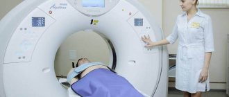
Computed tomography is used to examine the kidneys.
Such informative examination procedures as computed tomography and magnetic resonance imaging are not prescribed for young children and people with mental pathologies. During the kidney check, it is necessary to remain completely still for an hour, which these categories of patients are incapable of. The following studies are usually carried out at varying levels of complexity:
- ultrasonography. The procedure allows you to assess the condition of the calyces, pelvis and tubules, differentiate between benign and malignant neoplasms, and determine the localization of the infectious focus. The study allows you to detect stones in the kidneys or bladder and suggest their chemical composition. This is the only diagnostic method that has no contraindications and does not require special preparation;
- urography. The method is indispensable for establishing the degree of damage to the kidney vessels and assessing the blood supply to the organs of the urinary system. Before the procedure, patients are injected with a contrast agent. After it spreads through the veins, arteries and capillaries, the smallest vessels are visualized on the computer screen. Urography is contraindicated for people who have individual sensitivity to contrast agents;
- X-ray examination. During the procedure, the human body receives a dose of radiation that is considered safe. The photographs are not always informative, since the images are obtained only in one or two projections;
- scintigraphy. The static method allows you to determine the shape of the kidneys, their location relative to each other, and assess the degree of damage to the pelvis and calyces. During dynamic scintigraphy, patients are injected with a contrast agent. On the monitor screen, the specialist monitors the movement of blood through the pelvic vessels in real time, checks the integrity of the veins, arteries and capillaries;
- Magnetic resonance imaging. The procedure is contraindicated for patients with pacemakers, metal dental implants, and even tattoos. There are also some restrictions for pregnant and breastfeeding women. Modern diagnostic techniques make it possible to obtain three-dimensional images of the kidneys and evaluate the functioning of a single kidney after removal of the other. After completing the study, the patient receives a transcript of the results within 15-20 minutes;
- CT scan. This diagnostic method is used to study possible damage to the renal structural elements, assess the effectiveness of the prescribed treatment, and determine the area of surgical intervention. Using CT, you can evaluate the condition of the kidney before making a decision to remove the second one.
When examining the kidneys, the urologist always examines the bladder using cystoscopy. To do this, a thin catheter with a built-in camera is inserted into the hollow organ, and the diagnostic results are visualized on a computer screen.
Cystoscopy is an auxiliary method for recognizing a decrease in the functional activity of the kidneys. If blood or pus is found in the urine, then it is necessary to determine the location of the inflammatory focus.
Ultrasound is an effective method for checking the kidneys
Many modern diagnostic methods are contraindicated or are not very informative for some patients (pregnant women, people with prostheses containing metal). Using endoscopic techniques, the urethra and bladder are examined. By installing the built-in camera so that the opening of the ureters is in the field of view, you can find out whether the kidney is bleeding.
After all laboratory and instrumental studies have been completed, a biopsy sometimes becomes necessary.
This morphological diagnostic method is used to differentiate malignant and benign neoplasms of the kidneys, liver, and lungs. Using a special endoscopic instrument, a small piece of the biological sample is pinched off for further examination in the laboratory.
For those people who care about their health, unexpected diagnoses are not scary. They undergo all examinations on time and immediately make an appointment with a doctor if their general condition worsens or if pathological signs of inflammation appear. Diagnosing kidney disease at an early stage allows you to avoid long-term hospital treatment.
Instrumental
- Endoscopy:
What symptoms can be observed with kidney pathologies?
View gallery
Every doctor should know how to check the kidneys, besides palpation. If an inflammatory process is suspected, special functional tests are performed to assess its presence or absence. The most widely used method is the "tapping symptom". It is carried out by a general practitioner who wants to rule out kidney disease. In addition, this method is used in any hospital during the doctor’s daily rounds. The test is carried out with the patient standing or lying on his stomach. The doctor places one palm on the kidney area, and with the other makes light tapping movements on it. After this you need to change sides. The test allows you to assess the presence of pain in the right or left kidney. Pain indicates an inflammatory process. Most often, a positive reaction to the “tapping symptom” is observed with pyelonephritis, a pathological condition in the tubules.
View gallery
What tests are the most informative?
To identify kidney pathologies and find out everything about possible organ diseases, you should undergo general urine and blood tests - this is mandatory and the procedure is considered a classic one. In addition, the doctor will make a number of other recommendations based on your medical history and visual examination. But if a specific pathology is not identified or there are no prerequisites for clarifying the diagnosis, a biochemical study of blood and urine will be the first and last analysis. If necessary, the examination will be complete and as thorough as possible.
An extensive list of studies is aimed at clarifying the diagnosis and narrowing the range of causes of illness. And if a nephrologist asks you to take the same test several times, this means that the working methods have not yet given the desired picture and the results should be checked again to eliminate any inaccuracies.
After conducting an external examination of the patient and listening to his complaints, the urologist will tell you where to start the examination and what tests are necessary. As a rule, the results of laboratory and biochemical tests of urine and blood are needed to make an initial diagnosis. The content of white and red blood cells, as well as epithelial tissues, is determined in urine.
An informative method for studying the functioning of paired organs is the daily volume of urine. It is necessary to submit to the laboratory urine collected during the day, excluding the first emptying of the bladder. In this way, it is possible not only to establish the presence of an inflammatory focus, but also the location of its localization in one of the organs of the urinary system.
In order to determine the type of pathogenic pathogen, laboratory technicians inoculate a biological sample in a nutrient medium. This method reveals the sensitivity of microorganisms to antibacterial drugs that will be used in treatment.
Normal kidney ultrasound findings
- from a finger to establish or refute the inflammatory process and the extent of its spread;
- from a vein to determine the concentration of proteins and urea.
For reliable test results, you must not eat food 12 hours before the procedure. If you suspect an endocrine etiology of decreased functional activity of the kidneys, you should not drink any liquid or even brush your teeth. People with systemic diseases need laboratory tests every 6 months.
Changes in the qualitative composition of urine
If kidney disease is suspected, many tests are prescribed to detect changes not only in the quantity, but also in the quality of urine. Such laboratory tests include the Nechiporenko, Amburge, and Kakovsky-Addis tests. All these tests involve taking an average portion of urine. The material is then examined for the presence of leukocytes, red blood cells and casts. In all cases, an accurate count of the formed elements is carried out, and then a conclusion is given.
The samples differ from each other in that each of them has different normal values. The Nechiporenko analysis is considered good if there are less than 2000 leukocytes and less than 1000 erythrocytes in the field of view. Cylinders with normal indicators are rarely determined, the norm is up to 500. In the analyzes according to Amburge and Kakovsky-Addis, the shaped elements are the same. The difference is that in the first the norms are 200 and 100 units, and in the second - 2 million and 1 million.
Laboratory methods
General urine analysis
If it is not a daily test, you should take urine in the morning, having first released the first small portion - during the night, a certain amount of mucus and epithelial cells accumulate at the end of the urinary canal, which can mislead as to the composition of the urine itself. First you need to wash your genitals and perineum. If you need to take a test during your period, it is better to do it in a medical institution, because then a catheter and disinfection of the genitals are necessary. The dishes must be dry and clean; the required amount of urine for analysis is 200 grams. If for some reason you cannot immediately take it for analysis, then place the carefully closed container in the refrigerator to prevent the growth of bacteria, which like to do this at room temperature.
A general urine test determines a number of important parameters.
- Density assesses the ability of the kidneys to filter and reabsorb fluid from primary urine. It also depends on the level of salts, including stone-forming ones.
- The reaction (acidic or alkaline) indicates the qualitative composition of urine, i.e. what salts are present in it in greater quantities. In addition, acidification or leaching may be markers of many other pathological processes that occur in or outside the kidney.
- Protein content in urine . Normally it should not be there, and if it is, then there are problems with reabsorption. This is an alarm bell - the body should not lose protein in the urine.
- Glucose . If sugar appears in the urine, this means that either its concentration in the blood has exceeded a certain threshold, or reabsorption in the tubules is impaired. This symptom means mandatory further, deeper diagnosis.
- Bilirubin is usually an indication of problems with the liver or red blood cells (red blood cells)
- Actually red blood cells , as you might guess, indicate that blood is getting into the urine. If there are a lot of red blood cells, the urine takes on a reddish tint, which becomes visible to the naked eye. This phenomenon is called gross hematuria, and we described it in a previous article. If red blood cells can only be seen under a microscope, then this is microhematuria. In this case, the processes occurring are less threatening, but still require attention.
- White blood cells , if their number in the urine is higher than normal, may indicate inflammation or, if they are eosinophils, an allergic reaction. Sometimes, to determine exactly where the inflammation is localized, the Thompson test can be used - urine is collected in three containers and the number of leukocytes in each of them is determined. If there are more of them in the first vessel, then the problem is “low” - the urethra, prostate. If in the third, then the inflammation is higher, in the bladder. If it’s equal, the problem is in the kidneys.
- Epithelium is the cells lining the mucous membranes. During inflammation, the epithelium appears in the urine in large quantities, and it is possible to determine by the nature and shape of the cells where exactly this epithelium came from.
- Cylinders, conglomerates , consisting of various “bricks”. They are formed only in the renal tubules, and by their composition one can determine the nature of the pathological processes occurring in the kidneys.
- Actually salt . We discussed in detail what they are and what they can mean in the materials on kidney stones.
Normally, urine should not contain any protein, sugar, or bilirubin.
Functional urine tests
Functional tests are simple in nature, but at the same time they reveal the essence of the important nuances of the life of the kidneys. For example, the Zimnitsky test, in which urine is collected every three hours throughout the day. This way you can estimate both the total amount of urine excreted and the nature of urination depending on the time of day. All this allows you to determine possible renal failure or suspect a number of other diseases.
Qualitative urine tests
- If doctors need to know how many blood elements your body loses, they need an Addis-Kakovsky test , for which they need to collect all the urine in a day.
- A bacteriological study is needed in order to determine which pathogens provoked inflammation in the urinary system, for their further effective destruction (normally, urine should be sterile).
- Using sensitive reagents, it is possible to determine the presence in the urine of specific enzymes that accompany specific pathological processes (for example, lactate dehydrogenase appears in tumors, and leucine aminopeptidase in glomerulonephritis).
General and biochemical blood tests
A blood test allows you to directly assess kidney function and more.
- Doctors will be helped by the number of red and white blood cells , as well as the qualitative composition of the latter.
- Thickening or excessive thinning of the blood may indicate renal failure in its various stages.
- One of the main markers for kidney diagnostics is serum protein .
- Blood clotting parameters are indicative in connection with the participation of the kidneys in this process.
- One of the most informative indicators of filtration function are the levels of creatinine , total nitrogen and urea , as well as the electrolyte composition of the blood (the content of sodium, potassium, magnesium, calcium and chlorine).
Urine analysis according to Zimnitsky
View gallery
The Zimnitsky method test is used to determine changes in the quantitative composition of urine. Deviation of the analysis from the norm indicates a violation of the concentration function of the kidneys. The main indicator that is assessed when performing the Zimnitsky test is the relative density of urine, which should change throughout the day. When it decreases, you can think about a large loss of fluid, which is often observed with diabetes. If the density remains at the same level all the time, then one should suspect a disorder in which the kidneys lose the ability to concentrate urine, that is, the ability to reabsorb. The test consists of taking tests throughout the day, every 3 hours (8 servings). In conclusion, daily diuresis, the ratio of daytime and nighttime urination are assessed, and protein loss is calculated.
Functional diagnostics
Renal function testing methods play an important role in the diagnosis of kidney pathologies because they help recognize and predict the outcome of treatment for a particular renal pathology.
There are a large number of methods for functional examination of the kidneys, but the most popular are the following:
- Concentration and water tests, which together constitute an accommodation test.
- Detection of the presence of residual nitrogen or urea in the blood.
- Simultaneous detection of the presence of residual nitrogen and urea in the blood and subsequent comparison of the obtained indicators.
It is important to remember that even the simplest functional study of the kidneys, which consists of monitoring urination, the specific gravity of urine excreted per day with the patient’s usual diet and in his usual environment, helps to get an approximate idea of the quality of kidney function.
Rules for collecting urine for analysis
If there are changes in the qualitative or quantitative composition of urine, the doctor must develop a further diagnostic plan, that is, think: how to check the kidneys more thoroughly and what instrumental research methods to prescribe? In some cases, poor tests depend on incorrect sampling techniques. In order for a laboratory test to be accurate, it is necessary:
- Rinse urine container thoroughly.
- Toilet the external genitalia immediately before taking the test.
- Immediately after filling the jar with urine, you must close it to prevent bacteria from entering.
- After collection, take the urine to the laboratory within 1-2 hours.
How can chronic kidney disease be diagnosed?
At the initial stage, kidney damage is asymptomatic. However, disorders in the functioning of the kidneys are well identified with the help of simple tests and studies. This:
- general urine analysis;
- biochemical blood test to determine the level of creatinine (this is the name of one of the end products of protein metabolism in the body, which must be excreted through the kidneys; its level is used to judge the excretory function of the kidneys);
- Ultrasound of the kidneys;
- test for microalbuminuria (protein in urine). Normally, the protein remains in the body and does not enter the urine. The presence of protein (albumin) in the urine may indicate impaired renal function. Those suffering from diabetes and hypertension should have this test done at least once a year for timely diagnosis of chronic kidney disease at an early stage.
At the time of the initial visit to a nephrologist, more than half of patients already have late stages of CKD, when treatment is impossible and only dialysis or transplantation remains. However, these people, usually already suffering from diabetes, obesity or cardiovascular diseases, could have been helped by their attending physician - an endocrinologist, cardiologist or internist - if they had promptly ordered blood and urine tests, correctly interpreted their results and recommended supportive treatment.
Unfortunately, 60-70% of patients with CKD in Russia learn about their diagnosis in intensive care, where they are admitted with acute renal failure, hypertensive crisis, heart attack or stroke. At this point, as a rule, there is only one way out - renal replacement therapy.
Simple tests completed on time not only save and prolong the lives of many people, but also provide a huge economic benefit. A biochemical blood test costs the clinic only 100 rubles, and a hemodialysis session costs from 6,000 to 10,000 rubles. The cost of conducting 156 sessions per year (3 times a week for 4 hours) is more than 750,000 rubles. per patient, taking into account drug provision - up to 1.5 million rubles. in year.
The importance of instrumental research methods
The final diagnosis can be made after special research methods, which include kidney ultrasound, excretory urography, and biopsy. These methods make it possible to detect the incorrect location of the organ (nephroptosis), the presence of developmental anomalies (polycystic disease, duplication), various stones indicating their size and shape. It is possible to confirm the diagnosis of chronic pyelonephritis even in the absence of its manifestations (expansion of the cerebral palsy on ultrasound). How are the kidneys checked if a malignant process is suspected? A biopsy is taken followed by histological and cytological examination of the material.
Instrumental tests
How can you find out if the kidneys are healthy by “looking” inside the body and receiving complete information about the location, size and internal structure of the organs? Fortunately, this is available to modern medicine with the help of high-tech devices. Below we consider popular instrumental methods for examining the kidneys, which are informative for the doctor and safe for patients.
Kidney ultrasound
Ultrasound is a non-invasive and safe way to diagnose kidney diseases, allowing you to evaluate:
- location;
- contours;
- exact dimensions;
- condition of the pyelocaliceal system;
- internal structure of the organ.
An ultrasound examination confirms that the patient has acute and chronic inflammation of the renal tissue (pyelonephritis, glomerulonephritis), nephrolithiasis, cystic formations, abscesses, tumors.
Overview R-graphy
Plain radiography of the abdominal cavity allows you to visualize the kidneys, bladder and ureters, but their contours will be unclear, blurred due to intestinal loops. Therefore, at present, R-graphy of the kidneys is being replaced by more indicative diagnostic methods.
Excretory urography
Among x-ray examinations of the kidneys, excretory urography remains the most popular. It is based on the intravenous administration of contrast (sodium amidotriazote), which is excreted mainly by the kidneys, and the creation of a series of x-rays:
- overview image before the introduction of a radiocontrast agent;
- 5 minutes after administration - for visualization of the kidneys;
- after 20 minutes - for a detailed examination of the collecting system and the upper part of the ureters;
- after 45 minutes - to visualize the ureter;
- after urination – to diagnose bladder pathology.
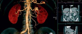
CT, MRI
Modern methods of layer-by-layer visualization and creation of a virtual 3D model of internal organs are used to clarify the results of ultrasound and excretory urography. Based on the action of X-ray radiation (computed tomography) or magnetic field (magnetic resonance imaging).
Angiography
Angiography is an x-ray method for examining vessels in the kidneys, in which a contrast agent is injected into the renal artery. Angiography is performed if an aneurysm, renal vessel stenosis, persistent bleeding, or renal hypertension are suspected.
Kidney biopsy
The method of puncture biopsy of the kidney involves percutaneous insertion of a thin needle into the organ and collection of biomaterial (for example, tumor cells) for further morphological examination. It is carried out for the differential diagnosis of cancer and other diseases requiring histology.
Thus, in diagnosing diseases of the urinary system, one cannot stop at just one analysis. Each of them - clinical, laboratory or instrumental - is important in its own way. In making the correct diagnosis, it is necessary to have a comprehensive examination of the kidneys, which will allow you to get a complete picture of the existing disease and draw up a plan for further action.
Related publications
- Renal sinus cyst - symptoms and treatment methods
- Chronic nephritis: symptoms and methods of treating the disease
- Causes of development of kidney hypoplasia and methods of its treatment
How to check your kidneys, what tests to take for diagnosis
The kidneys are a paired organ. Their function is to filter the blood, removing waste products and toxins in the urine. Impaired kidney function leads to poisoning of the body with decay products and diseases of other organs and systems.
Timely detection of deviations is easier to treat and leads to fewer negative consequences. How to check the kidneys is decided by a nephrologist or urologist.
The doctor prescribes several tests and hardware techniques that will reveal abnormalities in the functioning of the organ.
How to check your kidneys at home
Kidney pathologies have similar symptoms to gastroenterological diseases and problems of the reproductive system.
Main distinctive symptoms:
- Back pain above the waist. Shooting acute pain is a sign of renal colic. Aching pain indicates chronic pathology.
- High blood pressure.
- Edema. They arise due to poor functioning of the organ. The liquid is not filtered into the urinary system, but accumulates in the tissues of the body. Most often the legs swell.
- Change in urine color. Depending on the cause of the problem, the urine may change color to brown or orange, mucus, streaks of blood, and flakes may appear in it.
- The volume of urine excreted per day changes. Normally, a person produces about 1.5-2 liters of urine per day. If this amount increases or decreases without changes in the drinking regime, the kidneys are not working properly.
If disturbing symptoms appear, be sure to visit a doctor. It is impossible to treat any disease on your own, since without test results a correct diagnosis cannot be made. Kidney disease can only be suspected if most of the symptoms are observed.
What does a doctor do first during an appointment?
A kidney examination is carried out by a nephrologist - a doctor who treats pathologies of this organ. You can come to see him either independently or with a referral from a therapist/urologist.
After collecting anamnesis, the doctor examines the person using palpation and percussion. During this test, the area where the kidneys are located is felt and tapped.
With the help of palpation, you can determine the prolapse of the kidney, and pain when pressing or tapping will be evidence of problems with the organ.
After the examination, the nephrologist will tell you where to start the diagnosis. Its stages and methods depend on the diagnosis that the doctor suspects during examination.
Lab tests
Diagnosis of kidney disease is impossible without laboratory tests of blood and urine. Any disease disrupts metabolism; the composition and proportions of components in liquids change. These changes are clearly visible in the analyses.
What urine test should I take to check my kidneys?
Changes in the functioning of the filtering organs will affect the quantity, composition and consistency of urine released. Most often, the pathology can be recognized during a general analysis. But specific tests may be required to clarify the diagnosis.
General urine analysis
This is the main test needed to check your kidneys. Determines the amount of salts, urea and protein in urine. To obtain correct results, you should not take medications or eat coloring foods on the eve of the examination - they change the shade and transparency of the liquid. If the use of medications cannot be stopped, doctors must be warned about taking the medications.
For examination, it is necessary to take 30-50 ml of morning urine from the middle portion.
According to Zimnitsky
This test determines kidney function by measuring the concentration and dilution of urine. A daily volume of urine is provided to the laboratory for testing. The results are distorted in women during menstruation, so it is better not to donate urine in the first days of the cycle.
The same test determines the deficiency of urine production or excess urine. The results of the analysis should take into account the patient’s drinking regime, that is, the amount of liquid he drinks during the day. The norm is 2 liters, but deviations within half a liter are acceptable.
According to Nechiporenko
In kidney failure, urine production is intermittent. Nechiporenko analysis reveals this condition. For a reliable result, you will need to collect urine every hour for 12 hours. Sometimes patients falsify the result by collecting urine every few hours and dividing it into several parts. These actions lead to false test results, making it difficult to make a diagnosis.
Bakposev
Mandatory tests for kidney inflammation include bacterial culture. In the laboratory, a small amount of urine is examined under a microscope to determine the pathogenic microorganisms in it, as well as their variety and resistance to antibiotics.
Other techniques
A complete kidney exam includes testing for specific indicators of chronic disease. For example, people with diabetic nephropathy need to regularly check the level of albumin (protein) in their urine.
Another specific analysis for functional diagnostics is the Bence-Jones study. This is how kidney tumors are determined.
Diagnosis of kidney disease always includes a blood test.
It shows the concentration of different substances, which changes during diseases. Normally, the blood in the kidneys is cleared of urea, creatinine and uric acid.
In case of illness, the indicators of these substances in the general analysis will exceed the norm.
A biochemical study shows changes in the level of protein and kidney enzymes. This test does not show the condition of the kidneys and does not identify a specific disease.
Instrumental methods for kidney diagnostics
Hardware diagnostics provide information about organ damage and the localization of inflammatory and tumor processes. Non-invasively, such data can be obtained using ultrasound, x-rays, and tomography. Before examining your kidneys, you must obtain a referral from your doctor about the need for procedures.
Ultrasound
The method is based on the reaction of tissues of different densities to ultrasound. Thanks to ultrasound, you can determine the size of the kidneys, their location in the body, the presence of sand, tumors and inflammation. There are more accurate methods, but due to contraindications, they are prescribed only if it is impossible to determine the pathology using ultrasound.
X-ray
An X-ray examination shows the condition of the kidneys, the presence of tumors, displacement, clogged ducts. Most clinics are equipped with devices for X-ray examination.
The disadvantage of x-rays is the harm caused to the body by radiation. This one-time procedure will not cause problems, but its regular use is contraindicated. In this regard, x-rays are prescribed only if a serious pathology is suspected. As a preventive measure, it is preferable to use ultrasound.
During this test, a sample of kidney tissue is taken for analysis. A biopsy is prescribed if there is a suspicion of cancer or a cyst. By analyzing kidney tissue, you can find out the nature of the tumor.
Endoscopy
Non-traumatic or low-traumatic method of research. An endoscope with a camera placed on it is brought to the organ through the ureter or a small incision on the body. Using this method, you can visually examine the injured or inflamed area of the kidney. Endoscopic techniques are also used for minimally invasive surgical interventions.
Modern methods of examining the kidneys, which provide information about the condition of the organ in a three-dimensional projection. The disadvantage of these studies so far is their high cost. Magnetic resonance imaging cannot be performed on patients with pacemakers. Most often, these studies are carried out in private offices or in regional medical centers.
Chromocystoscopy
The patient is injected with a substance that colors the urine. After this, an X-ray examination of the kidneys is performed. Prescribed if tests have shown a violation of the excretory and secretory functions of the kidney.
A person is injected with a radioactive drug that accumulates in the kidneys. After this, a study is carried out using a gamma camera. The drug is harmless and is excreted in the urine within several hours after injection.
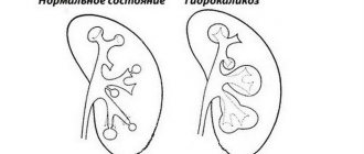
The examination reveals renal failure, abnormalities in organ development, hydronephrosis, metastases from tumors.
The method is based on the same technique as with scintigraphy. Instead of being examined in a gamma camera, after the person is injected with a radioactive substance, they are placed under an X-ray machine and pictures of the kidneys are taken every few minutes. The results show stagnation of urine, narrowing and blockage of the ureters, and kidney stones.
Another examination with a coloring pigment. The blood vessels of the kidneys are stained with a radioactive substance. After the injection, the person is taken pictures using an X-ray machine, magnetic resonance imaging or computed tomography scanner. Blood flow pathologies are clearly visible in the photographs. The analysis is prescribed if aneurysms, blood clots, stenoses, or internal bleeding are suspected.
Other methods
X-ray diagnostic methods require caution. Dye preparations are considered harmless, but their use requires indications for examination. Therefore, such methods are a last resort. Urologists mainly prescribe ultrasound and CT/MRI. If cancer is suspected, a biopsy is used.
Risk group: who needs to regularly check their kidney health
The risk group includes people who have a higher risk of developing kidney disease due to bad habits, being overweight or underweight, or having had kidney disease in the past. The danger comes from working in hazardous industries, for example, those associated with varnishes and paints. Such people need to check whether their kidneys are hurting every few months.
How much does it cost to check kidney function in Moscow clinics?
A general blood and urine test can be taken at municipal clinics if there is any suspicion of kidney disease. The cost of further diagnostics depends on the chosen methods and indications.
X-ray and ultrasound examinations are available in every institution. To conduct a tomographic examination, magnetic resonance imaging and some tests, you will need to go to private clinics.
The average cost of research is:
- General blood test – 200-500 rubles.
- General urine analysis – 175-200 rubles.
- 200-700 rub. – for laboratory tests of urine according to Zimnitsky, Nechiporenko, Reberg.
- 1200-3000 rub. for ultrasound examination, including the use of special reagents.
- MRI and CT scans cost 3-6 thousand.
Kidney diseases are dangerous and easier to treat in the early stages. As the disease progresses, it will be necessary to use more expensive and complex diagnostic and treatment methods. To prevent risks, it is worth checking your kidney health during an annual medical examination.
Source: https://tden.ru/health/kak-proverit-pochki
Indications for diagnosis
Indications for the diagnosis of various kidney diseases:
- changes in the organoleptic properties of urine, daily amount;
- pain in the back, lower back, groin area;
- detection of blood, sediment, flakes in urine;
- chronic internal diseases (diabetes mellitus, hypertension, endocrine disorders, anemia);
- injuries, poisoning with toxic substances;
- congenital anomalies, hereditary pathology;
- suspicion of a malignant or benign tumor;
- pregnancy with complications;
- obesity, anorexia, signs of dehydration;
- a history of genetic or autoimmune diseases.
In what cases is the examination carried out?
Anyone can have their kidneys checked, since almost all procedures are harmless, and some do not require special preparation.
This manipulation will also be useful for those people who want to check the condition of a powerful filter throughout the body.
But there are also medical indications for checking the kidneys. These include some diseases and pathological conditions. Which ones exactly:
- Increased blood pressure levels.
- Little urine output.
- Frequent urination, trips to the toilet at night.
- Unpleasant sensations, a feeling of heaviness and pain in the lumbar region.
- Feelings of pain when urinating.
- Urine gives off an unpleasant odor.
- The color of urine changes noticeably. The blood content in the urine should be especially alarming.
- Low hemoglobin levels, characterized by anemia, if there is no blood loss in chronic or acute forms.
- For diagnostic purposes for urolithiasis. Here, kidney diagnostics can be performed in two stages. In cases where the patient likes to indulge in chocolate products, pickles, meat, legumes and offal, a routine examination is carried out. If the presence of kidney stones is suspected, an emergency check is carried out.
- Dehydration may occur due to shortness of breath, diarrhea, or high fever. In this case, this procedure is required.
- Many drugs are toxic to the kidneys. This list includes diuretics, Aspirin, Biseptol and others. After treatment with these medications, you need to undergo an examination to check your kidneys. When the level of body temperature rises to 37.5 degrees, when the patient is daily worried about weakness and malaise in the evening, it is imperative to pass all the necessary laboratory tests to check the condition of the body’s main filter.
- Many diseases, such as diabetes mellitus, systemic lupus, also require kidney testing.
- If neoplasms are detected in the abdominal cavity by palpation, a kidney examination must be performed.
Before you check your organs, you should find out how the kidneys are examined and what methods exist.