Intravenous urography of the kidneys - indications, contraindications, technique and side effects
For various pathologies of the renal and urinary systems, intravenous urography is increasingly being used in medical clinics.
The modern examination method allows you to obtain highly accurate results.
However, this procedure has its limitations for use, and it is also important to know a number of rules for proper preparation before intravenous urography.
Indications for the procedure
Intravenous urography of the kidneys is prescribed by the attending physician in the presence of the following diseases and disorders:
- various pathologies of the genitourinary system;
- inflammatory process of the urinary tract;
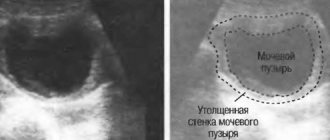
- disruption of the integrity of the bladder;
- abnormal change in bladder functionality;
- chronic kidney disease;
- urolithiasis disease;
- abnormal location (prolapse) of the kidneys;
- oncological neoplasms (both benign and malignant);
- failure and slowdown in the excretory functioning of the kidneys.
A fairly extensive list of pathologies in which intravenous survey urography will help to determine the patient’s condition as fully as possible.
If the patient is suspected of slowing the excretory functioning of the kidneys, he is prescribed intravenous excretory urography.
Also, intravenous urography is a mandatory procedure performed before any surgical intervention in the area of the genitourinary system (for example, if surgery directly on the bladder itself or removal of kidney stones is indicated).
Undergoing an intravenous urography procedure is a serious intervention in the human body. The decision to perform the procedure must be made by the attending physician. It is strongly not recommended to carry out this examination technique on your own initiative!
Contraindications
Like any medicinal method, this procedure has a number of contraindications in which it is strictly forbidden to carry out this examination procedure.
Contraindications to intravenous renal urography are presented in the following list:
- hyperfunction of the thyroid gland (hyperthyroidism);
- excess iodine in the body or intolerance to substances containing iodine;
- feverish condition.
However, if the patient’s health and life are at risk, the attending physician may decide (in exceptional cases!) to refer the patient for examination.
For the fair sex, there is another conditional contraindication - the menstrual cycle.
It is strongly recommended to reschedule the scheduled examination until the end of the menstrual period if the attending physician decides that this examination is not urgent and that it may be delayed for several days.
Women during pregnancy and lactation (breastfeeding) require special, increased attention and careful treatment. In case of pathology of the renal and genitourinary systems, the attending physician must decide to refer the patient for intravenous urography with special precautions!
Preparation for the procedure
Preparation for intravenous urography requires special attention.
If the patient has received a referral from the attending physician for this examination, he needs to familiarize himself with a number of rules for proper preparation:
- the patient needs to undergo a complete bowel cleanse. This is done with the help of an enema or by using special medications aimed at soft bowel movements. One of the most famous and effective drugs intended for this purpose is Fortrans. The enema must be performed in the evening, on the eve of the examination, and also early in the morning, three hours before urography. Any of these options is suitable for people of the older age group, but for children it is preferable to cleanse the intestines with the help of special preparations;
- the day before the procedure, you must refrain from consuming foods and drinks that increase gas formation in the intestines. Such products include all kinds of sweets, baked goods, fruits (especially those with a high sugar content), peas, cabbage, bread, fruit juices, carbonated drinks;
- On the day of the procedure, the patient is allowed to eat a small portion of morning breakfast. In addition, you need to significantly increase the amount of water consumed. Moreover, the water must be purified, non-carbonated. You should refrain from sweet drinks and give preference to spring water;
- Three hours before the start of the procedure, you must completely abstain from any food intake.
After following all the above recommendations, you can be sure that the examination will be as effective as possible, and the result will be impeccably accurate. It is worth noting that in different medical clinics, patient preparation for intravenous urography may differ slightly.
Also, immediately before the procedure, the patient must be fully informed about how the examination will be carried out and how the patient will feel.
The fact is that intravenous urography can cause very unpleasant symptoms and sensations in a person.
And human psychology is designed in such a way that all unusual and uncomfortable feelings can cause panic and fear. The patient may also experience obvious anxiety before an unknown procedure. Any nervous disorder or emotional stress of the patient can have an extremely negative impact on the results of the examination.
Some medical institutions provide for the administration of a sedative to the patient (intravenously or intramuscularly, or in tablet form). This will allow the patient to return to a normal psycho-emotional state and get rid of fears and neuroses.
With the help of intravenous urography, the healthcare professional monitors the shadows of the urinary tract during x-rays. If the patient is nervous and under emotional stress, the shadows may not be displayed correctly, which will ultimately lead to inaccurate results.
Methodology of the procedure
Having familiarized yourself with all the indications and contraindications, as well as preliminary preparation, it’s time to understand how intravenous urography of the kidneys is done.
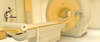
Equipment for urography
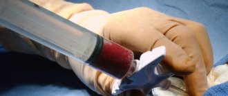
The procedure is carried out in several stages. The patient lies down on the X-ray table, after which several standard images are taken. After the first stage, the patient is administered a contrast agent intravenously.
It is usually inserted into a vein in the elbow. A contrast agent is a medicinal composition that, when conducting radiological studies, allows you to visualize the area being examined as accurately as possible and significantly increases the accuracy of the data.
The contrast is completely harmless and cannot cause negative consequences (such as an allergic reaction).
However, in some cases, a person who receives intravenous contrast may experience some discomfort such as headache, dizziness, nausea, and vomiting. This is quite rare and is of an exclusively individual nature. One of the most important points when performing intravenous urography of the kidneys is that the medical worker very slowly injects the contrast agent into the patient (the duration of administration takes about two minutes). This technique allows to minimize the occurrence of discomfort and unpleasant sensations in the patient.
Some time after the administration of the drug (within 5-10 minutes), the X-ray procedure begins. Several new images are taken at different time intervals, which are determined by an experienced urologist individually for each patient.
The injected contrast agent helps doctors monitor how long it will take for it to be excreted by the kidneys, and also makes it possible to most accurately determine the state of the renal and urinary systems, and to detect oncological tumors and kidney stones at an early stage.
In some cases, another stage of examination may be required at a later date after the administration of the contrast agent (on average an hour later). The doctor can also refer the patient for an x-ray in a standing position.
This will allow you to observe the work of the kidneys in dynamics and track their mobility, and in addition, detect pathology or anomaly regarding the location of the kidneys.
The procedure is absolutely painless; you may only experience slight discomfort when inserting a needle with a contrast agent. However, since intravenous procedures are quite common in medical practice and are familiar to almost every person, intravenous administration of the drug should not cause any concern.
Intravenous urography of the kidneys is a fairly safe procedure, especially if performed by experienced medical specialists. However, it is a prerequisite that the radiography room contains all the necessary equipment to provide first aid if the patient feels unwell when the drug is injected into a vein.
Side effects
Despite the fact that with proper preparation and under the close supervision of experienced physicians, the procedure is quite safe, side effects may occur after it is performed.
Side effects include the following:
- after the procedure, the patient may feel a taste of iron in the mouth;
- in some cases, a rash may be observed on the patient’s skin;
- after the procedure, the patient may feel very thirsty and dry mouth;
- slight swelling of the lips is a rather rare pathology after urography;
- a contrast agent can lead to tachycardia (rapid heartbeat), which soon stops and the person notices the rhythm of the heart muscle that is familiar to him;
- during urography, as well as after its completion, the patient’s blood pressure may drop significantly;
- the most severe and dangerous consequence after the procedure is the appearance of liver failure (even if the patient has never previously complained of problems with the body’s main barrier – the liver).
Since the side effects are quite significant, it is worth noting once again that intravenous urography must be carried out under the close supervision of experienced doctors and all prescribed recommendations must be followed. If you feel unwell or have complications after urography, you must immediately notify your doctor.
on this topic
How does it feel during and after intravenous urography? Feedback from one of the patients before you:
Source: https://mkb.guru/bolezni-pochek/diagnostika-i-analizy/vnutrivennaya-urografiya.html
How is the procedure performed?
The excretory urography procedure involves taking a series of targeted images of the kidney and pelvis area between 5 and 25 minutes.
Delayed images are rational only if there is a delay of contrast in the pyelocaliceal structures on the last radiographs.
This approach is used when the excretory function of the kidneys decreases.
To assess renal motility (nephroptosis), one image from the series is taken with the patient in an upright position. Pathological displacement is observed when the kidneys are displaced by more than 1 cm.
Intravenous urography provides doctors with information for several kidney diseases, but has serious contraindications and leads to complications.
Did you know that urate stones are easily detected using survey urography, but phosphate stones will have to be looked for in other ways? Survey urography: the essence of the procedure, indications, and how important preliminary preparation for the procedure is. In addition to urography, ultrasound remains a mandatory method in the study of kidney diseases. Read this article about how to properly prepare for it for adults and children.
Urography in Children: Preparation and Indications for Carrying Out
Excretory urography is an x-ray method for studying the genitourinary system.
This procedure allows us to identify pathological changes occurring in the organs of the genitourinary system and study the dynamics of urine output.
The combination of the method and the use of contrast agents helps to track the position of kidney stones, foci of infectious inflammation and neoplasms. Modern medical developments have made it possible to apply the method to children.
Pediatric urography: types and indications
Kidney urography in children is prescribed according to indications, after the rest of the genitourinary diagnostics and for really serious indicators.
Urography, according to the volumetric indicator of information and the types of implementation, is divided into 3 main types;
- Excretory urography.
- Survey urography.
- Intravenous urography.
Urography of the kidneys with the use of a contrast agent is prescribed for children with certain pathologies, and if other methods have not provided a complete clinical picture of the disease.
Main indications for the procedure:
- The course of urolithiasis in the organs of the urinary system.
- Diagnosis of various types of anomalies of the urinary system.
- Neoplasms of unknown origin.
- Various kidney damage after injury.
- Complex cancer tumors.
- Severe cases of kidney tuberculosis, pyelonephritis, glomerulonephritis.
When prescribing a procedure for children, all risks and contraindications are assessed, but in many cases this procedure is vital.
Methodology
To begin kidney urography in children, it is important to properly prepare the child physically and psychologically.
Before the procedure, the child’s gastrointestinal tract is prepared several days in advance, eliminating fried, salty, spicy and pickled foods, everything that contributes to distorting the results of the study. A laxative is prescribed one day before, and a cleansing enema is given immediately before the procedure.
Taking into account the development of modern medicine, the risks of allergic reactions to the contrast agent used are minimized. But the human body is individual, so a reaction test is carried out. Immediately before the procedure, an antiallergenic drug is administered as a preventive measure.
Parents should talk to their child about the procedure and explain all the stages. The room requires a temperature of more than 31 degrees. The child’s body must be motionless; due to circumstances, there are cases of using general anesthesia for the procedure.
Survey urography
Survey urography of the kidneys is a method of examining the genitourinary system without the use of a contrast agent.
It is not very informative, but helps to diagnose the presence of parasites, stones, and tumors.
With the help of such diagnostics, information is obtained about the exact location of the organ relative to others in the system and relative to the human skeleton. Most often prescribed before excretory urography.
The procedure is carried out with the patient standing, X-rays are directed to the area of 3-4 vertebrae.
Indications for survey urography of the kidneys will be: back injuries, urolithiasis of the kidneys, renal colic. As a result, the image will show the entire abdominal area, with outlines of organs, areas of various human tissues and bones.
During survey urography, some patients learn that they have only one kidney; approximately 5% of the world’s population has this pathology, but only a few know about it.
Excretory urography
Allows you to study the dynamics of bladder filling and the movement of urine through the urinary system. With the use of contrast agents, it is possible to track changes in the size of organs, their structure, the appearance of foci of inflammation and neoplasms. The diagnostic procedure shows all anomalies in the structure of the system.
The purpose of the procedure is a complete diagnosis of the functioning of organs, down to the smallest mechanisms. Helps track the structure of the renal pelvis, ureters and bladder. The presence of blockages and stagnant processes, insufficient filling of the renal pelvis is checked.
The absence of contrast movement may indicate severe inflammation or blockage of the ureter with a large stone.
The procedure is carried out with the patient lying down on a special table, a contrast agent is injected into a vein and after a few minutes the first images are taken when the contrast reaches the kidneys. Pictures are taken at a certain frequency (approximately 5-15 minutes).
Intravenous urography
Intravenous urography is a certain modification of excretory urography, but unlike it, contrast is administered intravenously by drip.
Diagnostics shows the speed of the filtration function, the volume of contrast accumulation, and the rate of excretion of the substance along with urine.
During the procedure, the doctor receives several images of the ureters, bladder, pyelocaliceal area and the entire length of the urethra.
Indications for such a procedure are pathologies in the anatomical structure of the urethra, neoplasms, disturbances in the functioning of the bladder, and slow excretory function of the kidneys.
Most often, a study is prescribed before surgery to study the full picture of the operated area.
Contraindications and complications
Any intervention in the human body, and especially a child, carries certain risks. Contraindications to the use of excretory urography using contrast are:
- Development of renal failure.
- Acute liver failure.
- Abnormalities of the thyroid gland.
- Tendency to bleed.
- The course of pyelonephritis and glomerulonephritis in acute form.
- Hormonally active tumor.
- Problems with blood clotting.
- Use of metformin (as therapy for diabetes mellitus).
- Sensitivity to iodine.
If there are contraindications, alternative, less informative research methods are used: ultrasound, computed tomography.
During the procedure, a burning sensation in the vein, dizziness, nausea, near fainting, a taste of iron may occur; these are side effects of contrast. Typically, these complications disappear after the contrast is removed and several hours after the procedure. To speed up the process, you need to drink more water, tea, fruit drinks and other healthy drinks.
Post-procedural complications, in addition to allergic reactions, which they try to minimize, include respiratory dysfunction and surges in blood pressure.
In most cases, this is due to psychological factors during the procedure. But it happens that such consequences arise spontaneously.
Therefore, urography rooms are located at hospitals, where they can provide first resuscitation care.
The risks of urography in children are the same as in adults. The only point is the precisely calculated dosage of the contrast agent, according to weight, age and condition of the urinary system.
This technique is not dangerous if all recommendations and requirements are followed. This research method helps to obtain the most accurate information about the condition of the child’s body, which will help the doctor build an effective treatment regimen.
Source: https://pochkam.ru/diagnostika/urografiya-u-detej-kak-delaetsya.html
System pathologies
Diseases can be of a congenital genetic or infectious nature. In the latter case, inflammation of specific components of the structure occurs. The pathological process predominantly affects the kidneys. Inflammation of other elements of the system, as a rule, poses less danger. However, in any case, pathological processes are accompanied by discomfort, pain, and pain. Genetic diseases are caused by abnormalities in the structure of one or another organ. As a result of these disorders, the formation and excretion of urine is difficult or impossible. Among genetic pathologies there are also anomalies in the formation of the body. These include, for example, the absence of one or two kidneys at once. In this case, death usually occurs immediately after birth. The ureter may also be missing or not enter the bladder. Developmental anomalies can also affect the urethra (excretory canal). Women are more at risk of contracting infections. This is due to the structural features of their urethra - they have it shorter than men. Due to this, the infectious agent quickly penetrates the system, rises to higher organs and provokes inflammation.
Kidney urography in children: when is it prescribed, how to prepare and how to do it
Urography of the kidneys in children is one of the most common studies. It provides an objective picture of the condition of the organs.
Based on this examination, the doctor can conclude whether there are damaged tissues, whether the functioning of the ureters and bladder is impaired. However, this type of diagnosis has its own characteristics.
They should definitely be taken into account in order to properly prepare children for research.
Characteristics of the survey
What is urography? This diagnosis is an x-ray examination that shows the condition of the child’s urinary system.
The essence of the method is based on the injection of a contrast agent into the kidneys. That is why the study is called contrast urography. The injected substance is capable of blocking X-rays.
Initially it accumulates in the kidneys. At this time, the doctor can assess their condition using images. The substance then passes into the urinary system. Thus, this diagnosis gives an idea of the functioning of the ureters and bladder.
When is the examination scheduled?
The child is recommended to undergo kidney urography if there are compelling reasons.
The main indications for this procedure are pathologies :
- stones in the kidneys, ureters;
- abnormal kidney development;
- hematuria, tumors of unknown nature;
- ailments - tuberculosis, acute pyelonephritis in children;
- kidney injuries;
- tumors.
There are several types of urography, each of which has its own indications, degree of information content and specificity of implementation.
Kinds
The following varieties are distinguished:
- Panoramic. With the help of this study, tumors, stones, foreign bodies in the kidneys, and parasitic diseases are detected.
If a child is suspected of having kidney pathologies, then this examination is prescribed. It gives an idea of the state of the organs. In addition, survey urography allows you to study the shadows of the kidneys, their location, shape, and skeletal bones. This study diagnoses the functioning of the ureter and bladder. - Excretory. This diagnosis allows you to study the time and intensity of filling of the bladder and pelvis with liquid.
The study gives an idea of the size, homogeneity, and shape of organs. It is used to detect the location of tumors, cysts, and stones. Diagnostics shows the structural features of the urinary organs. - Intravenous. In this procedure, a contrast agent is injected into a patient with an empty bladder.
The doctor takes pictures while the kidneys absorb contrast material from the blood and store it. The study allows us to identify stones, tumors, cysts, expansion of cavities, pathological stretching or shrinkage of organs.
Objectives of the study
It is quite difficult to identify various kidney lesions or urinary tract disorders in children, since the symptoms of some kidney diseases are very similar to other pathologies. Such signs may indicate colds, acute respiratory viral infections, problems with the gastrointestinal tract, heart, and even diseases of the spine.
Therefore, in order for the doctor to make a correct diagnosis, it becomes necessary to check the functioning of the kidneys. It is urography that can solve such a problem.
This event allows you to determine kidney parameters that are directly related to their functioning:
- dimensions;
- contours and shapes;
- presence of increments (urolithiasis);
- location in relation to internal organs;
- functioning of the ureters, bladder;
- functional state.
In order for the diagnosis to be carried out correctly and show the most reliable results, children should be carefully prepared for urography.
Preparation.
Before urography you will have to undergo blood biochemistry . Such an analysis allows us to exclude pathologies such as renal failure, in which diagnosis is strictly contraindicated.
Preparation for the study consists of the following activities:
- 3 days before the procedure, you should exclude foods rich in fiber and carbohydrates from your diet. Babies are transferred to dietary nutrition.
- Older children are recommended to take Activated Charcoal.
Children are given this remedy only as prescribed by a doctor. This measure allows for excellent gas absorption. - 1 day before urography you need to take a laxative. The doctor will prescribe it. Before going to bed, the child is given an enema.
- If the procedure is performed on teenagers, it is recommended to refrain from eating in the morning.
Young children are allowed to eat a small piece of meat with white fried bread. After eating, you should give the enema again.
Mechanism
Now let's look at how the procedure itself is done . Before urography, an ultrasound is initially performed. It allows you to clarify the individual characteristics of the body.
Various substances are used for diagnosis:
- triombrine,
- urotrast,
- verografin,
- triiodotrust.
All of them are strong allergens. Therefore, the doctor initially calculates the required amount of contrast agent. It takes into account the child’s age, weight, liver and kidney condition.
To protect the patient from allergic reactions, it is recommended to take an antihistamine. Children can receive an injection half an hour before the examination. The most commonly used drug is Suprastin.
The doctor will definitely explain how the diagnosis is made. During urography, 5 images are recorded. It is very important that they turn out right the first time.
Therefore, the parents are with the child. Older children are asked to lie still. Parents should take care of the immobility of their children. After all, the slightest movement can affect the quality of the pictures. It is quite difficult to determine kidney disease in such a situation. One-year-old children may be given anesthesia.
Consequences of the procedure
A child may experience negative reactions to a contrast agent:
- feeling of heat;
- nausea;
- slight dizziness;
- hives.
Such reactions most often go away on their own. Sometimes you may need to take an additional antihistamine.
More serious consequences of the procedure, which are observed extremely rarely, are:
- breathing problems;
- a sharp decrease in pressure.
Despite the fact that such symptoms occur only in isolated cases, the urography room is necessarily equipped with all the necessary tools to provide emergency anti-shock care.
The results of kidney urography allow an accurate diagnosis in children. X-ray examination with a contrast agent determines the degree of functioning of the kidneys, ureter, and bladder.
Source:
Source: https://ekran-stroka.ru/roscherk-01/1088-yrografiia-pochek-y-detei-kogda-naznachaut-kak-gotovitsia-i-kak-delaut
Diagnostics
To assess the condition of the urinary system, excretory intravenous urography is used. This method is an X-ray examination using a contrast agent. With conventional imaging, the urinary tract is not clearly visible. Survey intravenous urography allows you to get a more complete picture of the state of the system and its individual components. The contrast agent enters the bloodstream. From there it enters the kidneys and concentrates there. The contrast then exits through the urethra along with urine. Due to the presence of the substance, X-rays are blocked in accordance with the kidney structure. In the photographs it looks like white spots. The images obtained during diagnosis are called intravenous urograms or pyelograms.
Urography in children
Diagnosis of many urological diseases in childhood is undoubtedly stressful for the baby. Some procedures are quite unpleasant and painful, but it is simply impossible to do without them, for example, when it comes to urography.
The study fully reflects the condition of the urinary system: the presence of tumors, stones and sand, injuries. And parents must know how to properly prepare their child for it so that the pictures are the most informative.
- 1 Indications
- 2 Excretory urography
- 3 Complications
- 4 Editor
Indications
Urography is an examination of a child’s urinary system using x-rays. There are several types of it - survey, excretory, intravenous. Indications for urography in children are as follows:
- pathological changes in urine samples;
- hematuria (determination of blood in the urine);
- developmental anomalies of the urinary organs;
- chronic glomerulonephritis;
- chronic pyelonephritis;
- hydronephrosis;
- nephrogenic hypertension;
- urolithiasis disease;
- suspected tumors in the kidneys, ureters, bladder.
The study makes it possible to determine not only the disease, but also the degree of its progression, the reasons for its formation, and the level of organ damage. Based on the results of urography, the doctor selects an individual effective treatment regimen.
Complications
Urography of the kidneys using a contrast agent in children rarely causes negative reactions. In exceptional cases, the following complications may occur:
- allergic rash, angioedema or anaphylactic shock;
- phlebitis or infiltration at the site of contrast injection;
- indigestion;
- acute renal failure.
To provide emergency medical care to a child, the urography room must be equipped with all the necessary medications and instruments.
Intravenous urography in children is a very informative examination that allows you to quickly and accurately establish a diagnosis. The procedure is quite safe, because the doctor performing it calculates the dosage of the contrast drug as accurately as possible based on the general health of the child, his weight and age.
Source: https://uran.help/surveys/urografiya-u-detej.html
Purpose of the study
Intravenous urography of the kidneys is prescribed in the following cases:
Presence of stones. Stones can be very well visualized during the procedure. Infectious lesions. In case of recurrence of the pathological condition, intravenous urography makes it possible to detect the cause of obstruction or other abnormalities. The presence of blood in the urine. This phenomenon may be due to various reasons. In particular, there may be a kidney tumor, inflammation or infection. Obstruction or damage.
Urography of the kidneys using a contrast agent: preparation, how to do it, consequences
There are many ways to assess the condition of internal organs, including the genitourinary system: MRI, ultrasound, radiography, CT and others. Their disadvantage is that they often do not provide a complete picture.
To fully diagnose anatomical and functional pathologies, a narrow range of methods is used, one of them is kidney urography using a contrast agent.
What are the indications and contraindications for this procedure? How is it carried out and what consequences may arise after it?
What is this procedure and why is it needed?
A black and white image of organs located in the abdominal region and the space behind the peritoneum helps to determine their location.
Their outline may be blurred, this is due to the fact that the projections of other organs are layered on top of each other. This method does not make it possible to assess the full functionality of the urinary organs.
Contrast urography of the kidneys allows you to fully examine what cannot be seen with this (survey) method.
This procedure is carried out through the introduction of a contrast agent to the patient (against the background of the kidneys and other organs, the injected agent has greater contrast) and the production of x-rays. The essence of the method is based on the ability of this very substance to block X-ray radiation. It initially accumulates in the kidney and is then excreted by the excretory organs.
Types of procedure
| Infusion (a contrast agent is used) | Pictures are taken at the moment when the organ removes contrast. This method allows you to fully assess the shape, uniformity, size and intensity of filling of the bladder and pelvis with fluid, as well as the location of stones and various formations, and the general condition of the genitourinary system. |
| excretory | When the bladder is empty, contrast is injected, pictures are taken in the first minutes, then after 4, 7 minutes (while the kidneys knock the substance out of the blood). The method reflects the condition and presence of pathologies of the entire excretory system (prostate gland, kidneys, etc.). |
| Overview | It is prescribed mainly to identify pathologies in the kidneys. The study also helps in diagnosing formations, the presence of parasites and foreign bodies. The procedure has a very wide scale and large areas can be analyzed. |
These radiation methods are applicable to patients of different ages. There are some categories for which the procedure is contraindicated.
Indications and contraindications
The range of indications is very wide. Most often, urography is prescribed for:
- infections in the urinary tract and other organs of the genitourinary system;
- pain syndromes in the abdominal region and lower back;
- pathological changes in urine samples;
- the presence of malignant or benign formations;
- such diseases: pyelonephritis, hydronephrosis, nephrogenic hypertension, glomerulonephritis;
- hematuria (to identify the causes of the disease);
- collapse and shock;
- prolonged abdominal pain;
- arterial hypertension;
- active phase of tuberculosis;
- glomerulonephritis in the chronic stage;
- kidney prolapse;
- surgical interventions or injuries of the urinary, ureter or kidneys;
- urolithiasis disease;
- congenital anatomical pathologies.
This type of analysis is often prescribed before surgery. This study simultaneously uses radiation and exposure to chemicals. Accordingly, it will not be tolerable for every patient. The method is not applicable for/with:
- pregnant and nursing mothers;
- patients with an adrenal tumor (pheochromocytoma);
- tendency to bleeding or their presence;
- decreased blood clotting;
- fever;
- menstruation;
- Graves' disease;
- hypertension;
- diabetes treated with Glucophage;
- chronic and acute renal failure;
- patients with thyrotoxicosis;
- the patient has an allergic reaction to iodine (iodine-containing contrast);
- acute stage of glomerulonephritis (chemical exposure aggravates the condition of the renal glomerulus).
Usually, if the research is inadmissible, more gentle methods of analysis are used. Most often this is computer or magnetic resonance imaging, ultrasound diagnostics. But these methods are less informative.
The essence of the study
What should the patient do to be fully prepared for the study? Preparation for urography includes several steps:
- Donating blood for analysis. This is necessary to study blood biochemistry and exclude renal failure (this is one of the basic contraindications), allergic reactions, various background and chronic diseases and to collect a detailed medical history.
- Pre-diet. A couple of days before urography is done, you should not eat foods that increase gas formation (milk, legumes, baked goods and sweets).
- Also, for several days, activated carbon or other sorbents are taken 3 times a day.
- If the patient is too nervous and excitable, he first takes a course of sedative drugs.
- Pre-treatment measures. Before the procedure, do not eat anything for a couple of hours; the day before you can drink a laxative (as recommended by your doctor).
- If allergies are present, treatment with antihistamines is prescribed.
- After dinner the day before coming for the procedure, it is recommended to do an enema. To do this, take 15 grams of salt per 1.5 liters of water.
- On the day of going to the hospital, you should have breakfast. Food should be easily digestible. You can snack on unsweetened tea and cheese.
- Before the doctor begins the examination, the patient needs to remove all metallic objects.
If a person follows these points, the visualization of the kidneys will be of the highest quality. Typically, all actions take no more than 1.5 hours. The patient is either left standing or placed on a couch.
Contraindications
As with other diagnostic methods, there are conditions for which intravenous urography cannot be used.
This method is contraindicated if:
- severe kidney pathology, as a result of which their excretory function is significantly impaired;
- the patient's state of shock;
- diseases of various organs with severe course;
- aggravated allergic history;
- pregnancy;
- radiation sickness.
Tulpa Vladimir Viktorovich, doctor, medical observer
13, total, today
( 39 votes, average: 4.15 out of 5)
Lumbar puncture: preparation and contraindications
Sputum culture for microflora and sensitivity to antibiotics