Contraindications for the study
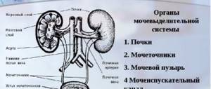
Urethrography is a type of contrast X-ray examination that is used to diagnose diseases of the urethra (urethra), such as narrowing or strictures, neoplasms, stones and foreign bodies, as well as injuries, injuries and fistulas of the urethra.
Cystography is also a type of contrast X-ray examination that is used to diagnose diseases of the bladder and prostate gland such as stones and foreign bodies, tumors, diverticula, chronic cystitis and pathological wrinkling (atrophy with a decrease in functioning volume), hyperplasia or excessive distension, fistulas Bladder.
Cystography can detect bladder function disorders, including abnormal contractions and so-called vesicoureteral reflux (pathological reflux of urine from the bladder into the ureter).
Urethrography and cystography can be ascending or descending, and can also be performed in static and dynamic mode (the so-called voiding urethrocystography or a study that is carried out during urination).
These studies are performed in the X-ray room, with the patient lying on the X-ray table. The tests are performed and/or supervised and interpreted by a urologist. Ascending urethrography (or retrograde urethrography) implies that a contrast agent (Urografin™, Ultravist™, Omnipaque™, etc.
) is injected by a urologist into the urethra and bladder with a special syringe through a catheter. At the command of the doctor performing the examination, an x-ray is taken. In descending versions of urethrography and cystography, an x-ray contrast agent is injected into a vein and after filling the bladder with x-ray contrast urine, an x-ray of it is taken.
Descending urethrography is always voiding, i.e. performed during urination. To obtain good quality images, it is sometimes necessary to take several photographs or radiographs.
Urethrography and cystography in all variants are little or completely painless and do not require anesthesia. For some particularly pain-sensitive male patients, as well as female patients suffering from interstitial cystitis, an anesthetic is injected into the urethra and bladder (we use Kategel™ or a 2% lidocaine solution).
Urethrography and cystography in some cases can be replaced or supplemented by urethroscopy and cystoscopy, ultrasound, computed tomography or nuclear magnetic resonance imaging of the pelvic organs.
The advisability of mutual replacement or combination of these procedures is determined by the attending physician. In any case, studies are performed only to obtain the most reliable information necessary to make an accurate diagnosis.
Well, an accurate diagnosis makes the treatment chosen on its basis as effective as possible. For our part, we guarantee the high professionalism of our specialists and the fact that the price of the service will be one of the best in St. Petersburg.
Reasons for prescribing urethrography
A urologist, oncologist, nephrologist or surgeon can refer you for urethrography when men come with complaints of discomfort in the lower abdomen, pain in the penis, especially during urination, to investigate the causes of infertility. Based on the results of urethrography, one can judge the presence of a number of pathologies relating to the prostate, kidneys, bladder, and urethra.
Urethrography
Our readers recommend
Our regular reader got rid of problems with potency using an effective method. He tested it on himself - the result was 100% - complete relief from problems. This is a natural herbal remedy. We tested the method and decided to recommend it to you. The result is fast. EFFECTIVE METHOD.
Pain and difficulty urinating may indicate the presence of the following diseases:
- Urethral diverticulum, which is an anatomical abnormality, bulge or protrusion of the wall. It may not be detected for a long time until complications associated with the development of infection and inflammation arise. Gradually, pain begins to bother you during any physical activity, including during sexual intercourse, blood appears in the urine, and the mechanism of urination is disrupted;
- malignant prostate tumor, adenoma. These growths can deform the ureter;
- tumor, foreign body directly in the urethra or bladder;
- bladder pathologies (cervical sclerosis);
- hydronephrosis;
- urethritis, cystitis;
- pelvic bone fractures;
- urolithiasis disease;
- fistula tracts of various localization, connecting the urethra with other organs, formed as a result of trauma, inflammation. In some cases, fistulas reveal themselves by splashing urine when urinating;
- strictures are narrowings of the urethra that appear when healthy tissue is replaced by scar tissue.
A urologist, oncologist, nephrologist or surgeon can refer you for urethrography
For all of the above diseases, urethrography is one of the most reliable diagnostic methods. The method also makes it possible to identify the cause of infertility associated with disturbances in the anatomy of the ureter.
It is possible to restore erectile function! Proven FOLK CARE ... Reviews My story spotenciey.ru
Treatment of PROSTATITIS using a new method To restore the functions of the prostate, you need every day... Website Interview with a doctor bezprostatita.ru
The problem of the small size of the dignity has been solved. The mechanism of erection has been revealed... Official website Recovery bigbigrazmer.ru
Contraindications to the procedure are active inflammatory processes in the urethral area, allergies to iodine-containing drugs, recent diagnostic or surgical interventions in the urethra.
Book an appointment in one click
| Name of service | Price in rubles |
| Urethrography | 9000 rub. |
| Cystography | 7000 rub. |
There are some contraindications for urethrography, in which the examination may lead to a deterioration in health. Absolute contraindications to urethrography include:
- acute purulent-inflammatory process in the external and internal genital organs, as well as in the pelvic organs;
- allergic reactions and intolerance to iodine-containing radiopaque substances.
In order to avoid the development of progression of inflammatory diseases of the genitourinary system and the occurrence of allergic reactions, the first step is to stop the acute process in the patient’s body or completely abandon this study.
What diseases can be detected
When performing urethrography, it is possible to determine the presence of most diseases associated with the genitourinary tract. The most common diseases that respond well to imaging include:
- congenital anomalies of the urethra;
- sclerosis of the bladder neck;
- oncological diseases of the organs adjacent to the urethra and bladder;
- primary and secondary deformations of the urethral canal;
- urethral injuries.
In modern urological practice, X-ray examination techniques, in particular urethrography, are one of the most effective examinations, which have a small number of contraindications.
Preparation for the procedure
Urethrography allows you to most accurately determine the degree and origin of injury to the urethra and provides information about the location of foreign bodies, if any. Thanks to the procedure, it is possible to obtain a detailed description of all present pathologies, as well as to identify congenital diseases. This will provide significant assistance in choosing the most effective treatment.
Most often, the study is carried out when there is inflammation of the urethra and the appearance of a number of negative symptoms. If the patient has urinary retention, urethrography is also required.
The procedure can be prescribed to achieve a number of objectives:
- accurate diagnosis;
- additional diagnostic method;
- identifying the necessary information to carry out the operation;
- assessment of the quality of treatment.
In this case, urethrography is of two types:
- voiding (descending urethrography);
- retrograde (ascending type of urethrography).
Descending urethrography is mainly performed on women. It is less informative than a retrograde examination, but the latter type of examination is much easier to carry out.
Ascending urethrography is more often performed in men, which is due to the structural features of their body. If you have the necessary equipment, it is sometimes possible to perform an ascending type of urethrography in women in this way. It is the most informative, therefore it is prescribed more often.
The urethroscopy procedure does not pose any significant health hazard, so no special preparation is required. On the eve of the procedure, the patient is recommended to perform hygiene of the external genitalia. Immediately before the urethroscopy itself, it is necessary to empty the bladder so that during the examination urine is not evacuated from the body.
Even before the examination, it is worth discussing with your doctor the possibility of taking pharmaceuticals. Some medications may distort the picture and results of the study. This includes insulin, acetylsalicylic acid.
It is not necessary to completely abandon these drugs, but the dosage will need to be adjusted. A significant role is played by the patient’s psychological mood and his attitude towards the examination.
At the end of the procedure, the doctor prescribes local antibacterial drugs for preventive purposes.
In addition to its diagnostic value, urethroscopy in men can also be used for therapeutic purposes: to introduce medicinal substances into certain areas of the urethra, to lubricate or cauterize them
Operating principle
X-rays of the urethra can be prescribed for both men and women. The essence of the urethrography method is to pass x-rays through the area of the body being examined. Since soft tissues transmit rays well, the urethra is necessarily filled with a contrast agent.
Given the anatomical features, in most cases, x-rays of the urethra in men and women are the only way to examine the condition of the urethra and establish the correct diagnosis.
There are two types of urethrography - descending and ascending. In the first case, a contrast enhancer is injected directly into the bladder and then spreads through the urethra. In this case, the anterior part of the urethra is clearly visible in the image. In ascending urethrography, contrast is injected directly through the urethra to help visualize the posterior part of this organ. To obtain a complete clinical picture, both types of diagnostic techniques are usually combined.
Does ureteroscopy hurt?
Patients who are indicated for undergoing the described study have a question: does it hurt? The answer is not so simple. Much depends on the gender of the patient, his sensitivity to pain and the skills of the endoscopist.
In women, the procedure takes place without any particular discomfort, since the urethra is short. Also, the female urethra has no physiological bends. Unpleasant sensations completely disappear due to anesthesia.
Urethroscopy in men is a completely different matter. In general, even despite pain relief, this is a painful procedure, since the male urethra is longer and has several physiological bends. However, it cannot be said that the pain is unbearable.
Possible complications
All complications can be divided into harmless, transient ones and those that require correction by a specialist.
Non-hazardous:
- Minor bleeding from the urethra.
Caused by mechanical damage to the walls of the urethra. If there is a significant amount of blood in the urine and acute pain when urinating, you should immediately consult a doctor! - Painful sensations. They go away on their own after a few hours or days.
Dangerous complications requiring medical consultation:
- Infection with characteristic symptoms: burning during urination, pain, discharge from the urethra.
- Increased body temperature (hyperthermia).
- Bleeding that does not stop for an hour or more.
- Reduced urine pressure.
- Intense pain in the urethra.
In such cases, you cannot hesitate. You need to see a doctor urgently.
- Is your beard not growing? Or is it not as thick and chic as you would like? All is not lost.
- Cosmetics and accessories for proper care of your beard and mustache. Log in now!
What will urethrography show?
There are many types of pathologies of the urethra. An X-ray of the urethra using a contrast agent allows the doctor to assess the condition of damaged organs and develop an effective treatment method. In the picture you can see stones, ruptures, cysts, saccular protrusions, fistulas, tumors, strictures and other congenital and acquired anomalies of the structure of the urethra.
An x-ray must be taken at the time of urination - in this way it is possible to achieve complete filling of the urethra with a contrast agent.
A referral for testing can be issued not only by a urologist, but also by a surgeon, oncologist or nephrologist. Interpretation of the received images is the task of the radiologist. The same specialist is obliged to issue a conclusion on the basis of which the attending physician will be able to establish a diagnosis.
Preparatory stage
Methods of preparation for the procedure vary depending on the expected diseases. An hour before the examination, you need to fill your bladder. To do this, it is recommended to drink 400-500 ml of water. And a few minutes before the test, the bladder is emptied.
If there is a suspicion of kidney stones, then the patient is contraindicated to eat and drink a lot of water for 8-12 hours before the procedure. This research methodology does not provide any further preparation requirements.
Process
Urethrography is performed differently in men and women.
During the examination, women should lie on their backs and a catheter will be inserted. After the catheter and contrast are inserted, a series of photographs are taken. The catheter is removed, after which the procedure can be considered complete. Ascending urethrography takes longer because it is technically more complex.
During the procedure in men, the patient must take a special position, lying down: the left leg is bent, the foot is at the level of the knee of the right leg, the thigh is abducted to the side. The bent knee is equipped with a weight for immobility. The genital organ straightens along the left thigh.
The opening of the urethra is moistened, and the surface of the head of the genital organ is treated with an antiseptic. After this, a contrast agent is injected. The bladder is filled and a picture is taken. With retrograde urethrography, additional images are necessary.
If the patient had previously undergone surgery, which caused complications and difficulty urinating, then the solution is administered through a tube in the bladder.
Diagnostic time is 10 minutes. The amount of substance administered is 200 ml or less.
Everything about urethrography in men: how it is performed, is it painful and how much does it cost?
Urethrography is a study of the condition of the urethra using x-rays taken after the injection of a contrast agent into its lumen. This non-invasive method was first used back in 1910, but still has not lost its relevance and is actively used in urology.
Preparation for urethrography
There is no need to prepare specially for urethrography. It is enough to wash your genitals and come to the treatment room at the appointed time. Patients with a tendency to allergic reactions should discuss the possibility of intolerance to the contrast agent with their doctor.
Execution technique
The man should be naked to the waist, lie with his back on the X-ray table, bend his left leg at the knee and move it to the side.
In some cases, the doctor fixes the bent leg with a weight so as not to spoil the pictures. The penis is pulled along the thigh of the left leg. The head and urethra are first treated with an antiseptic.
If pain relief is required, then Cathegel or a 2% lidocaine solution is administered.
Gel "Katejel" with lidocaine - an anesthetic drug with an antiseptic effect
The next step is to draw a contrast agent into a Janet syringe with a rubber tip and slowly inject it into the urethra.
Rush is unacceptable, since high pressure is created, which may damage the integrity of the canal walls. If there is damage to the pelvic veins, the contrast agent can be thrown into them.
Urethrovenous reflux will occur.
The process of introducing contrast occurs under X-ray television control. When the doctor sees that the bladder is full, he gives a command and the technician takes an image. The patient is then asked to urinate and the image is taken again.
If ascending urethrography is needed, then more drug is drawn into the syringe. One part is poured in to fill the bladder, and the rest is injected during urination.
It turns out a counter stream, which causes the urethra to expand. The patient then urinates freely and another series of images are taken at different stages. The entire procedure takes a maximum of 20 minutes.
A diagram of the contrast injection process and an x-ray image are shown below.
Prices and clinics where you can do urethrography
Urethrography is a simple procedure that can be performed in any medical institution that has X-ray equipment and a urology department.
Moscow:
- CELT: 3500 rub.
- “Best Clinic”: 4025 rub.
- “Dobromed”: 2000 rub.
Voronezh:
- Voronezh Regional Clinical Hospital No. 1: 1450 rub.
- Multidisciplinary clinic "Sova": 1500 rubles.
- "Russian Railways Medicine": 770 rub.
In Ufa, the procedure can be done inexpensively in the clinic of the Russian Railways Medicine system: ascending urethrography from 586 rubles.
In Ekaterinburg:
- Honey: ascending urethrography 1600 rub.
- European honey: 2250 rub.
- Ural Research Institute of Phthisiopulmonology: 1300 rub.
As part of compulsory medical insurance, the procedure is carried out free of charge upon doctor's prescription.
Conclusion
Urethrography is a first-line diagnostic method that allows you to identify most pathologies of the urethra. The study is inexpensive, non-traumatic, with a low risk of negative consequences.
Sources:
Source: https://muzhchina.info/diagnostika/uretrografiya
Decoding the results
Decoding urethrography shows data on the condition of the patient’s genitourinary system and determines most diseases of the urethra:
- deformations of the urethra, which arise due to injury, but can also be congenital;
- metastases and tumors;
- damage to the bladder neck;
- anomalies of the urethral canal, often congenital.
The attending physician must decipher the result.
Urethrography can be performed in Moscow. The cost of the service is from 700 rubles. The price varies depending on the location of the clinic and the professionalism of the doctors.
This research method is also convenient because it can be performed on a child, and no pain is felt during the procedure.
The essence of the technique
The term urethrography is made up of two Greek words: "urethra" and "grapho", which means to depict the urethra. Urethrography refers to a special x-ray examination method in which a special x-ray contrast agent is injected into the cavity of the urethra. After this, an X-ray procedure of the urinary system is performed. The main purpose of urethrography is to assess the patency of the urethral canal. With this study, it is possible to identify and confirm some serious diseases associated with the human genitourinary system. This research method is used more often in men than in women, and this is due to the more complex structure of the urethral canal in the male body.
The moment of introduction of a radiopaque substance with the ascending technique
Urethrography is performed for diseases and conditions such as:
- urethral canal injury;
- urethral stricture;
- anomalies and defects in the development of the urinary tract;
- prostate neoplasms;
- inflammatory diseases of the urethra, bladder and prostate;
- disturbances in the act of urination.
For all of the above diseases, this method turns out to be a very informative and valuable diagnostic study and allows you to confirm not only the disease itself, but also its form and degree.