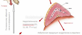
The adrenal glands are one of the most important elements of the paired organ, synthesizing corticosteroids and closely adjacent to the apical region of the kidney.
From an anatomical point of view, it can be divided into certain zones. The first will contain the cortex, which produces aldosterones, deoxycorticosterones, cortisol, corticosterones, and cortisone.
The medulla produces adrenaline and norepinephrine. In cases of abnormalities in the functioning of the adrenal glands, the body ceases to adapt to various situations created by severe stress. The working process of the adrenal glands is under the control of the nervous system.
In what cases is diagnostics necessary?
Ultrasound of the adrenal glands and kidneys is prescribed for:
- diagnosing pathology;
- assessing the level of changes from the disease;
- control of the treatment process.
Using this procedure, it is possible to assess the need for additional examinations and determine the subsequent specifics of the therapeutic course.
Ultrasound examination of the adrenal glands is prescribed for the following indications:
- There was a suspicion of the appearance of neoplasms.
- It is necessary to establish the real causes of arterial hypertension.
- It is necessary to determine the reasons that led to the increase in body weight.
- Organs should be checked for hyperfunctional or hypofunctional activity.
- The reasons leading to infertility are identified.
- The source of muscle tissue weakness is determined.
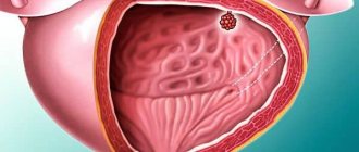
Ultrasound examination reveals tumors, cysts, hematomas, cancerous lesions in the glands, and inflammatory processes.
Treatment Basics
The approach to managing tumor diseases of different types is somewhat different. If a benign tumor up to 3 mm in size has been identified, does not produce hormones and has no signs of malignancy, then treatment will not be necessary. The patient will be required to undergo an ultrasound scan of the adrenal glands 2 times a year to monitor tumor growth.
If we are talking about hormone-active tumor diseases, especially malignant ones, then surgical intervention is performed. Its volume will depend on the size of the tumor and the degree of its growth into other tissues.
Note. Most often, the entire affected adrenal gland is removed, even if there is only a focal pathological process. For example, for a tumor of the right adrenal gland, the entire gland is usually removed. If there are no metastases, then such treatment often leads to complete recovery.
Surgical treatment is often the patient’s only chance for a normal life
In the presence of metastases to other organs from malignant tumors of the adrenal glands, surgical treatment is not always carried out or is carried out in combination with the introduction of special radioactive isotopes that reduce the rate of disease progression.
Important! Treatment of malignant neoplasms should be carried out in any case and as early as possible. The quality and duration of a person’s subsequent life will depend on this.
Adrenal tumors pose a great danger, but with timely treatment and proper treatment, it is possible to get rid of such diseases without complications.
A mass formation on the adrenal glands is a tumor process that occurs against the background of pathological proliferation of organ cells. Formations can be benign or malignant.
Pathology can lead to various complications. That is why it is necessary to promptly treat tumors on the adrenal glands.
Contraindications for ultrasound
The procedure is known for its safety and painlessness, and can be performed with virtually no restrictions. But there are still several factors that can create a ban on its appointment:
- Pregnancy . If there is a need to perform diagnostics, then during this period ultrasound is used, which has the least effect on the formation of the fetus.
- Skin diseases . When there is inflammation, this becomes an obstacle to contact of the sensors with the patient’s skin.
- Open wounds.
Experts believe that women during pregnancy need to undergo only routine examinations. But if the risk to the expectant mother exceeds the possible danger to the developing fetus, the doctor may prescribe an ultrasound examination.
What does hypoechoic formation mean?
Local hypoechoic formation in a particular organ, in contrast to hyperechoic formation, is the result of lower echogenicity of tissues - in comparison with the parameters of the acoustic density of healthy tissues of the organ. That is, this area weakly reflects the ultrasonic signal directed at it (in the frequency ranges 2-5, 5-10 or 10-15 MHz). And this is evidence that this formation - from the point of view of its structure - either contains liquid or has a cavity.
A hypoechoic formation is visualized on the screen as gray, dark gray and almost black zones (with hyperechogenicity the zones are light, often white). To decipher an ultrasound image, there is a scale of six categories of gray Gray Scale Imaging, where each pixel of the image of a hypoechoic formation obtained on the monitor - depending on the strength of the ultrasound signal returning to the sensors - represents a specific shade of gray.
The results of an ultrasound examination, deciphered by ultrasound diagnosticians (sonographers), are studied by doctors of a specific profile (endocrinologist, gastroenterologist, urologist, nephrologist, oncologist, etc.) and compared with the indicators of tests taken by patients and the results of other studies.
In many cases, differential diagnosis is required, for which, in addition to ultrasound, other hardware methods for visualizing pathology are used (angiography, color Dopplerography, CT, MRI, etc.), and histological examination of biopsy specimens is also carried out.
Preparation for the procedure
The patient will have to undergo a preparatory course, the main task of which is to reduce gas formation in the intestines. Espumisan helps with this; it must be taken for several days before the examination.
Special diets have proven themselves well. Black bread, beer, fried and fatty foods are excluded from their set. You can drink juices, teas, potatoes, cereals, fruits or vegetables. You should not drink large amounts of liquid on the day of the procedure.
As an addition to the preparation for the procedure, blood donation for hormones is prescribed, and a consultation with an endocrinologist is carried out. If the patient suffers from arterial hypertension, then the entire preparatory stage for him consists of two liters of natural water, which must be drunk before the ultrasound.
When performing an ultrasound examination of the adrenal glands, the specialist pays special attention to the contours of these elements. In normal condition, they should be even. In addition, the doctor checks the location and dimensions and examines the structure for uniformity. The most crucial moment of ultrasound is the identification of new formations.
How to prepare for an ultrasound with the help of medications?
Microenemas are performed or glycerin suppositories are placed. Use Pikolaps or Guttolax. It is recommended to start drinking Smecta, Sorbex, Espumisan in a few days. All meals should be accompanied by Mezim or Pancreatin.
What is a hypoechoic formation?
Hypoechoic formation is a formation localized in any organ and having echogenicity below the normal level. This area weakly reflects ultrasonic rays. The monitor is darker than other areas.
A hypoechogenic formation contains water or a cavity. On the monitor, the area is visualized as gray or black spots. With hyperechogenicity, the zones are light or even completely white.
To decipher the picture, a special scale with 6 categories of gray shade is used. The diagnosis is made by specialist doctors. Often hypoechoic formations are cysts. In this case, the patient is additionally referred for a biopsy.
You can decipher the image using a special scale
Conducting research
The patient is placed on his back or on his stomach, there is not much difference in this. The lumbar region and lower abdomen should be freed from things worn on the body. After this, the doctor applies a gel composition to the projection area of the organs, with which the examination can be performed.
The adrenal glands are not visualized - what does this mean?
The hope that a specialist will be able to see two adrenal glands is negligible. As follows from statistics, the organ located on the left can be seen in fifty percent of one hundred cases. It is believed that the adrenal gland in a healthy state remains invisible, since if we talk about its structure, then it is no different from the cells of the retroperitoneal cavity. In a normal situation, it is possible to consider only the area of the intended location.
Treatment, symptoms, drugs
An adrenal tumor is a serious diagnosis. Very important for predicting its outcome is the stage at which this pathology was identified. Tumor diseases of the adrenal glands are accompanied by quite characteristic symptoms, and at the first signs of it, you should immediately contact a specialist.
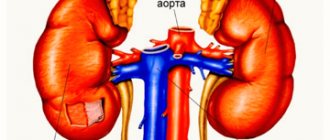
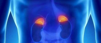
Anatomical location of the adrenal glands
What does an ultrasound scan of the adrenal glands show?
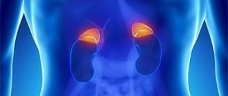
The research procedure evaluates the parameters and structural structure of the adrenal glands - paired organs located on the upper part of the kidneys.
Tumors are often found on the adrenal glands. Thanks to CT scans, it was possible to find out that this problem is typical for five percent of the world's population. But it is necessary to reassure that the maximum number of such tumors are considered benign and do not pose a risk of developing oncology.
But tumors on the adrenal glands can also have a negative effect on a person’s general condition:
- Having reached a certain size (from four to five centimeters), the tumor can push away nearby organs, interfering with their functioning.
- Some tumors produce hormones in excess, disrupting the functioning of the entire body.
- You should not assume that malignant neoplasms cannot exist. The likelihood of cancer depends on the size of the tumor, and this fact has long been proven.
Classification of adrenal tumors
Neoplasms arising in the adrenal gland are divided depending on the layer in which they appeared: medulla or cortex. Situations often arise when the tumor affects both layers, then this form is called mixed.
In addition, neoplasms are divided depending on the organ in which it developed:
- tumor of the right adrenal gland;
- tumor of the left adrenal gland;
- neoplasm on both adrenal glands.
But despite the location of the formations, they are all divided into benign and malignant. Benign tumors on the adrenal glands are usually small in diameter and do not contain cancer cells.
Such tumors usually do not produce pronounced symptoms and are often detected by chance during examination of internal organs.
Malignant neoplasms of the adrenal glands are dangerous to human health. They tend to rapidly grow altered cells in organ tissues and spread to other organs.
Malignant tumors include adrenal lymphoma. One of the symptoms of the disease is pathological enlargement of the lymph nodes. The disease is difficult to treat, especially if the metastases have penetrated into nearby organs.
Among primary gland tumors, hormonally active neoplasms are distinguished and hormonally inactive ones. Inactive formations of the adrenal gland are classified as benign.
This tumor can develop in any person, regardless of age and gender. Inactive formations are rarely of the malignant type and are better amenable to therapeutic treatment.
Normal size of the adrenal gland
Ultrasound examinations should be performed at normal resolution. When this type of examination is performed during a simple examination, the adrenal glands are most often not visible. It must be added that organs may differ in unequal sizes. And if the left adrenal gland remains invisible in half the cases, then the right one is visualized once out of ten examinations.
The longitudinal dimensions of the adrenal glands should not exceed two to three centimeters. When, during an ultrasound, an increase in excess of the permissible size is detected, it is imperative to undergo an additional check using MSCT, without paying attention to the large radiation exposure.
What reasons are provocateurs?
Neoplasms of the right or left adrenal gland appear due to various negative factors, which can be primary or secondary in nature. In the first case, the space-occupying formation is a consequence of congenital anomalies, as a result of which a pathological process develops. There are other reasons why hormonally active adrenal tumors first appear in a person:
- genetic factor;
- abuse of tobacco and alcohol products;
- unhealthy diet with excess fast foods and carcinogenic foods;
- age characteristics;
- endocrine pathologies;
- constant stress and nervous tension;
- hypertension;
- injury to internal organs.
According to statistics, tumors of the kidneys and adrenal glands are more often found in children or people 50 years of age or older.
What is a substitute for ultrasound of the adrenal glands?
Today, computed tomography using contrast agents administered intravenously is popular. This method of examination helps not only to clearly determine the structure of organs, but also to study its internal structure.
But you shouldn’t get a CT scan for everyone. According to experts, it is not as harmless as compared to conventional ultrasound. The patient manages to receive a certain dose of radiation, and the contrast injected into the vein contains a lot of iodine, which interferes with the normal functioning of the thyroid gland for a month, or even a month and a half.
Diagnostics: basic methods
Benign and malignant tumors of the adrenal glands are detected through laboratory and instrumental examinations, such as:
- General urine and blood tests.
- Ultrasound diagnostics. During manipulation, a hypoechoic formation is detected in the adrenal gland, the size of which is more than 10 mm.
- CT scan. It is used most often because it gives accurate results. Thanks to Godfrey Hounsfield, who invented such a diagnostic method, it is possible to determine the development of a tumor in an organ at an early stage. During the procedure, the native density of the lesion is detected, indicating the appearance of a cyst, myelolipoma or other formation.
- MRI. The examination reveals pathology of the medial pedicle of the internal organ. The doctor discovers a hypodense formation that has clear outlines.
- X-ray. This diagnostic method helps to exclude or confirm metastasis.
- Urine analysis to determine hormonal levels.
- Phlebography. During manipulation, blood obtained from the vessels of the adrenal glands is examined.
- Laboratory test for testosterone. If its norm is very high, then a tumor of the ovaries or adrenal glands is diagnosed.
- Biopsy. Allows you to explore solid education to its fullest extent.
Over the counter
“I have been doing ultrasounds of the abdominal organs for 25 years, and in the conclusion they always write that the adrenal glands are not visualized. What does it mean? What does ultrasound even show?
Olga, Dubrovno.
Ultrasound of the abdominal cavity is the fastest, simplest, highly informative and safe diagnostic method. Radiation passes freely through the internal organs, which are filled with air, and is reflected from all dense structures. This signal is recorded by a sensor, and the image of the organs (shape, density) is transmitted to a computer where a special program is installed to create a two- or three-dimensional image. For a better picture, the air gap is removed between the skin and the highly sensitive device using gel. At the same time, the abdominal cavity, pelvic area and kidneys can be examined in 20 to 30 minutes. Their structural changes, the presence of inflammation, pathological formations, and blood supply characteristics are determined. For example, in this way they can detect stenosis, hematoma, pancreatitis, cirrhosis, cyst, appendicitis, pyelonephritis and glomerulonephritis, rupture of the spleen and liver, kidney stones and gall bladder, and diseases of the adrenal glands. In addition to ultrasound, to examine the adrenal glands in the presence of symptoms of the disease, they examine the blood for hormones and perform a CT scan, which is supplemented with contrast for greater effectiveness. Ultrasound also helps to identify tumors, but does not provide information about the degree of their malignancy.
The stomach and intestines are the most difficult organs to diagnose using ultrasound. Due to the fact that they contain semi-liquid contents or air, they cannot be completely tested. It is better to use fibrogastro- and colonoscopy. Lymph nodes should not normally be visualized, as should the adrenal glands. This means that their size is normal and ultrasound simply cannot notice them. But an increase in these organs indicates either the presence of an infectious disease in the abdominal cavity, or that a tumor is developing or metastases have occurred. It is advisable to study the spleen and liver in case of injuries received in road accidents, peritoneal injuries, chronic and infectious diseases, and suspicion of the formation of neoplasms.
Classification of tumors
Depending on the location of the tumors, they are divided into two types and can be located in the cortex or medulla. Neoplasms can develop in the outer cortical layer of the organ and manifest themselves as:
- Aldosteromas.
- Corticoestroma.
- Corticosteromas.
- Andosteromes.
Quite often, mixed forms of neoplasms occur in patients. In the inner medulla, the development of ganglioneuroma or pheochromocytoma can be observed. The development of benign and malignant neoplasms is diagnosed in the medulla or cortical layer.
Benign tumors are small in size. In this case, there are no pronounced clinical manifestations. That is why the identification of adrenal tumors is completely random.
If the formation is malignant, then it is characterized by fairly rapid growth.
Primary adrenal tumors are distinguished, which are divided into producing and hormonally inactive. The second type of neoplasm in most cases is benign in nature.
They can develop in a person, regardless of his age and gender. In rare cases, malignant tumors are isolated in this group.
There is a wide variety of tumors of the left and right adrenal gland. When the first signs of a pathological process appear, the patient is advised to seek help from a doctor, who can not only determine the type of pathology, but also prescribe rational therapy.
Symptoms of pathology
Some neoplasms are asymptomatic and therefore are diagnosed completely by chance during examination of the patient. When calcified and other tumors appear in the adrenal gland, the presence of corresponding symptoms is observed.
It manifests itself in the form of increased blood pressure. In most cases, this symptom occurs with aldosteroma. This tumor is accompanied by muscle weakness, heart failure, and frequent urge to urinate, especially at night.
With pheochromacytoma, adrenaline and norepinephrine are produced in excessive quantities. This disease is accompanied by a paroxysmal increase in blood pressure.
With this pathology, patients complain of:
- Feeling of fear.
- Headache.
- Increased sweating.
- Excessive irritation.
If both adrenal glands are affected, delayed puberty is diagnosed. In some cases it may be premature. This symptom appears with neoplasms that produce sex hormones.
During the course of the pathological process, patients experience changes in appearance. In representatives of the fairer sex, the voice becomes rougher, and the breasts also decrease in size.
Some women complain of developing baldness or excessive hair growth in the chest and face area. The pathological process is accompanied by the absence of menstruation. The main symptom of the disease in men is breast enlargement.
The disease is accompanied by a decrease in potency and sexual desire. When sick, facial hair growth becomes less intense.
Most tumors that develop in the adrenal gland are characterized by the presence of corresponding symptoms. In this case, the patient is recommended to visit a doctor who will determine the type of tumor and develop an effective treatment plan.
Diagnostic measures
In modern endocrinology, there are a large number of diagnostic methods that help identify tumors. The type and location of the tumor is also determined.
After examining the patient and collecting anamnesis, the doctor makes a preliminary diagnosis. To confirm it, it is recommended to undergo laboratory and instrumental examinations.
In order to determine aldosterone, catecholamines, cortisol, vinylmandelic and homovanillic acid, a urine test is performed.
To determine the hormonal activity of tumors in the adrenal glands, phlebography is recommended. This is radiopaque catheterization of the adrenal veins. This makes it possible to determine hormone levels.
To determine the size and location of the adrenal glands - ultrasound, computed tomography and magnetic resonance imaging.
Diagnostic measures for suspected tumor processes should be carried out in a complex manner, which will guarantee their informativeness, and will also make it possible to determine the causes and methods of therapy.
Features of treatment
If a patient is diagnosed with a cystic formation or other types of tumors, this requires appropriate therapy. For hormonally active neoplasms and tumors whose size is more than 3 centimeters, it is recommended to use a surgical treatment method.
In other cases, it is recommended to ensure dynamic monitoring of the development of the tumor. Surgical intervention may involve the use of a traditional or laparoscopic method.
During the operation, the adrenal glands are completely removed. If the patient has a malignant neoplasm, then nearby lymph nodes are removed along with the organ.
The most difficult are surgical interventions for pheochromocytoma, which is explained by the possibility of developing undesirable effects. In most cases, after surgery, the patient's hemodynamics are impaired.
This requires proper completion of the preparatory period. In order to stop pheochromocytoma crises, it is necessary to correctly select anesthesia methods.
Also, for the treatment of this type of tumor, intravenous administration of radioactive isotopes is recommended. This will reduce the size of not only the tumor, but also the metastases.
Chemotherapy is widely used to treat some tumor processes. If a patient experiences a pheochromocytoma crisis, then to relieve it the following is administered intravenously:
- Nitroglycerine.
- Phentolamine.
- Sodium nitroprusside.
If the crisis cannot be stopped and the patient develops catecholamine shock, then emergency surgery is performed in accordance with vital indications. After it is completed, patients are prescribed replacement therapy, which uses adrenal hormones.
There are many methods for treating tumor processes, the selection of which is carried out by the doctor in accordance with their characteristics.
Anatomical features of the adrenal glands
The adrenal glands are located in the retroperitoneum directly below the diaphragm. Despite the fact that they have a complex structure and during pathomorphological examination they distinguish between the medulla and the cortex, they are quite difficult to see with ultrasound diagnostics. This is due to the fact that the density of the adrenal gland tissue is comparable to the fatty tissue of the retroperitoneal space.
Normally, they are located at the level of the 11th–12th thoracic vertebra, so the ribs, spinous processes of the vertebrae, and a dense layer of muscles prevent the penetration of ultrasound from the back. Anteriorly, even with good bowel preparation, the left adrenal gland is not visualized on ultrasound, this occurs because air usually remains in the colon. It is not detected in half of the patients, while the right one is visible in 90% of patients.
The right adrenal gland is surrounded by structures that transmit ultrasound well. It is visualized in a triangular area formed by the kidney, the right lobe of the liver and the inferior vena cava. The best image of the adrenal glands can be obtained from lateral projections along the mid and anterior axillary line. For better visualization, patients are examined on their side and asked to take deep breaths during the examination.
M16 - potency remedy: composition, release form, principle of action and advantages
Many men today are faced with poor potency. Moreover, people of different ages suffer from this deviation. According to statistics, 87% of representatives of the stronger sex of society have problems in their intimate lives.
Previously, men over 40 had poor potency, but today even 18-year-old boys have begun to suffer. The reason for this is many negative environmental factors, poor lifestyle, bad habits, unbalanced diet, etc.
However, do not be upset, because in most cases such intimate problems can be solved. There are many products that help improve male potency, and one of them is M16 potency product, which has proven itself exclusively on the positive side.
Compound
M16 is the best remedy for potency; the main advantage of this drug is that it is based on a special composition. Each component is selected in such a way as to bring the most positive effect from use. Moreover, the composition is exclusively natural and safe for health.
It includes:
- Guarana extract. It has a positive effect on the entire body of the stronger sex, allowing for blood flow to the penis due to its vasodilating effect.
- The amino acid L-arginine is responsible for the expansion of blood vessels in the penis. Helps nitrogen move actively in the male body.
- Glycine is a component that relieves stress at the mental and emotional level. It is known that sometimes it is anxiety that acts as a blockade for normal sexual life.
- Magnesium is a component that actively affects a healthy erection.
- Beaver musk is a natural element that enhances blood circulation, fills the cavernous bodies of the penis with blood, thanks to it the erection becomes long, and the problem with untimely ejaculation disappears.
- An extract taken from the antlers of an Altai deer. Works at the level of brain centers, activating arousal. Increases libido.
- Sainfoin extract. This is an extract from the herb of the same name, which has long been used to strengthen and increase a man’s strength in bed.
- The red root is used as a concentrate. Its task is the synthesis of testosterone production, which in turn normalizes the imbalance at the hormonal level.
Release form
Release of the medicine: a bottle of liquid for use in the form of a spray. The product is applied using a dispenser. You can buy by going to the official website of the manufacturer. No pharmacy sells this drug.
pharmachologic effect
When sprayed, the potency product M16 first enters the bloodstream, then passes into the adrenal glands, the body reacts by producing a hormone responsible for stabilizing potency, and libido is activated.
It is also important to note that not only men can get results when using the spray. The woman also feels the impact on herself.
So, when penetrating the vagina, arousal increases due to improved blood circulation in the female reproductive system.
The influence lies in the following factors:
- Increased sexual desire.
- Increased duration of sexual intercourse.
- Improving the quality of sperm, increasing its quantity.
- Discovering new sensations during sex.
Those couples who wanted to conceive a child were also able to experience the power of the drug for themselves. The vast majority of them wrote a review that after using the M16 they were able to achieve their goal. This is because the structure affects both increased libido and sperm activity.
By going to the manufacturer’s website or to a forum where intimate problems are shared, in particular, deteriorated erectile function, you can see that the vast majority of men consider the result worthy of the set price.
The advantage of the drug over analogues
In order to understand why the M16 male potency product is so widely used, it is worth understanding its advantages:
- It can be used as an additional remedy for getting rid of prostatitis and infertility.
- Due to the fact that the drug increases libido, sexual intimacy becomes brighter, even if the man is tired.
- The duration of an erection is an important factor. It is observed 12 hours after spraying M16.
- The quality of intercourse improves due to improved blood circulation in the vessels of the penis.
- Daily use of the spray is possible without side effects or addiction.
- Not only the man, but also his partner gets pleasant sensations from sex. Orgasms become many times stronger, their number increases.
- After applying the product, excitement occurs within 5-7 minutes.
- It has no side effects due to its natural composition. The sale is made only on the manufacturer’s website, therefore, any counterfeits can be excluded.
- M16 is easy to use, minimal contraindications.
- Does not cause withdrawal syndrome.
For the relevance of using the spray in a specific situation, a doctor can prescribe or cancel it.
Spray M 16 for potency: indications, contraindications, instructions for use
The fact that the M16 potency spray has the most correctly selected composition makes it guaranteed that the desired effect will be achieved.
The product can be used from the age of 18 and has no restrictions or side effects.
It affects not only the male genitals, but also gives an amazing experience to the woman.
Indications for use
Every man can experience disorders in the sexual sphere; the disease does not choose based on status, age or physical fitness. Often the stronger sex experiences a lack of erection after shocks, neuroses, or experiences, because not only the circulatory and hormonal systems, but also the brain and nervous system are responsible for strength in bed.
Therefore, it is worth using a spray to improve potency in such cases when:
- the penis does not erect after drinking alcohol or smoking cigarettes;
- a normal erection is observed, but as soon as a condom is put on, its decline is noted;
- during sexual intercourse, the penis falls before reaching the end;
- there is psychological impotence at the level of experiences and neuroses;
- there is excess weight;
- age over 45 years.
Contraindications
Just as every drug has contraindications, this drug also has them. M16 should not be used by young people under 18 years of age, and those who have allergic reactions to the components included in the composition are also prohibited from spraying it.
To test the body's reaction before use at the destination, you need to spray a small area of skin, for example, on your arm.
The medicine cannot be used along with other supplements and medications that are used for the same purposes. It is not recommended for epileptic seizures, heart failure, kidney and liver diseases. It is also important to consult a doctor when there is deformation of the penis or inflammatory processes in the organ.
Instructions for use (method and dosage)
In order to use the drug, you do not need to be an ace. It is enough to perform a few simple manipulations, namely:
- carry out hygiene of the groin area and organ;
- shake the spray container;
- spray it along the entire length of the penis, moving from the base to the head. It is not recommended to do more than 5 sprays;
- in 5-7 minutes you can enjoy amazing sex.
The spray has been developed for regular use or in required situations. The aerosol should not be used more than once a day, especially since once sprayed, arousal and erection will last up to 12 hours. You can safely spray your penis after drinking alcohol, medications, or fatty foods. This does not reduce the effect of the drug on the male organ.
Interaction
Due to the fact that M16 spray for improving potency consists only of herbal components, it can be applied together with the use of any medications. Do not worry when taking medications to improve the functioning of the heart and blood vessels, antidepressants, vitamins, dietary supplements.
The only limitation is the combined use of the aerosol with drugs that are aimed at improving erection.
Potency remedy M16: reviews of the drug
After conducting a survey among men suffering from sexual problems, we can conclude that the M16 potency remedy has extremely positive reviews.
Some patients note that this drug helped not only improve and diversify their intimate life, but also avoid divorce.
Analogs
Among the analogues, there are no drugs exactly the same as the product for male potency M16, reviews fully confirm this.
In this case, the following can be called similar in their effect:
- Dominator.
- MachoMan.
- Aerosol 999.
- TheHardGoldGun.
- Stud100.
However, all of the above has its drawbacks. So, for example, Stud100 cannot be used repeatedly; with long-term use it leads to a negative consequence, which is strong addiction, and spray 999 begins to act after a longer time than the M16 potency product; consumer reviews fully prove these facts.
Reviews
Anatoly, 42 years old, hotel manager:
“If I say that the spray helped me, I will say absolutely nothing. My age is far from small, but my wife is 10 years younger, so I need to satisfy her body. Lately, I have become less and less able to resolve this intimate issue. That is why I decided to try the miracle cure on myself. I’ll say right away that I didn’t believe in its capabilities. But I was stunned when the sex became so great that even my wife admitted that she had never had anything like this. I am completely satisfied with everything. Don't listen to what they say, just try it. You will definitely remember me with a kind word.”
Lena, 19 years old, student:
“I want to write about my first experience using the M16. Despite our young age, my boyfriend and I were virgins, so we had a lot of worries and excitement. In general, he didn’t get hard, they tried a couple of times, but nothing. Then we read about the drugs on the Internet, everything could be used when one is already getting older. And M16 is allowed from 18 years of age. I liked that there was nothing contraindicated in the composition, and I could get an unreal orgasm. In short, we decided. After 5 minutes everything went as if we were trying this not for the first time. We relaxed, had great fun, and just the two of us! Enough action for the whole night. Now we have full-fledged sex, we use it occasionally when he gets tired after school and work. Don’t be afraid to try, the effect is amazing!
In what cases is diagnostics necessary?
Ultrasound of the adrenal glands is prescribed to identify certain diseases and pathologies:
- hyperplasia, which often causes endocrine infertility;
- tumors, mostly benign (adenoma, aldosteroma), grow slowly, cause changes in hormonal levels, but with large formations, cancer can be suspected;
- an inflammatory process, usually associated with infections (syphilis) causes insufficiency of function, a decrease in the production of hormonal substances;
- trauma, hematoma formation, observed during difficult births in newborns;
- cysts are rare.
The symptoms that require testing are:
- muscle weakness;
- fast fatiguability;
- darkening of skin color;
- high blood pressure;
- psychoneurological disorders;
- change in appetite;
- a sharp increase or decrease in body weight;
- hirsutism (excessive hair growth, hair growth disorder that does not correspond to gender);
- sexual dysfunctions;
- infertility.
This variety of symptoms is due to the fact that the adrenal glands produce various hormones that affect different organs and systems of the body. Failure to function properly can lead to life-threatening conditions.
Hormonally active neoplasms
An adrenal tumor is a neoplasm in the adrenal glands that causes excessive production of one of the hormones, which leads to malfunctions in the functioning of the body.
Such formations cause pronounced changes in the body, which can often be seen with the naked eye.
Hormonally active tumors include:
- aldosteroma;
- androsteroma;
- corticoestroma;
- corticosteroma;
- pheochromocytoma.
Aldosteroma is a tumor of the adrenal gland that produces the hormone aldosterone. The formation develops in the cortical zone of the adrenal glands. Aldosteroma causes disruption of the water-salt balance in the body. This leads to muscle weakness, headaches, interruptions in heart function, hypoglycemia, and increased blood pH.
If the level of the hormone sharply increases in the body, then a crisis occurs, which is manifested by vomiting, shallow breathing, blurred vision, and the onset of flaccid paralysis. Neoplasms of this type can be single or multiple, but most often they are benign. Malignant aldosteroma occurs in only 2-5% of all patients.
Androsteroma is a formation that develops in the cortical layer of the gland and produces an excess amount of androgens. When androsteroma appears, early maturation begins in boys, signs of hermaphroditism appear in girls, and male-type changes in appearance are observed in women.
If the tumor is malignant, it quickly metastasizes to other organs: liver, lungs and lymph nodes. This disease is quite rare, accounting for no more than 3% of all neoplasms. But the probability of a malignant process in this case is 50%.
Corticosteroma, or glucosteroma, is a formation that develops in the cortical layer of the gland and produces glucocorticoids. This type of neoplasm is the most common. With glucosteroma, patients experience obesity, frequent headaches, increased fatigue and muscle weakness, early puberty or, conversely, premature decline of sexual function.
Hemorrhages and stretch marks appear on the abdomen and chest; in women, male-pattern hair growth begins and the timbre of the voice decreases. Such tumors can be either benign or malignant. A benign neoplasm is called “adenoma”, and a malignant one is called “corticoblastoma”, “adenocarcinoma”.
Corticoestroma is a neoplasm that produces an excessive amount of estrogen, which leads to the development of a man’s body according to the female type (increase in fat mass in the abdomen, hips and chest), sexual impotence. This neoplasm develops in the adrenal cortex and is common mainly in men. Most often it is malignant.
Pheochromocytoma is a neoplasm in the adrenal medulla that produces catecholamines. This type of tumor is most often benign; signs of malignancy are observed in only 10% of patients.
What does an ultrasound scan of the adrenal glands show?
In many patients, the adrenal glands are often not located, which means that the diagnostician cannot detect them during examination. This is due to constitutional features and the small size of the organs. The examination begins with a search for the right adrenal gland in the triangular zone formed by the renal, hepatic edge and the inferior vena cava.
The best visualization is achieved with the patient positioned on the left side, the ultrasound sensor is located along the anterior right axillary line. The left one is better visualized if the patient turns on the right side, and the Sensor is located in the midaxillary line. The study is also used while standing, at the height of inspiration.
During the examination, the doctor evaluates:
- dimensions;
- homogeneity of structure;
- location of organs;
- capsule structure.
On ultrasound, the adrenal glands may have the shape of a pyramid (right), crescent (left). The capsule itself looks like a hyperechoic thin line surrounding a homogeneous structure from the outside, visible only on devices with high sensitivity. Its intermittency and uneven thickness in different areas indicate an inflammatory process. When a cyst or hypoechoic formation is detected, it should be differentiated from subcapsular renal pathology. With hyperplasia, a uniform increase in size is usually detected.
Benign tumors are oval in shape, their echogenicity is lower than the adrenal gland itself. They can reach 4–6 cm in diameter. Pheochromocytomas are more often found in children; they can reach large sizes and are characterized by the formation of several foci. In conclusion, the doctor indicates the exact size, structure, and presence of tumors.
Complications of adrenal tumors
When the adrenal glands develop, serious disturbances occur in the body, which often lead to crises, expressed in a sharp increase in blood pressure, dizziness, the skin becomes pale, the heartbeat quickens, sweating increases, attacks of vomiting, panic attacks, and increased body temperature begin.
The main complications of tumors depend on their type. For benign tumors, this is the probability of their degeneration into malignant ones. And with a malignant neoplasm, metastases that affect healthy organs pose a greater threat.
They primarily affect the bones, uterus, liver and lungs.
In what pathologies are the adrenal glands not visualized?
Due to poor visualization, careful preparation is required before the examination in order to reduce gas formation in the intestines. It includes:
- Diet for 2-3 days. Hardly digestible foods that cause flatulence (legumes, sweets, baked goods, cabbage), and carbonated drinks are excluded from the diet. Porridges and vegetables are recommended, only after heat treatment.
- Purgation. It is carried out with the help of laxatives; an enema is not always effective.
- The examination is carried out on an empty stomach. A light dinner is recommended the evening before.
Even with such preparation, it is not always possible to examine the adrenal glands. They are most clearly visible on expert-class equipment in thin, asthenic people. The causes of poor visualization are the following pathologies:
- obesity, even with a low degree of adipose tissue deposition;
- adhesions in the abdominal cavity after surgery;
- flatulence, which accompanies many diseases of the gastrointestinal tract.
Changes in the structure of the spleen
The echogenicity on ultrasound of the spleen is uniform and slightly higher than that of the liver, but this is a variant of the norm. The hypoechoic inclusion in this case is:
- Intraparenchymal hematoma in acute form. It is a consequence of abdominal trauma and occurs when an organ ruptures.
- Benign vascular formation (hemangioma).
- Lymphoma of the spleen.
- Organ infarctions.
- Various metastases (osteosarcomas, sarcomas, etc.).
According to experts, in the spleen cystic formations of dermoid, tapeworm and echinococcal nature have a mixed echostructure.
Pathology in the ovaries
Typically, a hypoechoic formation on ultrasound of the ovaries can be:
- cyst;
- unruptured follicle;
- luteal body;
- carcinoma (rare).
If the patient is of reproductive age, a heterogeneous organ structure is normal. During menopause, everything is exactly the opposite.
In order to promptly diagnose a hypoechoic formation in the ovary, as well as other pathologies of the organ, it is necessary to conduct an ultrasound scan annually.
Changes in liver tissue
A hypoechoic formation in the liver is often detected. It may indicate the following pathologies:
- Knots. They appear in cirrhosis, have a rounded shape, with a possible bumpy structure at the edges.
- Cysts. There is hemorrhage, the boundaries are smooth and clear.
- Thrombosis. The intrahepatic region of the portal vein is affected. The formation is oval, round or elongated, its structure is heterogeneous (loose).
- Abscesses. Areas of varying echogenicity are observed, and gas bubbles occur. The outlines are blurry.
- Nodular hyperplasia. Rounded shape, clear and even borders. Echogenic projections extend to the periphery. Ultrasound of the liver is not normal.
- Adenoma. The contour is clear, the structure is homogeneous. Signals are detected inside the inclusion.
- Carcinoma. Heterogeneous, has areas of hemorrhage, deposits of calcium salts. Local lymph nodes are deformed, ascites cannot be excluded.
- Metastases. The borders are variable and hypoechoic. The organ tissue is not deformed.
Important: A hypoechoic area can occur in any human organ. Tumor-like formations and cysts are more common, but other pathologies cannot be excluded. Some of them can be eliminated by drug therapy, while others will require surgical intervention. To obtain the most accurate results and specify the condition, the patient needs to properly prepare for the ultrasound procedure.
Forecast
Further prognosis depends on the type and nature of the tumor. If the formation is benign, after surgery to remove it, the patient can return to normal life. Complications occur in rare cases if treated promptly.
But the lack of therapy, especially when a malignant neoplasm is diagnosed, leads to the development of complications. Patients experience dysfunction of the cardiovascular system, kidneys and liver.
In the presence of a malignant tumor, the prognosis is most often unfavorable, especially in the last stages of the disease.
Pathologies in the kidneys
In the kidney, an area of low density is either a cyst or a tumor-like formation. Only the cyst has the same, uniform structure and evenly outlined contour. The characteristics of the tumor are completely opposite, and the situation is complicated by enlarged retroperitoneal lymph nodes. It is not possible to detect blood flow during a Doppler study.
In order to accurately determine the nature of the tumor, it is not enough to just do an ultrasound examination of the kidneys. Angiography, computed tomography, and possibly a biopsy will also be required.