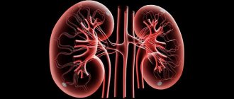
Hydronephrosis code according to ICD-10 is a progressive disease in which shaped and structural changes in the kidneys occur due to disturbances in the proper removal of urine from the body. It was found that boys in childhood and women after reaching 20 years of age are most susceptible to this kidney pathology. Hydronephrotic transformation of the left or right kidney is observed equally often, and the number of pathologies of both organs at once does not exceed 10% of cases of this disease.
Classification
Doctors divide the disease according to various criteria. As hydronephrosis appears, it is divided into congenital and acquired; depending on the number of affected organs, unilateral and bilateral hydronephrotic transformation is distinguished.
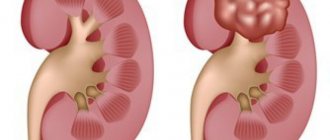
Based on how the disease progresses, there are three stages of the disease. In the first stage, pyeelectasis increases slightly without disrupting the functioning of the body. In the second stage of the disorder, the kidney grows by an average of 20% by expanding the renal calyces and disrupting the normal flow of urine. At the third stage, the kidney increases in size up to 2 times and can completely atrophy.
Aplasia (agenesis) of the kidney
ICD-10 code
Epidemiology
Ultrasound screening of 280,000 school-age children determined that renal aplasia occurs with a frequency of 1 in 1,200 people (0.083%).
Causes of kidney aplasia (agenesis)
Both renal agenesis and renal aplasia are considered anomalies of the urinary system, congenital malformations. Severe anatomical abnormalities can cause fetal death in utero; more compensated cases do not manifest clinical signs and are discovered during clinical examinations or randomly. Some anatomical abnormalities may progress gradually throughout life and be diagnosed in old age. Also, agenesis or aplasia can provoke kidney stones, arterial hypertension or pyelonephritis.
Kidney agenesis has been known since ancient times; Aristotle, in one of his works, described cases when a living creature could exist without a spleen or one kidney. In the Middle Ages, doctors were also interested in anatomical renal pathology, and an attempt was even made to describe in detail the aplasia (underdevelopment) of the kidney in a child. However, doctors did not conduct large-scale full-fledged studies, and only at the beginning of the last century by Professor N.N. Sokolov established the frequency of renal anomalies. Modern medicine has specified statistics and presents data on this indicator - agenesis and aplasia among all pathologies of the urinary system is 0.05%. It was also found that underdevelopment of the kidney most often affects men.
Risk factors
The following provoking factors are considered clinically established and statistically confirmed:
Symptoms of kidney aplasia (agenesis)
Where does it hurt?
- Bilateral anomaly (complete absence of kidneys) - bilateral agenesis or arena. As a rule, the fetus dies in utero, or the born child dies in the first hours or days of life due to renal failure. Modern methods make it possible to combat this pathology with the help of organ transplantation and regular hemodialysis.
- Agenesis of the right kidney is unilateral agenesis. This is an anatomical defect that is also congenital. A healthy kidney takes on the functional load, compensating for insufficiency as much as its structure and size allow.
- Agenesis of the left kidney is an identical case of agenesis of the right kidney.
- Aplasia of the right kidney is practically indistinguishable from agenesis, but the kidney is a rudimentary fibrous tissue without renal glomeruli, ureter and pelvis.
- Aplasia of the left kidney is an anomaly identical to the underdevelopment of the right kidney.
Variants of agenesis are also possible, in which the ureter is preserved and functions quite normally; in the absence of the ureter, the clinical manifestations of the pathology are more pronounced.
As a rule, in clinical practice a unilateral anomaly is encountered for obvious reasons - bilateral agenesis is not compatible with life.
Agenesis of the right kidney
If the left kidney takes over the function of the missing right kidney, then agenesis of the right kidney practically does not manifest itself symptomatically and is discovered randomly. The diagnosis can be confirmed using computed tomography, ultrasound examination and urography. Also, the pediatrician, as well as parents, should be wary of excessive puffiness of the child’s face, a flattened wide nose (flat bridge of the nose and a wide bridge of the nose), strongly protruding frontal lobes, and ears that are too low, possibly deformed. Ocular hypertelorism is not a specific symptom that indicates renal agenesis, but it often accompanies it in the same way as an enlarged abdomen and deformed lower limbs.
Agenesis of the left kidney
Visually pronounced agenesis of the left kidney can be determined by the same parameters as the agenesis of the right kidney, which are formed as a result of congenital intrauterine defects - oligohydramnios and fetal compression: a wide bridge of the nose, excessively wide-set eyes (hypertelorism), a typical face with Potter syndrome - a puffy face with underdeveloped chin, low-set ears, with prominent epicanthic folds.
Aplasia of the right kidney
Aplasia of the left kidney
An underdeveloped kidney does not have a leg or pelvis and is not able to function and produce urine. Aplasia of the left kidney, just like aplasia of the right kidney, is called in urological practice a solitary kidney, that is, a single kidney. This refers to only the kidney that is forced to function, compensatory to do double work.
Aplasia of the left kidney is detected randomly, since it does not manifest itself with clinically significant symptoms. Only functional changes and pain in the collateral kidney can give rise to a urological examination.
The right kidney, which is forced to perform the work of the aplastic left kidney, is usually hypertrophied and may have cysts, but most often it has a completely normal structure and completely controls homeostasis.
Diagnosis of kidney aplasia (agenesis)
Diagnosis of the disease is carried out using X-ray renal angiography, as well as spiral CT or MSCT and magnetic resonance angiography. Agenesis and renal aplasia are not diagnosed using other diagnostic methods.
What needs to be examined?
Who to contact?
Causes of the disease
Most often, hydronephrosis occurs as a result of a blockage in the urinary tract. Kidney stones or the passage of a kidney stone can disrupt the natural functioning of the kidneys and impede the natural flow of urine.
In men, two common causes of pathology are prostate carcinoma and non-cancerous prostate tumor, in which pressure on the ureters increases, thereby blocking the normal flow of urine.
Women often have the following reasons:
- pregnancy - during which the uterus may increase in size and put pressure on the ureters;
- cancers of organs such as the kidneys, ovaries, uterus and bladder;
Less common causes include diseases such as endometriosis, tuberculosis, ovarian cysts, as well as infections or various types of trauma that cause a narrowing of the ureter.
Aplasia of the right and left kidney: ICD code, causes, symptoms, disability
Medicine knows many anatomical anomalies, which can be congenital or acquired. Among congenital kidney defects, aplasia is sometimes found - a rather serious disease that can be fatal.
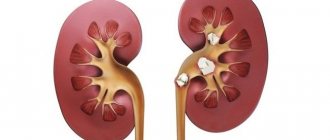
Kidney aplasia
Renal aplasia is a congenital anomaly in which one of the kidneys does not develop fully and instead of it, an underdeveloped rudiment is located in the place of the organ; it lacks a normal structure, which is why the kidney is unable to perform its functions. In such a situation, a healthy kidney assumes the responsibilities of an underdeveloped one, resulting in hypertrophy of its tissues due to compensatory activity.
In the international classification of diseases according to ICD-10, the code for renal aplasia is Q60. The pathology occurs twice as often in male children. In general, the incidence of renal aplasia is 1 case in 700-1200 newborns. Aplasia is characterized by the absence of the stalk and pelvis, or they are not fully formed.
In some sources, aplasia is equated with agenesis, however, these conditions are somewhat different.
If aplasia is the underdevelopment of the kidney with the presence of a ureter, then agenesis is the complete absence of the organ and its legs. Since these conditions have identical etiology and clinical manifestations, they are often combined into a common disease.
Aplasia can be unilateral, most often affecting the left kidney. Although there are cases of bilateral damage. In this case, the child is not viable. Such newborns are acutely underweight at birth and soon die.
Reason for development
It is impossible to determine why the fetus’s kidney stopped developing during a certain period of intrauterine development. For unknown reasons, the metanephric canal does not grow to the metanephrogenic blastema. Usually, the ureter with renal aplasia remains formed normally, although sometimes it is also underdeveloped or completely absent.
In boys, along with the absence of a ureter, there are problems such as a shrinkage or absence of a testicle, cystic formations on the testes and the absence of a vas deferens.
Experts note that the development of pathology is influenced by factors such as:
- Genetic predisposition to pathology;
- Radiation therapy for a pregnant woman;
- If a pregnant woman has diabetes;
- The presence of pathologies of infectious origin such as rubella or influenza in the first trimester of pregnancy;
- The pregnant woman has a history of syphilis and other pathologies of sexually transmitted origin;
- Alcohol addiction;
- Uncontrolled use of hormonal contraception.
In fact, renal aplasia is a congenital urinary anomaly. In severe cases, the fetus may die before birth. In compensated forms, aplasia may not manifest itself in any way, and pathology is detected only during examination. Sometimes aplastic changes progress gradually and are detected already at a fairly mature age.
Symptoms
There are several types of renal aplasia:
- The bilateral form of the anomaly is considered absolutely incompatible with life; children with such a lesion die soon after birth. The child can be saved through kidney transplantation or continuous hemodialysis;
- Aplasia of the right kidney is considered the most favorable from a prognostic point of view. Usually in such a situation, the left kidney assumes all the responsibilities of the right organ, so there may be no negative symptoms. Sometimes right-sided aplasia is detected during the diagnosis of nephropathology or persistent hypertension;
- Aplasia of the left kidney is considered more unfavorable, since with such underdevelopment of the function of the left kidney, the right one, which is considered less functional and more mobile, takes over. Renal aplasia on the left in men is usually accompanied by underdevelopment of adjacent structures such as the bladder, prostate, vas deferens or testicle. A feature of left renal aplasia is its frequent combination with underdevelopment of the lung. In women, such an anomaly is detected much less frequently, but is also accompanied by deviations of neighboring structures, for example, a bicornuate uterus or aplasia of the uterine body, absence of the ureter or appendages, vaginal duplication, underdevelopment of the septa inside the uterus.
Aplasia of the left kidney Aplasia is less pronounced than agenesis. In some cases, underdevelopment of the kidney does not manifest itself at all, only sometimes the patient may feel some pain in the lumbar region. Left-sided aplasia manifests itself somewhat more clearly than right-sided aplasia.
In general, with such a defect the following manifestations are noted:
- Oligoanuria – a decrease in the amount of urine and even its complete disappearance;
- Underdevelopment of the ureter;
- Adrenal gland abnormalities;
- Renal colic on the affected side, caused by the proliferation of fibrous tissue;
- Nagging pain in the lower abdomen;
- Persistent hypertension;
- Sometimes pain from a normal kidney can be bothersome, which doctors associate with an increase in its size.
If aplasia is severe, then renal failure develops or the body becomes intoxicated with its own metabolites. Then the patient’s condition worsens, almost all body systems begin to function intermittently.
Diagnostics
Diagnostic procedures to detect renal aplasia, regardless of the side on which the anomaly is located, include:
- Ultrasonography;
- X-ray of the kidneys;
- Renal angiography;
- Laboratory tests of urine;
- Spiral computed tomography;
- Magnetic resonance angiography, etc.
Treatment as such is not required for renal aplasia. The patient is necessarily prescribed a lifelong diet, the purpose of which is to reduce the load on the healthy kidney. If the pathology is accompanied by persistent hypertension, then treatment with mild diuretics is necessary.
It is also necessary to take preventive measures against bacterial kidney infections. The immune system also requires support so that the likelihood of viral agents or pathogenic bacteria entering the body is minimal. In particularly severe cases, transplantation is necessary.
The forecasts are generally favorable. If the necessary diet and prevention are followed, patients live their entire lives without any particular problems.
In patients with renal aplasia, disability can be established when the functionality of a single kidney decreases, which is accompanied by severe symptoms of renal failure. Although disability for such a pathology is not always granted, patients are released from the army and immediately enlisted in the reserves.
Rate this article:
gidmed.com
Treatment methods
For patients suffering from uncomplicated hydronephrosis of the first stage, which does not impair the body’s performance and well-being, while all kidney functions are still normal, medications with analgesic and anti-inflammatory effects are used. In other cases, only surgical intervention can restore the patency of the urinary tract and resume normal kidney function.
Despite the fact that doctors classify hydronephrosis as a serious disease, timely diagnosis and effective treatment will help restore the kidneys and normalize the natural functions of removing fluid from the body. Preventive examinations by a urologist using kidney ultrasound and prevention of urinary tract diseases can prevent the development of hydronephrosis.
Methods of therapy
Treatment of kidney hypoplasia is complex, determined and corrected only by a doctor. There are no methods for complete recovery; therapy is usually symptomatic.
Traditional remedies
The prescription of any medications depends on the characteristics of the pathology. If patients are diagnosed with arterial hypertension, the selection of therapy is carried out in accordance with the standards.
The antihypertensive treatment regimen is individual, established and adjusted by the doctor. Commonly used drugs:
- groups of ACE inhibitors (enalapril, perindopril, lisinopril);
- beta blockers (bisoprolol, nebivalol);
- sartans (losartan, valsartan);
- diuretics (indapafone, spironolactone).
It is important to note that antihypertensive therapy is rarely successful, and patients usually require kidney removal to normalize blood pressure.
If patients are diagnosed with pyelonephritis, they are prescribed antibacterial therapy using drugs from the fluoroquinolone group. Often such patients are also indicated for nephrectomy.
Preparations with a high content of vitamin D and uroseptics are useful. Patients should also adhere to recommendations for dietary correction - the amount of salt, liquid and protein should be limited.
You should eat easily digestible foods, cooked or steamed. Fatty, fried, canned foods, and carbonated drinks are prohibited. You can drink clean water and fruit drinks.
In what cases is surgery necessary?
In addition to resistant arterial hypertension and bilateral lesions, surgery may be necessary for organ dystopia. Patients undergo removal of one or two kidneys. It is carried out through abdominal or laparoscopic nephrectomy. Tactics are selected individually.
Traditional methods
Traditional therapy methods are based on the use of herbs. Herbal medicine is carried out in conjunction with traditional medicine; before doing this, you should consult a doctor.
Herbal infusions from juniper, parsley, bearberry, horsetail, chamomile, calendula, mint, St. John's wort, and marshmallow are useful.
These plants help cleanse the body of toxic metabolic products and have an antiseptic effect on the organs of the urinary tract.
They can be brewed either separately or in various combinations. Drink decoctions 3-4 times a day half an hour before meals.
Treatment of children
Treatment of pediatric patients is in many ways similar to treatment of adults. A significant difference lies in cases where bilateral kidney damage is diagnosed.
In such situations, it is necessary to remove the affected organs, otherwise life expectancy will be short.
Patients after double nephrectomy require a transplant. Before transplantation, they need to attend hemodialysis sessions to cleanse the body of toxic substances.
If one kidney is affected and the functions of the second are at a satisfactory level, patients do not need surgery; it is enough to constantly visit the doctor and follow all lifestyle recommendations.
If the affected organ does not function well, it should be removed.
Nutrition and lifestyle tactics for children are the same as for adults. You can periodically take uroseptic drugs of plant origin.
Diagnostics
The main diagnostic method was ultrasound of the kidneys. The examination reveals how much the organ has decreased in thickness and volume.
To detect discrepancies in the structure of the urinary tract, excretory urography is performed. This x-ray testing method involves injecting a contrast agent, urografin, into the blood.
Immediately after entering the body, it begins to be removed by the kidneys, which allows it to envelop the pelvis, ureter, bladder and urethra. X-rays are taken three times at seven-minute intervals.
An MRI may be performed for quick diagnosis. This procedure gives an idea not only of the size of the kidney, but also allows you to obtain a 3D image of the organ. This procedure is quite expensive, and few clinics can afford such equipment.
CT result for hypoplasia
The next method is ureteropyelography. The contrast agent is injected into the ureter through a catheter. A rather unpleasant procedure, which, if carried out inappropriately, can cause minor trauma to the mucous membrane, pain when urinating and minor bleeding.
Another type of study is nephroscintigraphy. This is an examination of an organ using radioactive material. A similar method is radioisotope examination through intravenous injection of a radioactive composition.
The disease can also be detected through angiography - an examination using a contrast agent introduced into a large vessel.
Urolithiasis (UCD): measures for the development of pathology
Urolithiasis is not a disease. It is classified as a borderline state when uric acid accumulates in the body. The pathological condition provokes the development of many diseases: gout, urolithiasis and others. Representatives of the male gender aged 40 years and older and women during menopause are susceptible to pathology. The code for the International Classification of Diseases (ICD 10) is E79.
What causes the disease
We have found out what MCD is, now let’s look at the most common causes of its occurrence:
- genetic predisposition;
- overweight, obesity;
- sedentary lifestyle;
- lack of proper nutrition;
- alcohol abuse;
- prolonged hypothermia;
- kidney function problems;
- autoimmune, endocrine diseases;
- various types of injuries.
The development of pathology is also caused by a violation of the daily volume of fluid consumed, radiation therapy for cancer, and fasting for a long time. This is not a definitive list of all the factors that cause the accumulation of uric acid.
Signs of uric acid accumulation
Signs of urolithiasis are clearly expressed. The main one is severe pain in the lumbar back. When the stone moves, the pain spreads to the entire abdomen. Therefore, the pathology can be confused with an exacerbation of appendicitis or ulcers.
Symptoms accompanying the phenomenon:
- painful bowel movements;
- the presence of blood and urates in urine;
- lack of desire to eat and sudden weight loss;
- poor sleep;
- weakening of the body, nausea, constipation;
- colic in the kidney area, tachycardia;
- fever, severe headache.
With urolithiasis, a person does not control his emotions and is often irritated. If you do not see a doctor promptly, seizures may occur. Signs may not appear all the time, but only when the condition worsens.
Consequences of urolithiasis
If a person has kidney MCD, and what it is, how the condition manifests itself, he does not know, the disease may not be detected in time. This will lead to the development of complications: acute nephropathy, urolithiasis, problems in the functioning of the gastrointestinal tract system, as well as the risk of developing uric acid infarction.
With advanced renal pathology, all organ systems suffer, especially the central nervous system, which is responsible for a person’s psychological health. The positive thing is that all consequences are eliminated without surgical treatment.
The folk way to cleanse the kidneys! Our grandmothers were treated using this recipe...
Cleaning your kidneys is easy! You need to add it during meals...
Ways to detect uric acid accumulation
Before treating this pathology, the patient is fully examined. He is interviewed by a nephrologist and examined by a urologist. It is important that the person explains to the doctor exactly where it hurts and what other symptoms have appeared. Next, the patient is sent for an ultrasound of the kidneys, urine and blood tests, and, if necessary, an x-ray.
Ultrasound examination reveals the presence of sand or stones even at an early stage of the formation of this condition. Therefore, ultrasound is the main diagnostic method. Sometimes additional research is used to determine whether inflammatory processes are developing in other parts of the urinary organs.
Treatment for MCD
Urolithiasis is treated in three stages: symptomatic therapy, reducing the severity of symptoms, and correcting the diet.
To maintain metabolic processes, a saline solution (Regidron, Disol) is administered. Cleansing of the body is carried out using an enema, enterosorbents (Atoxil, Polysorb, activated carbon). For severe pain, No-Shpu and Novalgin are prescribed. Dissolve salt compounds with Canephron, Cyston, Urolesan. Anti-inflammatory drugs are also needed here: Hexicon, Betadine and the like.
Antibacterial medications are prescribed at the discretion of the treating doctor. With this pathological phenomenon, the use of Levomycetin, Erythromycin, and Penicillin will be effective. Your back should always be kept warm. To do this, use a heating pad or a warm scarf or belt. Warm baths are helpful.
Diet food
ICD of the kidneys can also be treated with proper nutrition. Chocolate, sausages, canned food, as well as meat, legumes, and offal are excluded from the diet. There is no need to eat mushrooms, processed foods, dairy products, pickles, or drink alcoholic beverages. It is worth removing fats and broths, hot and spicy snacks, and canned vegetables.
Eggs, prunes, potatoes, as well as nuts, citrus fruits, cabbage, including sea cabbage are recommended for consumption. Fill your daily diet with fruits, wheat bran, various cereals and eggplants, freshly prepared juices, fruit drinks, and compotes. Grapes, dried apricots, and vegetable oil will be useful. The standard drinking volume is two liters of liquid per day.
Clinical picture
Common manifestations of the disease are considered to be:
- the presence of streaks of blood in the urine;
- pain in the lumbar region;
- rapid weight loss;
- increased body temperature;
- Constantly bad mood, general weakness.
Attention! Cancer of the left kidney in ICD 10 is listed under code C64.1, but its symptoms exactly coincide with those that appear when the right side of the urinary organ is affected. And one of the main things is to palpate the painful tumor in the lumbar area.
Most patients complain of pain not only in the lumbar region, but also in the abdomen. Moreover, renal colic may be accompanied by hematuria. But if this phenomenon is one-time or short-term, then it is simply not given importance, which is very dangerous. As the disease progresses, metastases appear. If the left kidney is diseased, secondary lesions are located in the para-aortic lymph nodes; if the right one is affected, then the aortocaval, precaval, and laterocaval lymph nodes are primarily affected.
Content
Definition and general information [edit]
A hypoplastic kidney is distinguished from aplasia by the presence of functioning (albeit in a reduced volume) parenchyma, a ureter that is passable along its entire length, and visualization of the vascular pedicle during aortography. This is a congenital reduction of the kidney, mainly associated with impaired development of the metanephrogenic blastema as a result of insufficient blood supply.
The anomaly occurs with approximately the same frequency as renal aplasia.
Pathomorphology and classification
Macroscopically, a hypoplastic kidney is a normally formed organ in miniature. On its section, the cortical and medulla layers are clearly defined. However, histologically, changes are revealed that make it possible to distinguish three forms of hypoplasia: simple hypoplasia, hypoplasia with oligonephronia, hypoplasia with dysplasia.
1) The simple form of hypoplasia is characterized only by a decrease in the number of calyces and nephrons.
2) In the second form, a decrease in the number of glomeruli is combined with an increase in their diameter, fibrosis of interstitial tissue, and dilation of the tubules.
3) Hypoplasia with dysplasia is manifested by the development of connective tissue or muscle couplings around the primary tubules. Glomerular or tubular cysts are detected, as well as inclusions of lymphoid, cartilage and bone tissue. This form of hypoplasia, unlike the first two, is often accompanied by abnormalities of the urinary tract.
Etiology and pathogenesis[edit]
Clinical manifestations[edit]
Unilateral hypoplasia may not manifest itself in anything throughout life, however, it is noted that a hypoplastic kidney is often affected by pyelonephritis and often becomes a source of development of nephrogenic hypertension.
Unilateral kidney hypoplasia: Diagnosis [edit]
Unilateral hypoplasia is usually detected by X-ray examination undertaken for pyelonephritis. Excretory urograms show a decrease in the size of the kidney with a well-contrasted collector system. The contours of the kidney may be uneven, the pelvis is moderately dilated.
Differential diagnosis[edit]
With hypoplasia of the kidneys, the calyces are not deformed, as with pyelonephritis, but are only reduced in number and volume. Urograms show compensatory hypertrophy of the contralateral kidney.
Renal angiography is of great help in differential diagnosis. With hypoplasia, the arteries and veins are evenly thinned along their entire length, while with a secondarily shriveled kidney, the angiogram resembles the picture of a “burnt tree.” Kidney biopsy may also be useful, but its practical diagnostic value for hypoplasia is limited.
Unilateral kidney hypoplasia: Treatment[edit]
In cases of unilateral hypoplasia complicated by pyelonephritis and hypertension, treatment usually comes down to nephrectomy.
Prevention of kidney hypoplasia
To prevent the development of congenital pathologies in the fetus, a pregnant woman needs to take a responsible attitude towards her health, as a source of new life. It is necessary to protect the fetus as much as possible from adverse factors, both external and internal. No smoking, alcohol or drugs! The first trimester is the most important stage, since the child’s systems and organs are formed. During this period, even medications should not be taken, if possible. Self-medication is unacceptable; all your actions must be coordinated with your doctor.
Viral infections that cause rubella, measles, influenza, herpes and diseases transmitted during sexual intercourse become dangerous for a child. To protect the baby, a woman should lead a healthy lifestyle, avoid hypothermia, and avoid crowded places during influenza and ARVI epidemics. A pregnant woman's diet must be balanced so that the child receives all the necessary elements. Fruits, vegetables, juices - all this should be in abundance in the diet. Solarium and excessive tanning on the beach are prohibited, since radiation exposure can be very harmful.
Your attending physician will tell you more about how to prevent the development of pathological conditions in an infant. By following his recommendations, you can avoid complications and give birth to a healthy baby.