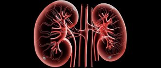
The structure of the nephron - how the main structural unit of the kidney works
The kidneys are a complex structure. Their structural unit is the nephron. The structure of the nephron allows it to fully perform its functions - filtration, the process of reabsorption, excretion and secretion of biologically active components occur in it.
Primary urine is formed, then secondary urine is excreted through the bladder. Throughout the day, a large amount of plasma is filtered through the excretory organ. Part of it is subsequently returned to the body, the rest is removed.
The structure and functions of nephrons are interrelated. Any damage to the kidneys or their smallest units can lead to intoxication and further disruption of the functioning of the entire body.
The consequence of irrational use of certain drugs, improper treatment or diagnosis can be kidney failure. The first manifestations of symptoms are the reason to visit a specialist.
This problem is dealt with by urologists and nephrologists.
What is a nephron
The nephron is the structural and functional unit of the kidney. There are active cells that are directly involved in the production of urine (a third of the total amount), the rest are in reserve.
Reserve cells become active in emergency situations, for example, during injury, critical conditions, when a large percentage of kidney units are suddenly lost. The physiology of excretion involves partial cell death, so reserve structures are able to be activated in the shortest possible time to maintain the functions of the organ.
Every year, up to 1% of structural units are lost - they die forever and are not restored. With a healthy lifestyle and the absence of chronic diseases, loss begins only after 40 years.
Considering that the number of nephrons in a kidney is approximately 1 million, the percentage seems small.
With old age, the functioning of the organ can deteriorate significantly, which threatens to impair the functionality of the urinary system.
The aging process can be slowed down by changing lifestyle and consuming enough clean drinking water. Even in the best case, over time only 60% of active nephrons remain in each kidney. This figure is not at all critical, since plasma filtration is impaired only with the loss of more than 75% of cells (both active and those in reserve).
Some people live after losing one kidney, and then the second one performs all the functions. The functioning of the urinary system is significantly impaired, so it is necessary to prevent and treat diseases in a timely manner. In this case, you need to regularly visit your doctor to prescribe maintenance therapy.
Nephron anatomy
The anatomy and structure of the nephron is quite complex - each element plays a specific role. If even the smallest component malfunctions, the kidneys cease to function normally.
Nephron structure:
- capsule;
- glomerular structure;
- tubular structure;
- loops of Henle;
- collecting ducts.
The nephron in the kidney consists of segments communicating with each other. The Shumlyansky-Bowman capsule, a tangle of small vessels, are components of the renal body where the filtration process takes place. Next come the tubules, where substances are reabsorbed and produced.
The proximal portion begins from the renal corpuscle; Then the loops extend into the distal section. The nephrons, when unfolded, are individually about 40 mm long, and when folded together they are approximately 100,000 m long.
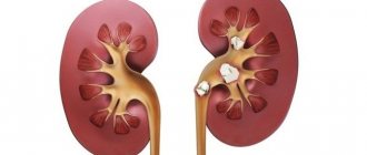
The nephron capsules are located in the cortex, are included in the medulla, then again in the cortex, and finally into the collecting structures that exit into the renal pelvis, where the ureters begin. Secondary urine is removed through them.
Capsule
The nephron originates from the Malpighian body. It consists of a capsule and a tangle of capillaries. The cells around the small capillaries are arranged in the shape of a cap - this is the renal corpuscle, which allows retained plasma to pass through. Podocytes cover the wall of the capsule from the inside, which, together with the outside, forms a slit-like cavity with a diameter of 100 nm.
Fenestrated (fenestrated) capillaries (components of the glomerulus) are supplied with blood from afferent arteries. They are otherwise called “magic mesh” because they do not play any role in gas exchange. The blood passing through this mesh does not change its gas composition. Plasma and dissolved substances enter the capsule under the influence of blood pressure.
The nephron capsule accumulates an infiltrate containing harmful products of blood plasma purification - this is how primary urine is formed. The slit-like gap between the layers of the epithelium acts as a filter operating under pressure.
Thanks to the afferent and efferent glomerular arterioles, the pressure changes. The basement membrane plays the role of an additional filter - it retains some blood elements. The diameter of the protein molecules is larger than the pores of the membrane, so they do not pass through.
Unfiltered blood enters the efferent arterioles, which pass into a network of capillaries that envelops the tubules. Subsequently, substances enter the blood and are reabsorbed in these tubules.
The nephron capsule of the human kidney communicates with the tubule. The next section is called proximal; primary urine then passes there.
Mixed Lot
Proximal tubules can be straight or curved. The surface inside is lined with cylindrical and cubic epithelium. The brush border with villi is the absorptive layer of the nephron tubules. Selective capture is ensured by the large area of the proximal tubules, the close dislocation of peritubular vessels and a large number of mitochondria.
Fluid circulates between cells. Plasma components in the form of biological substances are filtered. The convoluted tubules of the nephron produce erythropoietin and calcitriol. Harmful inclusions that enter the filtrate using reverse osmosis are removed with urine.
Nephron segments filter creatinine. The amount of this protein in the blood is an important indicator of the functional activity of the kidneys.
Loops of Henle
The loop of Henle involves part of the proximal and part of the distal section. At first, the diameter of the loop does not change, then it narrows and allows Na ions to pass out into the extracellular space. By creating osmosis, H2O is absorbed under pressure.
The descending and ascending ducts are the components of the loop. The descending region, 15 µm in diameter, consists of epithelium where multiple pinocytotic vesicles are located. The ascending portion is lined with cubic epithelium.
The loops are distributed between the cortex and medulla. In this area, water moves to a downward section, then returns.
At the beginning, the distal canal touches the capillary network at the site of the afferent and efferent vessels. It is quite narrow and is lined with smooth epithelium, and on the outside there is a smooth basement membrane. Ammonia and hydrogen are released here.
Collecting ducts
The collecting ducts are also called “ducts of Belline”. Their internal lining consists of light and dark epithelial cells. The former reabsorb water and are directly involved in the production of prostaglandins. Hydrochloric acid is produced in the dark cells of the folded epithelium and has the ability to change the pH of urine.
The collecting ducts and collecting ducts do not belong to the nephron structure, as they are located slightly lower, in the renal parenchyma. Passive reabsorption of water occurs in these structural elements. Depending on the functionality of the kidneys, the amount of water and sodium ions in the body is regulated, which, in turn, affects blood pressure.
Types of nephrons
Structural elements are divided depending on their structural features and functions.
Types of nephrons:
- cortical;
- juxtamedullary.
Cortical ones are divided into two types - intracortical and superficial. The number of the latter is approximately 1% of all units.
Features of superficial nephrons:
- low filtration volume;
- location of the glomeruli on the surface of the cortex;
- the shortest loop.
The kidneys mainly consist of nephrons of the intracortical type, of which more than 80%. They are located in the cortex and play a major role in filtering primary urine. Due to the greater width of the efferent arteriole, blood enters the glomeruli of intracortical nephrons under pressure.
Cortical elements regulate the amount of plasma. When there is a lack of water, it is recaptured from the juxtamedullary nephrons, located in greater quantities in the medulla. They are distinguished by large renal corpuscles with relatively long tubules.
Juxtamedullary ones make up more than 15% of all nephrons in the organ and form the final amount of urine, determining its concentration. Their structural feature is the long loops of Henle. The efferent and afferent vessels are of equal length. Loops are formed from the efferents, penetrating into the medulla in parallel with Henle. Then they enter the venous network.
Functions
Depending on the type, kidney nephrons perform the following functions:
- filtration;
- reverse suction;
- secretion.
The first stage is characterized by the production of primary urea, which is further purified by reabsorption. At the same stage, beneficial substances, micro- and macroelements, and water are absorbed.
The last stage of urine formation is represented by tubular secretion - secondary urine is formed. It removes substances that the body does not need.
The structural and functional unit of the kidney is the nephron, which:
- maintain water-salt and electrolyte balance;
- regulate the saturation of urine with biologically active components;
- maintain acid-base balance (pH);
- control blood pressure;
- remove metabolic products and other harmful substances;
- participate in the process of gluconeogenesis (production of glucose from non-carbohydrate compounds);
- provoke the secretion of certain hormones (for example, those that regulate the tone of the vascular walls).
The processes occurring in the human nephron make it possible to assess the condition of the organs of the excretory system. This can be done in two ways. The first is to calculate the content of creatinine (a protein breakdown product) in the blood. This indicator characterizes how well the kidney units cope with the filtration function.
The work of the nephron can also be assessed using a second indicator - glomerular filtration rate. Blood plasma and primary urine should normally be filtered at a rate of 80-120 ml/min. For older people, the lower limit may be the norm, since after 40 years the kidney cells die (there are significantly fewer glomeruli, and it is more difficult for the organ to fully filter fluids).
Functions of some components of the glomerular filter
The glomerular filter consists of fenestrated capillary endothelium, basement membrane and podocytes. Between these structures is the mesangial matrix. The first layer performs the function of coarse filtration, the second filters out proteins, and the third cleanses the plasma of small molecules of unnecessary substances. The membrane has a negative charge, so albumin does not penetrate through it.
Blood plasma is filtered in the glomeruli, and their work is supported by mesangiocytes - cells of the mesangial matrix. These structures perform contractile and regenerative functions. Mesangiocytes restore the basement membrane and podocytes, and, like macrophages, they engulf dead cells.
If each unit does its job, the kidneys function like a well-coordinated mechanism, and urine formation occurs without toxic substances returning to the body. This prevents the accumulation of toxins, the appearance of swelling, high blood pressure and other symptoms.
Nephron function disorders and their prevention
If the functioning of the functional and structural units of the kidneys is disrupted, changes occur that affect the functioning of all organs - the water-salt balance, acidity and metabolism are disrupted.
The gastrointestinal tract ceases to function normally, and allergic reactions may occur due to intoxication. The load on the liver also increases, since this organ is directly related to the elimination of toxins.
For diseases associated with transport dysfunction of the tubules, there is a single name - tubulopathies. They come in two types:
The first type is congenital pathology, the second is acquired dysfunction.
Active nephron death begins when taking medications whose side effects indicate possible kidney disease. Some drugs from the following groups have a nephrotoxic effect: nonsteroidal anti-inflammatory drugs, antibiotics, immunosuppressants, antitumor drugs, etc.
Tubulopathies are divided into several types (based on location):
- proximal;
- distal.
With complete or partial dysfunction of the proximal tubules, phosphaturia, renal acidosis, hyperaminoaciduria and glycosuria may occur.
Impaired phosphate reabsorption leads to the destruction of bone tissue, which is not restored with vitamin D therapy.
Hyperaciduria is characterized by a violation of the transport function of amino acids, which leads to various diseases (depending on the type of amino acid). Such conditions require immediate medical attention, as well as distal tubulopathies:
- renal water diabetes;
- tubular acidosis;
- pseudohypoaldosteronism.
Violations can be combined. With the development of complex pathologies, the absorption of amino acids with glucose and the reabsorption of bicarbonates with phosphates may simultaneously decrease. Accordingly, the following symptoms appear: acidosis, osteoporosis and other bone tissue pathologies.
Kidney dysfunction is prevented by proper diet, drinking enough clean water and an active lifestyle. It is necessary to contact a specialist in a timely manner if symptoms of kidney dysfunction occur (to prevent the transition of the acute form of the disease to the chronic form).
It is not recommended to take medications (especially prescription drugs with nephrotoxic side effects) without a doctor's prescription - they can also disrupt the functions of the urinary system.
Source: https://propochki.info/anatomiya/nefron-stroenie-vidy-funkcii
Structural and functional unit of the kidney
The main structural and functional unit of the kidney is the nephron. The anatomy and physiology of the structure is responsible for the formation of urine, reverse transport of substances and the production of a range of biological substances.
The structure of the nephron is an epithelial tube. Next, networks of capillaries of various diameters are formed, which flow into the collecting vessel.
The cavities between the structures are filled with connective tissue in the form of interstitial cells and matrix.
The development of the nephron begins in the embryonic period. Different types of nephrons are responsible for different functions. The total length of the tubules of both kidneys is up to 100 km. Under normal conditions, not the entire number of glomeruli is involved, only 35% work. The nephron consists of a body, as well as a system of canals. It has the following structure:
- capillary glomerulus;
- glomerular capsule;
- near tubule;
- descending and ascending fragments;
- distant straight and convoluted tubules;
- connecting path;
- collecting ducts.
The structural and functional unit of the kidney is the nephron (there are more than a million nephrons in one kidney alone). This means that the nephron of the kidney performs the main renal work of the urinary system. Nephrons, as the functional units of the kidneys, perform the task of removing waste products from the body in a timely manner (before toxins reach toxic levels).
The main parts of the nephron are the glomerulus and the tubular system. The glomerulus is a network of intertwining capillaries collected in a cup-shaped structure called Bowman's capsule. The blood is filtered in the capillaries of the glomeruli, and the filtered liquid (filtrate) collects in the space of Bowman's capsule, passing through the filter membrane.
MORE: Nephroptosis of the right kidney, 1st degree - Kidneys
Types of nephrons
Based on the layer in which the nephron capsules are located, the following types are distinguished:
- Cortical - nephron capsules are located in the cortical ball; they contain small or medium-sized glomeruli with the corresponding length of bends. Their afferent arteriole is short and wide, and their efferent arteriole is narrower.
- Juxtamedullary nephrons are located in the medullary renal tissue. Their structure is presented in the form of large renal corpuscles, which have relatively longer tubules. The diameters of the afferent and efferent arterioles are the same. role - concentration of urine.
- Subcapsular. Structures located directly under the capsule.
In general, in 1 minute both kidneys purify up to 1.2 thousand ml of blood, and in 5 minutes the entire volume of the human body is filtered. It is believed that nephrons, as functional units, are not capable of repair.
The kidneys are a delicate and vulnerable organ, so factors that negatively affect their functioning lead to a decrease in the number of active nephrons and provoke the development of renal failure. Thanks to knowledge, the doctor is able to understand and identify the causes of changes in urine, as well as carry out corrections.
READ MORE: How to clear oxalate stones from your kidneys
etopochki.ru
Since the renal corpuscles of most nephrons are located in the cortical layer of the kidney parenchyma (in the outer cortex), and their loops of Henle of short length pass in the outer renal medulla along with most of the blood vessels of the kidney, they are usually called cortical, or intracortical.
The rest of them (about 15%), with a loop of Henle of greater length, deeply immersed in the medulla (up to reaching the apices of the renal pyramids), is located in the juxtamedullary cortex - the border zone between the medulla and the cortical layer, which allows them to be called juxtamedullary.
Less than 1% of nephrons located shallowly in the subcapsular layer of the kidney are called subcapsular, or superficial.
You now know that the structural and functional unit of the kidney is the nephron. But it turns out that there are several types of nephrons, differing in functional purpose and structural features:
- Juxtamedullary.
- Cortical, namely intracortical and superficial.
Cortical
There are two types of nephrons located in the renal cortex. Of these, superofficials account for only 1%. Their differences are a low filtration volume, a shortened loop of Henle, and superficial localization of the glomeruli in the cortex.
The share of intracortical nephrons accounts for 80%. They are localized in the middle part of the cortex. These nephrons perform the main functions of filtering urine. At the same time, the blood in such nephrons flows under high pressure. This is due to the expansion of the adductor artery.
Juxtamedullary
This is a small group of nephrons, accounting for only 20%. Most of the nephron is located in the medulla, and the capsule is located at the border of the medulla and the cortex. In such nephrons, the loop of Henle descends almost to the renal pelvis.
READ MORE: Symptoms of kidney prolapse (right or left), signs of pathology
These nephrons are important for the concentrating function of the kidneys, that is, the organ's ability to concentrate urine. This type of nephron has the longest loop of Henle, and the efferent and afferent arteries have the same diameter.
Since the renal corpuscles of most nephrons are located in the cortical layer of the kidney parenchyma (in the outer cortex), and their loops of Henle of short length pass in the outer renal medulla along with most of the blood vessels of the kidney, they are usually called cortical, or intracortical.
Source: https://pocki.top/kamni/edinitsa-pochki/
Glomerular filtration: the rate of processes and their structure
Kidney function is characterized by the filtration rate provided by the glomeruli. It is capable of reaching almost 170-200 l/day. This figure is 16-18 times greater than the total volume of blood circulating in the body. The high speed of the processes ensures increased filtration of fluids, due to which it passes through the nephrons about 18-20 times during the day.
The rate of renal filtration makes it possible to judge the performance and general condition of the kidneys. At the same time, a decrease in such values indicates the presence of any disorders or pathologies.
Therefore, determining the intensity of blood supply to functional units is an important parameter that allows for timely detection of renal failure.
The glomerular filters of the nephrons ensure the separation of fluid and other substances with a small specific gravity of molecules, helping to create blood plasma. During filtration, a barrier is created through which no substance of large mass or high molecular structure can penetrate. In this case, the membrane that directly filters blood consists of several layers:
potocytes;- basal tissues;
- vascular endothelial cells.
Passing through these balls of tissue, the crude liquid penetrates the glomerulus and undergoes filtration. This structure ensures that protein and other larger impurities are filtered out. In this case, plasma and liquid freely leak through the filtration membrane - this is the fundamental function of the nephron and the kidney itself.
The nephron, as a structural and functional unit of the kidney, has a rather complex structure, consisting of several sections and forming an isolated system. Each of them has microscopic dimensions and performs its own specific functions, ensuring high performance of nephrons. The renal nephron provides blood filtration, participates in excretory processes and promotes the formation of urine, ensuring the balance of substances in the body and its timely cleansing.
vsepropechen.ru
What is a nephron: structure, types and main functions – Don’t worry, liver!
- The general structural plan of all nephrons is similar, however, depending on the location, the parameters of individual fragments of the structural unit may differ (the length of the tubules and loops, as well as the dimensions of the vascular glomeruli formed by the branching afferent arteriole and the efferent vessel).
- From an anatomical point of view, the structure of the structural and functional unit of the kidney is as follows:
- Shumlyansky capsule. Inside this formation there is a glomerulus formed by the smallest capillaries - the Malpighian corpuscle, also called the “renal corpuscle”. The Shumlyansky-Bowman capsule has a diameter of 0.2 mm. Its epithelium is tightly adjacent to the capillary glomerulus.
- The proximal tubule extends directly from the capsule containing the renal corpuscle. The proximal tubule has the following structural feature: the cellular elements of the epithelium lining its inner surface have a brush border - a system of microvilli that face the lumen of the proximal tubule.
- Loop of Henle. The loop of Henle has thin and thick sections. The thin one is represented by the descending part of the loop and part of the ascending part. The thick section is represented by part of the ascending section of the loop, the convoluted tubule and the connecting section.
- Distal convoluted tubule. Its beginning necessarily comes into contact with the glomerulus formed by the afferent and efferent arterioles.
- A connecting tubule that connects the distal tubule to the collecting ducts.
- Collecting tube. From a functional point of view, it is a continuation of the distal segment of the nephron. These anatomical formations originate in the renal cortex and, merging with each other, subsequently form excretory ducts. Passing through the renal medulla, which lies under the cortex, the excretory ducts open into the cavity of the renal pelvis.
The total length of the nephron tubules is from three to five centimeters.
How does a nephron work?
Urine formation occurs in the nephrons. This process takes place in several stages.
Filtration of blood plasma occurs in the glomerulus formed by high-pressure capillaries (70 millimeters of mercury). The pressure gradient in these microvessels is determined by the following factors (anatomical reasons):
- the renal arteries, which supply blood to the entire organ, arise directly from the abdominal aorta;
- small length of these arteries;
- a significant difference in the diameter of the afferent and efferent arterioles: the lumen of the efferent artery is twice as narrow as the afferent arteriole.
As a result of pressure, the mechanism of a semi-permeable membrane is activated, which helps filter out the liquid component of the blood circulating through the capillaries. After passing through the glomerulus and freed from substances to be excreted, the blood enters the efferent vessel, through which it flows away from the nephron.
Blood plasma filtered through a specialized membrane enters the capsule. The resulting liquid is called primary urine.
It includes:
- water;
- microelements;
- organic nutrients.
Its quantity reaches 100−150 liters per day.
Next, the liquid moves through the tubule, the function of which is reabsorption (reverse absorption) of water and substances dissolved in it (microelements, glucose and other organic compounds).
As a result of this process, so-called secondary urine is formed. It differs from the primary one in having a higher concentration of salts. Its amount reaches about 2 liters per day.
The process of tubular secretion occurs in the distal tubule. This is the process of transition through the epithelial cells of the distal tubule into the urine of substances that must be excreted from the body (in particular, potassium ions, hydrogen, ammonia, toxic substances entering the body from the outside, including as part of medications). In this case, the following mechanisms are triggered:
- Active (the movement of chemicals is carried out using the energy of biochemical reactions).
- Passive (due to the energy of cellular osmosis).
The result of tubular secretion is the restoration of the acid-base balance of the blood.
Subsequently, secondary urine enters the renal pelvis through the collecting duct, from where it is transported through the ureters to the bladder.
Briefly, in the form of a table, the diagram of the process of blood filtration by the nephron can be presented as follows:
| Anatomical education | Processes |
| Shumlyansky-Bowman capsule | Blood plasma filtration |
| Proximal tubule and loop of Henle | Reabsorption |
| Distal tubule | Secretion to form the final composition of urine |
The number of nephrons is limited
For normal life, approximately a third of the nephrons present in the kidneys are sufficient. The rest are reserve, in case of death of those functioning (as a result of injury or illness).
The structural and functional unit of the kidney cannot recover, therefore, as a result of any damage, their number in the kidneys decreases.
Over time, if such processes progress, renal failure may develop, negatively affecting the functioning of all organs and systems.
With the invention of means that help restore the filtering structures of the kidney, a lot of problems arising as a result of diseases affecting this organ will be solved.
In the meantime, experts say, the only measure to prolong the functional viability of the kidneys is the prevention of diseases of the urinary system and timely comprehensive treatment of acute diseases, which does not allow them to become chronic.
Source:
What functions do kidney nephrons perform and their structure - ODSIS Medical portal
The nephron is the structural unit of the kidney responsible for the formation of urine. Working 24 hours, the organs pass up to 1700 liters of plasma, forming a little more than a liter of urine.
Nephron
The work of the nephron, which is the structural and functional unit of the kidney, determines how successfully the balance is maintained and waste products are eliminated.
During the day, two million nephrons of the kidneys, as many as there are in the body, produce 170 liters of primary urine, condensed to a daily amount of up to one and a half liters.
The total area of the excretory surface of the nephrons is almost 8 m2, which is 3 times the area of the skin.
The excretory system has a high reserve of strength. It is created due to the fact that only a third of the nephrons work at the same time, which allows them to survive when the kidney is removed.
Arterial blood flowing through the afferent arteriole is cleansed in the kidneys. Purified blood comes out through the exiting arteriole. The diameter of the afferent arteriole is larger than that of the arteriole, due to which a pressure difference is created.
Structure
The divisions of the nephron of the kidney are:
- They begin in the cortex of the kidney with Bowman's capsule, which is located above the glomerulus of capillaries of the arteriole.
- The nephron capsule of the kidney communicates with the proximal (closest) tubule, directed to the medulla - this is the answer to the question in which part of the kidney the nephron capsules are located.
- The tubule passes into the loop of Henle - first into the proximal segment, then into the distal segment.
- The end of the nephron is considered to be the place where the collecting duct begins, where secondary urine from many nephrons enters.
Nephron diagram
Proximal tubule
This species consists of cells covered on the outside with a basement membrane. The inner part of the epithelium is equipped with outgrowths - microvilli, like a brush, lining the tubule along the entire length.
Outside there is a basement membrane, assembled into numerous folds, which straighten when the tubules are filled. At the same time, the tubule acquires a rounded shape in diameter, and the epithelium becomes flattened. In the absence of fluid, the diameter of the tubule becomes narrow, the cells acquire a prismatic appearance.
Functions include reabsorption:
- H2O;
- Na – 85%;
- ions Ca, Mg, K, Cl;
- salts - phosphates, sulfates, bicarbonate;
- compounds - proteins, creatinine, vitamins, glucose.
From the tubule, reabsorbents enter the blood vessels, which encircle the tubule in a dense network. In this area, bile acid is absorbed into the cavity of the tubule, oxalic, para-aminohippuric, and uric acids are absorbed, adrenaline, acetylcholine, thiamine, histamine are absorbed, and drugs are transported - penicillin, furosemide, atropine, etc.
Here, the breakdown of hormones coming from the filtrate occurs with the help of enzymes in the epithelial border. Insulin, gastrin, prolactin, bradykinin are destroyed, their concentration in plasma decreases.
Loop of Henle
After entering the medullary ray, the proximal tubule passes into the initial part of the loop of Henle. The tubule passes into the descending segment of the loop, which descends into the medulla. The ascending portion then ascends into the cortex, approaching Bowman's capsule.
The internal structure of the loop initially does not differ from the structure of the proximal tubule. Then the lumen of the loop narrows, through which Na is filtered into the interstitial fluid, which becomes hypertonic.
This is important for the operation of the collecting ducts: due to the high concentration of salt in the washer fluid, water is absorbed into them. The ascending section expands and passes into the distal tubule.
Source: https://ahcrb.ru/bolezni-pochek/chto-takoe-nefron-stroenie-vidy-i-osnovnye-funktsii.html
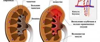
Damage to the renal glomeruli
Thus, it is obvious that any pathology of the renal glomeruli leads to serious problems. The pathophysiological mechanisms of damage to the main structural unit of the kidney, the glomerulus, are explained using three models:
- Whole nephron theories.
- Theories of hyperfiltration.
- Theory of complex deposits.
The whole nephron theory is explained as follows. Each nephron is a miniature kidney. Therefore, damage to one of its components leads to damage to the entire nephron. This may occur due to defects in the peritubular capillary network, changes in the composition of the fluid flowing through the tubules, reduced oxygen supply and resulting metabolic deficits.
The consequences of damage to the nephron are a decrease in protein filtration and a decrease in the synthesis of hormones, primarily erythropoietin. The result is necrosis of the tubular epithelium and insufficient filtration.
Sometimes the nephron can repair itself. But the opposite picture also happens – necrosis of the nephron. In this case, hypertrophy or hyperfunction of the nephrons that surround the dead unit may occur as compensation. This is followed by fibrosis of the affected parts of the kidney, followed by vascular failure of the remaining nephrons and progressive kidney damage.
The second hypothesis is the theory of hyperfiltration, when increased filtration leads to damage to the renal glomeruli due to increased blood pressure, which puts more intense pressure on their tissue. This may be the result of drugs that are toxic to the kidneys.
The complex deposit theory suggests that the problem occurs when immune complexes, which are clumped clumps of antibodies, cannot pass into the tubules due to their large size. Therefore, they are deposited in the glomerulus, causing sclerosis and tissue scarring.
In any case, no matter what damage the nephrons cause, the situation is dangerous not only for health, but also for human life. Therefore, if you have any suspicions about kidney failure, you should consult a doctor and undergo examination.
2pochku.ru
Source: pochki5.ru
The structural and functional unit of the kidney is the nephron
The renal unit is called the nephron. It is responsible for filtering blood and forming primary urine. The functional unit of the kidney removes toxins and metabolic products from the body. Nephrons work around the clock, filtering up to 1.7 thousand liters of blood plasma. In this case, a little more than a liter of excreted urine is formed.
In this case, about 170 liters of primary urine are formed per day. Subsequently, this volume is condensed to the daily urine norm. There are about 2 million nephrons in our kidneys. If we calculate the total surface area of the nephrons performing the excretory function, it will be approximately 8 m².
This is three times the area of skin.
Distal tubule
The distal tubules are shorter sections consisting of low epithelial cells. The inner surface of the canal is no longer lined with villi. On the outside, the folded basement membrane is still present. In this part, the nephron, as a structural unit of the kidney, functions on the principle of reabsorption of water, sodium, and also releases ammonia and hydrogen ions into the lumen.
Types of nephrons
There are several types of nephrons, differing in functional purpose and structural features
You now know that the structural and functional unit of the kidney is the nephron. But it turns out that there are several types of nephrons, differing in functional purpose and structural features:
- Juxtamedullary.
- Cortical, namely intracortical and superficial.
Functions of renal nephrons
the task of these renal nephrons is the formation of urine and the reabsorption of important and useful substances and compounds
Since the nephron is the functional unit of an organ, the main tasks of this organ are as follows:
- regulation of vascular tone;
- concentration of urine;
- blood pressure control.
The process of urine formation consists of several stages:
- The glomeruli filter the blood plasma that enters the organ through the arteries. As a result, primary urine is formed.
- Beneficial substances are reabsorbed from the resulting filtrate.
- Urine concentration occurs.
Functions of cortical nephrons
The task of these renal nephrons is the formation of urine and the reabsorption of important and useful substances and compounds - amino acids, proteins, glucose, minerals, hormones.
These nephrons are involved in the process of urine filtration and reabsorption, since they have some features of the blood supply.
All reabsorbed beneficial substances and compounds immediately enter the blood through the capillary network of the efferent artery, which is located nearby.
Functions of juxtamedullary nephrons
The task of these kidney elements is to concentrate urine. This is achieved due to certain features of blood transportation through the efferent artery. The artery does not pass through the capillary node, but immediately flows into venules, which transform into veins.
Important: this type of nephron is involved in the formation of substances that regulate blood pressure. The complex of these nephrons produces renin, which is needed for the formation of a special vasoconstrictor substance - angiotensin 2.
Functional disorders in the activity of nephrons
If malfunctions occur in the functioning of the nephrons, this affects the activity of all organs and systems. Among the disorders that arise due to nephron dysfunction, the following disorders can be mentioned:
- water and salt balance;
- acidity;
- metabolism.
All diseases that develop against the background of impaired transport activity of nephrons are usually called tubulopathies. Among them the following varieties are distinguished:
- Primary tubulopathy occurs against the background of congenital nephron dysfunction.
- Secondary forms of the disease arise due to acquired disorders of the transporting activity of the organ.
Common causes of secondary tubulopathy are damage to the nephron due to toxic damage to the body, malignant neoplasms or heavy metal poisoning. According to the location, all tubulopathies are divided into distal and proximal, depending on which tubules are affected (distal or proximal).
Source: https://LecheniePochki.ru/anatomiya/edinica-pochki.html
Varieties
The location on the kidney, the size of the glomeruli themselves, and the depth of penetration into the cortex allow us to distinguish three types of nephrons:
- superficial (superficial);
- intracortical;
- juxtamedullary.
They also differ from each other in such characteristics as the size of the segments, as well as the structural features of the loops present. In particular, superficial ones have short loops, and juxtamedullary ones have long ones.
This is due to the fact that the latter type of nephrons, according to their functional tasks, must reach the part of the kidney located under the cortical substance.
The parts of the kidney equipped with tubules, regardless of their location, carry out the most important functional work associated with the process of filtration and formation of urine.
Functional significance lies in the direct location of the nephron units themselves in the kidneys, which affects the functioning of the renal organ as a whole, as well as the process of concentration of urinary fluid.
If we calculate the functionality of all units of nephrons, it turns out that their excretory surface reaches about 8 m2, which is almost five times the surface of the body.
Such calculations were carried out taking into account the fact that there are more than a million of their units. Of course, the body does not need such “overfulfillment” of the plan, so only a third part works, the rest are a functional reserve.
When a forced nephrectomy has been performed and a person loses one renal organ, life does not stop there, since the functional performance of the second renal unit is quite sufficient for further normal life.
This becomes possible thanks to the use of units that are still in the functional reserve.