The article talks about calicoectasia, a pathology of the pyelocaliceal system of the urinary organs. The causes of occurrence and characteristics of symptoms are described.
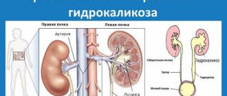
Renal calicoectasia, or hydrocalycosis, is a pathology involving the pyelocaliceal system. It is characterized by expansion of the renal calyces, leading to disruption of the outflow of urine. The disease is not life-threatening, but can provoke the development of other kidney diseases.
People of all ages are susceptible to the disease
Cause
Its main cause is defects in urinary outflow. The disease does not always have an independent character, and in some cases it can develop along with one or another ailment of the urinary system. In such a situation, a comprehensive study is required, which includes x-rays, ultrasound, and MRI. Thanks to ultrasound, it is possible to examine the picture of the pathology comprehensively and identify the factors of its development. It is a sign of the occurrence of even more serious diseases, for example, tumor formations in the calyx and tuberculosis. The symptoms of kidney hydrokaliosis resemble other pathologies, so in some cases there are difficulties in making a diagnosis.
Since the pathology arose recently, it is not absent from ICD 10. However, the classification includes hydronephrosis, which is a consequence of renal hydrokaliosis.
Causes
It is worth saying right away that renal calicoectasia cannot be an independent disease. It is provoked by a number of other reasons:
- urolithiasis disease,
- cancer of the urethra or bladder,
- pyelonephritis,
- prostate adenoma,
- pregnancy,
- pathological changes in the vessels that are located near the ureter,
- injuries of the genitourinary system.
Also one of the causes of calicoectasia is injuries to the lumbar region. There are often cases when this disease began to progress against the background of congenital pathologies of the kidneys or renal arteries. The blow is also quite dangerous for the paired organ.
All of the above reasons only prove once again that a violation of the outflow of urine can not only appear as a result of illness, but also exist from the moment of birth.
Interesting fact! Functional caliectasis affects only pregnant women. It occurs due to compression of the urinary tract by the uterus.
Kinds
There are three types of the disease: calicoectasia of the right kidney, left, and also a bilateral disease. One of the varieties is hydrocalycosis of the right kidney, which is most often diagnosed. With such a disease, urine accumulates, which first stretches the tissue of the renal pelvis, and then the renal pelvis. Due to constant compression of the canals, atrophy of the renal papilla may occur. Based on the symptoms, the pathology of the right lobe can be confused with colic of the gallbladder or appendicitis.
Calicoectasia of the left kidney is characterized by chills accompanied by pain in the lower back. In this case, the patient’s body temperature rises, nausea appears, and with it vomiting. Cloudy urine is produced, urination is frequent, but the volume is small. Hydrokaliosis of the left kidney is a rare type that is rarely diagnosed in medical practice.
Calicoectasia of both kidneys is a dangerous disease. This is due to the fact that when the kidneys stop functioning, the entire body fails, since they are vital. Development factors include both congenital and acquired characteristics. Moderate renal caliectasis is characterized by an increase in its symptoms, pain spreading to the entire lower back, which is why its comprehensive diagnosis is necessary.
We'll talk about this later.
Diagnostics
Like many diseases, caliectasis of both kidneys is quite easy to diagnose using a detailed urine test. Its results make it possible to see the full picture of damage to kidney function.
But you should not rely only on a urine test, because its results can be affected by many other factors that are not at all related to the disease.
Diagnosis of kidney pathologies can be carried out using modern equipment. The most effective methods include:
- excretory urography,
- angiography,
- Ultrasound of the kidneys,
- computed tomography,
- pyelography.
During excretory urography, a specialist injects a special contrast agent into the patient, after which images are taken. In this way, the condition and functioning of the organs of the urinary system are studied.
The next test is called angiography, during which contrast is injected directly into the kidney through an artery. Angiography allows you to study the functioning of the kidney vessels.
During an ultrasound examination, enlarged pelvises, calyces, and the degree of their filling with urine are clearly visible. You can also find out the true cause of the disease (tumor, calculus, narrowing of a vessel).
To diagnose unilateral dilatation of the renal pelvis, it is best to do pyelography. In this case, a contrast agent is administered using a catheter.
Instrumental examination is also quite informative for making a diagnosis of calicoectasia. But, most importantly, the urologist must determine the initial cause of kidney inflammation.
During the examination, the specialist must examine the patient. The urologist pays attention to the intensity, duration of symptoms, what diseases of the paired organ were previously suffered, etc.
If a patient has structural defects of the kidneys (duplication, hypoplasia), we can assume that he had calicoectasia from birth.
Causes of the disease
In medical practice, the main causes of pathology are identified as:
- neoplasms in the pelvic area that put pressure on the urinary tract;
- diseases characterized by the formation of stones in the excretory system;
- late periods of bearing a child, in which the uterus puts pressure on the kidney (most often on the right), after childbirth the pathology disappears;
- anomalies caused by the functioning of blood vessels in the kidney area;
- prolapse of the kidneys, due to which the ureter is twisted;
- pelvic injury;
- the appearance of tuberculosis and its development;
- inflammation in the lymph nodes;
- obstructions in the ureter, which appear when scars form after surgical intervention;
- abnormalities in the structure of the urinary tract.
Stages of development and characteristic symptoms of calicoectasia of the kidneys: treatment and prevention
The development of pathological processes of kidney inflammation entails a possible failure in urinary functions.
Thus, urine begins to stagnate in the calyces and pelvis.
This condition is called calicoectasia. What are the main reasons for its development and are there optimal treatment methods?
Basic information about the disease
Calicoectasia (hydrocalicosis) is a condition in which the cups in the kidneys become filled with urine and become dilated.
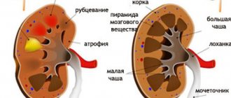
Because of this, the normal functioning of the kidneys is disrupted. As the disease progresses, atrophy of the renal papillae may occur.
As a rule, this pathology develops against the background of a concomitant disease of the urinary system.
It is very important to start treatment of calicoectasia on time, since delay can lead to complete atrophy of the organ.
Forms of the disease
Most often, the right kidney is affected by this disease. There are also left-sided and right-sided forms. Sometimes caliectasis in the right kidney is mistaken for inflammation of the appendix or colic.
The lesion on the left side is accompanied by severe lumbar pain; this is a rather rare form of calicoectasia, it can be rarely encountered.
The bilateral form is very dangerous, since in this case the functioning of both kidneys fails. Against the background of this process, other organs of the patient are also affected. If unpleasant symptoms intensify, you should definitely go to the hospital.
Causes
As mentioned above, a factor must be present in the body that will trigger the development of hydrokaliosis. All causes are divided into 2 main groups: organic and functional.
Functional factors are present in women during pregnancy. This is due to compression of the urinary tract by the ever-growing uterus.
That is why pregnant women need to especially closely monitor their health and avoid inflammatory and infectious diseases of the urinary system.
If this fails, then you need to carry out the correct treatment, which is selected by the doctor individually and to avoid complications.
Organic factors that provoke the development of hydrokaliosis include:
- kidney tuberculosis;
- inflammatory processes of the urinary system;
- pelvic organ injuries;
- change in the shape of the ureters;
- disruption of blood vessels in the kidneys;
- a large number of scars after surgical operations;
- genetic predisposition.
Stages of development and characteristic symptoms
In the early stages of development of renal caliectasis, there are practically no clear symptoms (this is especially typical for the congenital form of the disease).
Sometimes right- or left-sided caliectasis is mistaken for diseases of the liver, intestines or pancreas.
As it progresses, the patient may experience the following symptoms:
- severe increase in body temperature (up to 40 degrees);
- nausea and vomiting;
- frequent urge to urinate (as a rule, most of them are false);
- pain in one of the sides or groin area (in men, pain symptoms are much stronger than in women or children);
- change in the color and smell of urine;
- severe chills.
We must remember: the sooner treatment begins, the more effective it will be. Therefore, when the first unpleasant symptoms appear, you should definitely consult a doctor for diagnosis.
In infants, this disease can appear due to disruption of many blood vessels in the kidneys. There are no symptoms and caliectasis is diagnosed during one of the examinations with a pediatrician. As the disease progresses, the temperature may rise, severe pain may appear, and changes in the color and nature of urination may occur.
Diagnostic methods
To begin with, the doctor conducts a thorough examination with palpation of painful areas, and orders blood and urine tests. Additional research methods are:
- Excretory urography, during which a special substance, urografin, is injected into the patient through an artery on the elbow. With its help, X-ray images of organs located in the pelvis are taken.
- During angiographic diagnosis, a contrast agent is injected into the arteries of the kidneys with further study using radiography and x-ray examination. This method is considered the most informative.
- Multislice tomography allows for a detailed examination of areas in the renal parenchyma.
- In retrograde pyelography, a contrast agent is injected directly into one of the kidneys.
After receiving the results of a comprehensive examination, the doctor can select an effective treatment for hydrokaliosis.
Therapy methods
As mentioned above, the treatment of renal caliectasis is complicated by the fact that in the early stages the patient does not have characteristic symptoms, so he goes to the hospital with complications. Depending on the reason why hydrokaliosis was caused, the correct treatment is selected.
Eliminating the cause that caused this condition
In this case, the patient is selected the necessary drug therapy. If calicoectasia is triggered by an infection in the body, a course of antibiotics is prescribed. They are prescribed after precise identification of the pathogen.
https://www.youtube.com/watch?v=ZjJ-D0praao
At the same time, analgesics and anti-inflammatory drugs are taken; at high temperatures, an antipyretic is prescribed.
In the case of a congenital form of pathology, treatment is not carried out until the patient’s condition noticeably worsens. To do this, the progression of the disease is monitored through regular blood and urine tests.
After eliminating the main cause that caused caliectasis, treatment begins. In some cases, surgery is required.
During its implementation, diseases that cause blockage of the ureter and urine retention in the body are eliminated. As a rule, laparoscopy is considered the most common method. It is less traumatic and does not require the patient to stay in a hospital for a long time.
Restoring normal kidney function
After eliminating hydrokaliosis, it is imperative to take measures to restore full kidney function.
The most popular drugs are “Omnic”, “Taxulosin”, diuretics of plant origin (help improve urine flow), “Canephron”, “Cyston”, “Fitolysin”.
The duration of treatment depends on the degree of the patient’s disease; as a rule, on average it ranges from 1 to 3 months.
For example, herbal diuretics are taken 3 times a day for a course of 30 days (“Fitolit”). For a more accurate dosage, and to avoid side effects, it is better to consult your doctor.
Various folk remedies are widely used to treat this disease. Infusions or decoctions are prepared from medicinal plants (chamomile, calendula, St. John's wort, etc.). Ginseng root, which is infused in alcohol or vodka for a month, is considered very effective.
Drink 1 tablespoon before meals, the course of administration is up to 3 months.
Regular consumption of cow's milk helps with severe edema. This product should not be used by people suffering from lactose intolerance.
To restore the urine excretion function, add fresh vegetables and fruits (watermelon, pumpkin, cucumber, raspberries) to the diet; they help speed up the process of eliminating urine accumulated in the body.
It must be remembered that before starting any treatment there must be a mandatory consultation with a doctor. There is no need to self-medicate, as this can lead to serious complications.
Special instructions after treatment
An important fact is the adjustment of the patient’s diet. To do this, you need to exclude any fried, spicy, salty foods or foods that put increased stress on the kidneys.
It is better to eat boiled or steamed dishes, fresh vegetables and fruits in season.
It is not recommended to drink a lot of fluids until the end of treatment for caliectasis. You should definitely drink herbal teas, which help strengthen your overall immunity.
If timely treatment is not carried out, the patient may experience complications in the form of renal failure, pyelonephritis or organ atrophy.
In this case, constant hemodialysis will be required to remove toxins accumulated in the body.
Relapse Prevention
To prevent a possible relapse of the disease, you need to carefully monitor your health, eat right, and engage in moderate physical activity.
You need to regularly take the necessary tests and undergo diagnostics of the pelvic organs. Fulfillment of all these conditions significantly reduces the risk of developing hydrokaliosis.
Conclusion
Calicoectasia is a very dangerous disease that does not occur for independent reasons, but is a concomitant disease of the urinary system (inflammation of the urinary tract, kidney stones, cystitis, etc.).
Its insidiousness lies in the fact that in the early stages there are no symptoms, so treatment becomes more complicated.
To reduce the risk of developing this disease, you need to carefully monitor your health and avoid inflammatory and infectious diseases of the genitourinary system.
At the first unpleasant signs, consult a doctor immediately, since delaying treatment is fraught with serious complications (complete disruption of the functioning of one of the kidneys).
We recommend other articles on the topic
Source: https://UroHelp.guru/pochki/anomalii/kalikoektaziya.html
The main signs of this pathology
How does the disease manifest itself? With calicoectasia of the kidneys, the patient feels severe chills, accompanied by high body temperature, which can reach forty degrees. An increase in temperature is observed in cases where urine is not excreted from the body in a timely manner. The appearance of vomiting and nausea is characteristic, which does not bring relief to the patient. With this disease, he feels severe pain, which spreads to other areas, including the groin area. In men, pain becomes stronger with the development of calicopyeloureterectasia, that is, inflammation in the urethra is added to the main pathology. Due to calicopyeloureterectasia, minor bleeding occurs because stones damage the walls of the canal through which urine flows.
What other signs of renal caliectasis exist?
The patient often feels the urge to empty, but the fluid comes out in a small volume. In this case, the urine is cloudy, and small amounts of blood may be present. Urination is accompanied by cutting pain. With bilateral hydrokaliosis, the signs of the disease appear even more intense and brighter.
Symptoms of the disease
Calicopyelectasia of the kidneys may not make itself felt not only for several months, but also for years. A person may not be aware of the presence of the disease, or that an irreversible inflammatory process is progressing in his body.
When the disease enters the active stage, the patient begins to worry about a systematic increase in temperature to 38-40 degrees. The body shows signs of intoxication.
However, these symptoms can indicate many other diseases. Suspicion of calicoectasia arises when frequent urination with a constant urge appears. The process itself is accompanied by cutting and burning.
There are times when doctors confuse calicoectasia with renal colic, since the clinical picture is very similar.
In addition to the above symptoms, the patient notes regular severe pain in the lower back. The pain is so strong that it radiates to the groin area.
Particular attention should be paid when all the signs include the following:
- nausea and vomiting,
- disruption of the gastrointestinal tract (diarrhea),
- general weakness.
Also, urine may take on a pink or reddish tint, caused by damage to the walls of the ureter.
Such symptoms indicate that the inflammatory process has spread to both the urethra and the abdominal organs. In this case, you should urgently seek professional medical help.
Manifestations of this pathology in children
Renal hydrokaliosis in newborn babies appears due to various reasons, including the presence of additional vessels inside the kidneys. The disease, in fact, develops in a child due to ailments that appeared earlier. It is a deviation from proper functioning that occurs without symptoms. The disease is most often diagnosed during a preventive examination of babies after birth.
Symptoms of renal caliectasis in newborns have different manifestations, depending on the degree of damage. The child becomes weak, blood pressure and temperature rise, and constant nausea is noted. The color of the urine is much darker, and the newborn feels pain when urinating. At first, the pain is mild, but over time its intensity increases, and the child begins to experience discomfort.
The essence of pathology
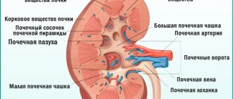
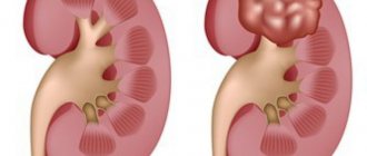
What is calicoectasia of the kidney and how does this pathology develop? Calicoectasia or hydrocalicoectasia of the kidneys is a pathological condition of the organ in which the accumulation of urine causes expansion of the renal calyces, and as a result, compression of the remaining tissues. Thus, kidney function is impaired.
The ICD 10 code for this disease is Q62.3.
Accumulation of urine in the pelvis and calyces leads to their expansion
Calicoectasia of the kidneys during pregnancy
During pregnancy, renal hydrokaliosis is characterized by a wide range of symptoms. The main symptom is hormonal imbalances in the female body, which appear from the first trimester. In addition, the development of the disease is facilitated by infections that either enter the body or arise as a result of the inflammatory process. It happens that during pregnancy, a woman’s uterus puts pressure on one side on the kidneys and other organs (mostly on the right, less often on the left), causing discomfort in one area or another. In the absence of timely treatment, the patient develops complications, which may include either an infection in the area of inflammation or the appearance of kidney stones. A significant complication is chronic liver failure.
Prognosis and prevention
Timely diagnosis and treatment allow us to hope for a favorable prognosis. Lack of therapy leads to irreversible atrophic changes in the organ. Prevention consists of annual examinations, tests, timely treatment of concomitant diseases and adherence to the principles of proper nutrition.
Renal calicoectasia is a disease that occurs due to stagnation of urine in the renal pelvis and calyces and is characterized by a latent course in the initial stages. It can be congenital or acquired, and can affect the left, right and both kidneys. The pathology is dangerous due to its complications, but can be easily treated in the early stages of development.
Methods for diagnosing the disease
To identify the main signs of pathology, the patient must undergo a full examination, which includes several methods. Thanks to this, it is possible to determine the correct diagnosis and identify the factors that provoked the development of the disease. The doctor first draws up an anamnesis, which describes the patient’s complaints and analyzes his previous illnesses. Then laboratory research methods are prescribed, including biochemical and general analyzes of urine and blood.
After this, an X-ray examination is carried out. This method requires the use of radiopaque agents. Thanks to MRI, radiography of the peritoneal cavity and ultrasound, it is possible to determine the true picture of the pathology. The development of abnormalities is monitored using ultrasound. Ultrasound is also used to establish the characteristics of the course of the disease, the causes of blockage of the pelvis and defects in the sections of the excretory system.
The following instrumental methods will also help to establish the correct diagnosis:
- Excretory urography. The procedure involves the introduction of a special liquid into the body, which is contrasting and can be clearly seen in special photographs of the urinary system. This method helps to identify structural pathologies of the kidneys and monitor the performance of urinary function.
- Renal angiography. It differs from the previous method in that the contrast agent is injected directly into the renal artery.
- Retrograde pyelography. It is carried out to examine the affected kidney. Provides for the introduction of a contrast agent through a catheter into the desired organ (right or left kidney).
- Multislice tomography.
What is the treatment for renal calicoectasia?
Treatment methods
Treatment for calicoectasia should begin as early as possible. Since stagnation of urine in the kidneys can lead to the development of complications - hydronephrosis, urolithiasis, renal failure.
The method of treatment - conservative or surgical - is selected depending on the root cause of the pathology and its severity. It is known that renal calicosis is a disease that develops due to an existing pathology of the organ.
Therapy is aimed at eliminating the underlying disease and normalizing renal function. The conservative method involves taking medications and adjusting nutrition. A specialist will talk about surgical methods for treating calicoectasia in the video in this article.
Basic therapeutic measures
A conservative treatment method is the use of medications, which is carried out under the supervision of a specialist. If an infection is the provoking factor of the disease, the doctor prescribes antibiotics to the patient. Thanks to urine culture, it is possible to identify the degree of sensitivity to drugs of microorganisms that cause renal hydrocalycosis. To reduce body temperature and eliminate pain, anti-inflammatory drugs and analgesics are prescribed.
Hydrocalycosis, also known as calicoectasia of the kidneys: causes and current methods of combating the disease
Due to a violation of the outflow of secondary urine, a pathological condition such as renal calicoectasia, otherwise called hydrocalycosis, can develop.
With this disease, the kidney calyces are in an expanded state due to overcrowding with urine, as a result of which the remaining tissues of the kidneys are compressed, their normal functioning is disrupted, and the kidneys themselves shift to the periphery.
Calicoectasia of the right kidney is more common, much less common is the left one, and in some patients a bilateral process is also detected.
- 1 Reasons
- 2 Symptoms
- 3 Diagnostics
- 4 Treatment
- 5 on topic
on this topic
About the symptoms and treatment of urolithiasis in the program “Live Healthy!” with Elena Malysheva:
Patients diagnosed with calicoectasia of the kidney require annual monitoring by a urologist, nephrologist, and timely conservative and sometimes surgical treatment throughout life.
Source: https://mkb.guru/bolezni-pochek/kalikoektaziya.html
Folk remedies
For renal calicoectasia, in addition to surgery and treatment with various medications, traditional medicine is also used. Treatment through the use of such methods includes the use of herbal infusions and decoctions by patients. Despite the naturalness of any folk remedy, in some cases an allergic reaction may occur, so before using this method, consultation with a specialist is necessary. The following herbs are used as folk remedies: burdock, St. John's wort, cornflower, rose hips, etc. They can help the patient follow a proper diet, maintain a healthy lifestyle and exercise.
Calicoectasia of the kidneys - what kind of pathology is it, is it dangerous?
The article talks about calicoectasia, a pathology of the pyelocaliceal system of the urinary organs. The causes of occurrence and characteristics of symptoms are described.
Renal calicoectasia, or hydrocalycosis, is a pathology involving the pyelocaliceal system. It is characterized by expansion of the renal calyces, leading to disruption of the outflow of urine. The disease is not life-threatening, but can provoke the development of other kidney diseases.
People of all ages are susceptible to the disease
The essence of pathology
What is calicoectasia of the kidney and how does this pathology develop? Calicoectasia or hydrocalicoectasia of the kidneys is a pathological condition of the organ in which the accumulation of urine causes expansion of the renal calyces, and as a result, compression of the remaining tissues. Thus, kidney function is impaired.
The ICD 10 code for this disease is Q62.3.
Accumulation of urine in the pelvis and calyces leads to their expansion
Causes
This disease is not an independent pathology, but develops against the background of diseases that cause persistent disruption of the outflow of urine or injury. The pathology can also be congenital.
The main reasons leading to the development of the disease:
- intrauterine anomalies in the development of the kidneys, arteries and vessels of the organ;
- urolithiasis, tuberculosis, nephrolithiasis, prostate adenoma, cancer;
- prolapse of the kidney and torsion of the ureter;
- postoperative adhesions;
- organ injuries.
Pregnancy is one of the factors predisposing to the development of the disease. In pregnant women, the kidneys are subject to double stress; in addition, in the third trimester, the urinary tract begins to be compressed by the enlarging uterus.
Mechanical impact on organs and increased stress can cause disease. Calicoectasia of the right kidney mainly occurs during pregnancy, due to pressure on the ureter on the right.
The cause of calicoectasia in children is a congenital anomaly - an additional vessel inside the organ.
Compression of the ureter by the uterus can cause disease during pregnancy
How it manifests itself
Calicoectasia of the right kidney occurs many times more often than in the left. Calicoectasia occurs against the background of other diseases, so the symptoms of the underlying disease are more pronounced. In the early stages, there are virtually no symptoms; there may be only moderate pain over the area of the affected kidney or in the groin.
As the disease progresses, the symptoms intensify, and the following signs appear:
- fever;
- general intoxication;
- painful vomiting;
- intense pain on the right or left in the lower back, less often on both sides;
- increased frequency of urination with decreased volume of urine excreted;
- cutting pain during urination;
- change in urine.
Calicoectasia of both kidneys - what is it? This is a simultaneous enlargement of the calyces of both kidneys. Usually has a congenital origin or is a consequence of an autoimmune disease.
With unilateral damage, symptoms appear after about 2-3 months, as the healthy organ compensates for the work of the affected one. Bilateral renal caliectasis is less common, its manifestations are more pronounced and appear a month after the onset of the disease.
Lower back pain from the affected organ is one of the first signs of the disease
Diagnostics
To establish a diagnosis, the patient must undergo a complete examination. First of all, the doctor collects an anamnesis, where he describes complaints and analyzes previous diseases and injuries.
Then additional diagnostic methods are carried out.
- Blood analysis. It will show the presence of inflammation in the body - an increase in ESR, an increase in the number of leukocytes.
- Analysis of urine . An increase in the number of leukocytes is detected, protein and blood impurities appear.
- Ultrasound . Pathology is indicated by the expansion of the boundaries of the pyelocaliceal system and deviations in the structure of the organ.
- Angiography, excretory urography. They are carried out using a contrast agent, which makes it possible to track the distribution of the substance through the vessels and urinary tract and determine the cause of the disturbance in the outflow of urine.
- CT. The most accurate, but also the most expensive method, the price of which varies in different medical institutions. Using the procedure, the exact location and nature of the obstacle is determined.
Since the disease is characterized by nonspecific symptoms, it must be differentiated from acute appendicitis, cholecystitis, pancreatitis, and urolithiasis.
Enlargement of the renal calyces is diagnosed using ultrasound
Treatment methods
Treatment for calicoectasia should begin as early as possible. Since stagnation of urine in the kidneys can lead to the development of complications - hydronephrosis, urolithiasis, renal failure.
https://www.youtube.com/watch?v=tnB-K02E3ew
The method of treatment - conservative or surgical - is selected depending on the root cause of the pathology and its severity. It is known that renal calicosis is a disease that develops due to an existing pathology of the organ.
Therapy is aimed at eliminating the underlying disease and normalizing renal function. The conservative method involves taking medications and adjusting nutrition. A specialist will talk about surgical methods for treating calicoectasia in the video in this article.
Prognosis and prevention
Timely diagnosis and treatment allow us to hope for a favorable prognosis. Lack of therapy leads to irreversible atrophic changes in the organ. Prevention consists of annual examinations, tests, timely treatment of concomitant diseases and adherence to the principles of proper nutrition.
Renal calicoectasia is a disease that occurs due to stagnation of urine in the renal pelvis and calyces and is characterized by a latent course in the initial stages. It can be congenital or acquired, and can affect the left, right and both kidneys. The pathology is dangerous due to its complications, but can be easily treated in the early stages of development.
Questions for the doctor
I am 32 weeks pregnant and have been diagnosed with kidney disease. How dangerous is caliectasis of the kidney in a pregnant woman for a woman and her unborn child? Anastasia U. 30 years old, Belgorod.
Hello, Anastasia. Calicoectasia is not uncommon in pregnant women, when the kidneys are under increased stress. If detected in time, the disease is not dangerous if treated correctly. Ignoring symptoms can lead to the development of urolithiasis in a woman, complications during childbirth and problems in the postpartum period.
What folk remedies can be used to treat caliectasis of the right kidney? Victor S. 47 years old, Volzhsky.
Hello, Victor. Not only medications, but also traditional medicine recipes will help alleviate the condition. It is useful to eat watermelons, melons, and drink non-concentrated juices from red currants and raspberries.
Traditional healers advised eating fresh, boiled, or baked pumpkin. Collections are available in large quantities in pharmacies. Conveniently, each pharmacy package contains detailed instructions for making decoctions and infusions.
Source: https://bolyatpochki.ru/bolezni/drugie-zabolevaniya/kalikoektaziya-pochek-246.html
Prevention
To restore kidney function, improve blood circulation and get rid of stagnation of urine, you need to use the recommendations of traditional medicine:
- Eating stewed pumpkin will help get rid of caliectasis;
- for edema you need to drink milk;
- raspberries, currants, watermelon have a cleansing effect on the kidneys;
- Pour boiling water over black currant leaves, leave, strain and squeeze. Heat the infusion and add fresh or dried currants to it. Drink as tea 4 rubles/day, eat berries;
- Grind the rosehip root and pour 30-50 g into 1 liter of boiling water, leave for half a day, strain and drink like water for at least a month;
- a collection of equal parts of horsetail, knotweed, birch buds, carrot seeds, cranberries, rose hips and juniper, crush, 2 tbsp. Brew the collection with a glass of boiling water, leave for 1 hour and strain. 3 months drink 3 times a day before meals.
To restore kidney function, you can use herbs
Before using decoctions and infusions of medicinal herbs, you should consult a doctor.
Symptoms
In cases where renal calicoectasia in children is congenital, it is practically asymptomatic, and the patient may not even be aware of the presence of problems. In this case, this condition is considered a variant of the norm.
If the disease is caused by certain reasons, it manifests itself with the following symptoms:
- severe pain in the lumbar region during palpation and without any impact, most often on the right;
- cloudy urine;
- chills and fever;
- traces of blood in the urine;
- increased urge to urinate simultaneously with a decrease in the volume of urine produced;
- nausea, sometimes vomiting.
Since this pathology is usually a consequence of another disease, the symptoms of this underlying disease come first.
The development of renal pathology in this case can be suspected only by observing the dynamics. At first, the kidneys can still function normally, and this period can last from several months to several years, but then a period of decompensation sets in, and the kidneys can no longer cope with the normal outflow of urine.
What signs will indicate the development of pathology?
Sharp pain in the lower back may indicate kidney disease.
The disease most often affects the right kidney, but sometimes calicoectasia of the left kidney is diagnosed. There is also a bilateral nature of the lesion. The severity of clinical manifestations depends on the speed of development of the pathological process and the nature of the underlying pathology. Congenital pelvicalyceal dilatation often does not bother a person and is an individual feature of the body. Symptoms appear after the end of the period of compensation of renal functions. The signs of caliectasis differ little from other diseases of the urinary system:
- chills, sweating;
- during prolonged urinary retention, body temperature rises to 40° and falls after the outflow normalizes;
- severe pain in the lumbar area, radiating to the stomach and groin area;
- increased pain when moving and touching the lower back;
- a feeling of nausea, and vomiting does not alleviate the patient’s condition;
- frequent urination, but small amounts of urine;
- urine is cloudy, with an unpleasant odor and streaked with blood;
- pain during urination.
Calicoectasia of both kidneys has intense symptoms and a high risk of accelerated development of renal failure.
Transient calicoectasia during pregnancy
In women during late pregnancy, moderate calicopyelectasia of the right kidney is often detected; left-sided pathology is less often detected. The reason is an increase in the size of the uterus, which deviates to the side and puts pressure on the bladder and other structures of the urinary system. The outflow of urine becomes difficult and the kidney tissue is pushed to the periphery of the organ. A woman feels discomfort in the lower back on the right or left, and difficulty urinating. The advantage of right-sided expansion is caused by the higher mobility of the right kidney and its location below the left. After childbirth, the condition of the cups returns to normal.
The peculiarity of manifestations in a child
It is important to monitor the amount of your baby's urine to prevent dangerous kidney diseases.
In a newborn, the disease is provoked by intrauterine pathologies in the structure and functioning of the urinary system and kidneys, trauma and inflammation. These factors lead to improper passage of urine and stagnation. The tissues of the pelvis and calyces in children are not fully developed, and the kidneys are larger. Hydrocalycosis of the kidneys can develop for a long time without giving any signals and be detected during a routine examination. In a child, the pathology may show signs that are unusual for it, but in most cases the manifestations are similar to adults, but more intense. The bilateral form of hydrocalycosis is especially dangerous. If suspicious symptoms appear, the child should be immediately taken to a nephrologist.
Treatment of calicoectasia
There are several treatment options for calicoectasia, which the doctor must discuss with the patient to make the best decision.
Taking antibiotics.
The preferred treatment option, which should only be carried out under the strict supervision of a physician. Antibiotics can strip the body of healthy bacteria, so it is recommended to add probiotics to your diet during treatment. In more severe cases, with severe physical pain, vomiting or nausea, intravenous antibiotic treatment is performed.
Kidney drainage.
The procedure is aimed at removing urine from the kidney through the skin through a minor surgical intervention and is usually performed if a patient has urolithiasis.
Installation of nephrostomy.
The procedure for restoring normal urine flow from the kidney by installing a drainage tube. Recommended as a treatment for acute urinary tract obstruction.
Surgery.
The procedure is recommended for patients whose kidney stones or various malignant neoplasms were found to be too large during diagnosis. The operation not only helps to get rid of calicoectasia, but also saves lives.
Calicoectasia is a serious medical problem that, if left untreated, can lead to kidney atrophy and chronic renal failure. In the latter case, patients become dependent on dialysis and their quality of life is seriously impaired.
Possible concerns
If, based on the primary signs of calicoectasia, the doctor cannot make an accurate diagnosis, he will prescribe additional laboratory tests and studies. Many patients begin to panic about confirmation of the diagnosis and the development of possible complications. In this case, the most important thing is not to self-medicate with folk remedies - it causes more harm than good.
Diagnostic methods
To begin with, the doctor conducts a thorough examination with palpation of painful areas, and orders blood and urine tests. Additional research methods are:
- Excretory urography, during which a special substance, urografin, is injected into the patient through an artery on the elbow. With its help, X-ray images of organs located in the pelvis are taken.
- During angiographic diagnosis, a contrast agent is injected into the arteries of the kidneys with further study using radiography and x-ray examination. This method is considered the most informative.
- Multislice tomography allows for a detailed examination of areas in the renal parenchyma.
- In retrograde pyelography, a contrast agent is injected directly into one of the kidneys.
After receiving the results of a comprehensive examination, the doctor can select an effective treatment for hydrokaliosis.
Calicoectasia of the kidneys (left, right, both): what is it, symptoms
Renal calicoectasia is a kidney pathology in which the calyces of the organ become enlarged due to the accumulation of urine in them. Because of this, other organs become compressed, and the body cannot function fully.
Pathology develops due to blockage of the urinary tract. As a result, deformation of the renal papillae occurs. This disease is also called “hydrocalicosis.”
Symptoms
The classic signs are:
- Terrible pain in the lumbar area. They can occur on their own, as well as during palpation;
- Chills;
- The color of the urine darkens;
- Discharge of urine along with blood;
- Fever;
- Frequent urination with a small amount excreted;
- Nausea and vomiting.
The intestinal walls may begin to contract, leading to diarrhea. Also, if the intestinal walls are damaged by a stone, bleeding may begin. A sign of this will be urine that is reddish or pink in color.
Calicoectasia of the kidneys is not immediately detected. The kidneys still try to function normally for some time. The duration of symptoms depends on the disease that caused the calicoectasia.
In some patients, symptoms are mild. The symptoms are similar to a number of other ailments, such as acute appendicitis, various diseases of the intestinal tract and biliary tract.
Therefore, the doctor must make a diagnosis and select the necessary therapy. The sooner hydrocalycosis is identified, the more effective the therapy will be.
If treatment is delayed, changes in the structure of the kidneys may be irreversible.
There are no special symptoms for the disease of the left organ. The symptoms are the same as described above. The pain syndrome is “zoned” in nature, which is why the pain is felt on the left side. Disease of the left kidney is an extremely rare case in medical practice.
Pathology on the right side is much more common than on the left. The pain occurs on the right side. Due to the concentration of urine, the pelvis enlarges and expands the tissue. As mentioned earlier, the renal papillae become deformed. This occurs due to compression of the channels.
Damage to both kidneys
Calicoectasia of both kidneys is the most dangerous form of the disease. Kidneys are a very important organ in the human body. Damage to both kidneys at once can completely disrupt the functioning of the entire body.
With this type of disease, the symptoms will be much more pronounced. Pain will occur throughout the entire lumbar region.
Diagnosis of the disease
The doctor takes a clinical blood test, which indicates inflammatory formations in the body. Unfortunately, this examination does not indicate where exactly the inflammation began. An increase in ESR, as well as the content of leukocytes with the majority of young cell forms, are the main signs for diagnosis.
A general urine test helps to more accurately identify the disease. This analysis is aimed specifically at the urinary system. The presence of inflammation will be indicated by a high content of leukocytes, protein and the appearance of blood in the urine.
To more accurately determine that you have calicoectasia of the kidneys, it is necessary to conduct an ultrasound examination, angiography, computed tomography and retrograde pyelography.
Of the above types of examination, the most popular is ultrasound examination of the kidneys and ureters. The study is aimed at identifying disorders in the body and determining the causes of their occurrence. Even infants can be subjected to this type of examination and used during pregnancy.
On ultrasound you can see the expansion of the calyx and the pelvis
In pregnant women, disease of the right kidney is more often detected. This is due to the fact that it is more energetic and is located below the left organ.
During gestation, the uterus deviates to the right side, thereby pinching the ureter and causing expansion of the pyelocaliceal system. The disease usually progresses favorably.
Treatment after childbirth is usually not prescribed.
With urography, pyelography, angiography, a special contrast agent is injected into the affected organ and an x-ray is used. In pictures taken at different times, you can see changes in the structure of the urinary tract and blood vessels. This allows you to determine the disease with 100% accuracy.
Computed tomography is a more innovative method. It is possible to view the disease in a cross-section. This makes it possible to determine exactly where the obstacle to the outflow of urine has arisen and its nature.
Treatment of the disease
The specialist prescribes treatment depending on:
- Level of kidney inflammation;
- Factors of expansion of the renal papillae;
- Age category of the patient;
- The presence of other diseases.
To treat the disease, as a rule, either conservative methods are used or surgical intervention is used.
The drugs are widely used at the initial stage of the disease
If the disease is infectious-inflammatory in nature, the doctor prescribes anti-inflammatory or antibacterial treatment. The doctor prescribes antispasmodic drugs such as Spazmalgon and No-shpa.
- Painkillers: Ibuprofen, Ketanov;
- Natural medicines: Urolesan, Canephron;
- To prevent infectious complications: Palin, Nitroxoline;
- To supply blood flow: Pentoxifylline, Trental;
- Treatment of infections: Levofloxacin, Cefazolin;
- As well as geodialysis and dietary table No. 7.
Surgery is used to remove formations that interfere with the outflow of urine. For example, removal of coral stones, strictures. The following operations are used: nephrotomy, nephrectomy, nephrostomy and kidney resection (complete removal). The listed interventions are used only in the presence of large formations.
Small formations are eliminated with the help of diuretics. They are used to dissolve and remove tumors. Medium-sized formations are removed using ultrasound or laser. For a congenital disease, if it does not develop over a long period of time and does not interfere with urination, the specialist does not prescribe treatment. A medical examination will be sufficient.
Impact with traditional methods
Let's consider the methods offered by traditional medicine:
- We take equal amounts of ginseng and eleutherococcus roots, fill them with forty-proof alcohol and let them brew in a dark, dry room for several weeks.
- Sick people need to drink milk until the swelling disappears.
- A good remedy is stewed pumpkin.
- Watermelon, red currant and raspberry juice have a beneficial effect on the organ.
- We take dried rosehip and remove ten shavings from it, the length of which is ten centimeters. Fill with a liter of liquid and leave overnight. In the morning, strain the broth and consume throughout the day. The course is a month.
- Take blackcurrant leaves and pour 500 ml of boiling water over them. After fifteen minutes, the broth should be strained and the leaves should be squeezed out. Place the resulting broth on the fire until it boils. Add two tablespoons of dried or fresh blackcurrants. This course should be carried out four times a day: drink the liquid and eat the remaining fruits.
- We take knotweed, parsley, birch buds, cranberry fruits, carrot seeds and rose hips in equal parts and chop them. Pour the resulting powder into 250 ml of water. After an hour you need to strain. This decoction should be consumed three times a day before meals. The course is three months.
Do not self-medicate under any circumstances, so as not to aggravate the situation. Before using folk remedies, be sure to consult a specialist. Observation by a nephrologist or urologist, as well as conservative or surgical treatment for patients with renal caliectasis are necessary throughout their lives.
ethnoscience
Calicoectasia of both kidneys is dangerous. Its clinical manifestation is not visualized at the initial stage of disease development. It is diagnosed only when the kidney tissue has significant damage.
Traditional healers have not ignored this disease and offer its treatment with folk remedies:
- The roots of ginseng and eleutherococcus are combined in equal parts. Fill the container filled with the composition with vodka and let it brew in a dark place for a week.
- Milk perfectly saves from edema when consumed regularly.
- Calicoectasia of the kidneys is effectively treated with stewed pumpkin.
- For the infusion, use 10 cm of shavings from rosehip root in the amount of 10 pcs. It is filled with 1000 ml of water and put away in a dark place overnight. The straining process will have to be done in the morning. The resulting volume of the folk remedy is drunk during the current day. The general course lasts 1 month.
- Bilateral failure in the functioning of the urinary system is well restored by a mixture of leaves and grass of knotweed, parsley, horsetail, birch buds, bear ears, carrot seeds, rose hips, juniper, cranberries. They are used in equal proportions. For infusion you will need 2 tbsp. l. powder and ¼ liter of boiling water. After an hour, the drink is filtered and consumed 3 times a day, 60 ml before meals. The course is designed for a quarter.
An objective assessment of the disease was given. Calicoectasia of the kidneys, what is it - a question that today has received a very accessible description.
The main task of the patient is to have time to diagnose at an early stage, then complications can be avoided.
Treatment
For a disease such as renal calicoectasia, treatment is prescribed based on the results of research and the patient’s complaints, it takes into account the general state of health, the patient’s age, and the duration of the disease.
In cases where the disease is congenital and is recognized as a normal variant for a given patient, treatment is not prescribed, but the patient must be examined periodically; he is advised to be examined by a nephrologist once a year.
You should not take it lightly and forget about preventive examinations, since prolonged stagnation of urine and circulatory disorders of the kidneys can cause diseases such as pyelonephritis, hydronephrosis, glomerulonephritis.
In addition, due to an increase in the concentration of urine, the likelihood of the appearance of sand particles from oxalates and urates increases, which become centers of crystallization and provoke the appearance of urolithiasis.
The doctor’s tactics, depending on the underlying cause and severity of the disease, can vary significantly: from observation and waiting to emergency surgery.
Since this pathology is almost always a consequence of other diseases, the main goal of treatment is to combat the problem that provoked calicoectasia.
For example, if deterioration in kidney function is associated with pyelonephritis, the doctor will prescribe antibiotics, diuretics, antispasmodics, and physiotherapeutic procedures. If there is an obstruction in the urinary tract that causes a deterioration in the outflow of urine, you will have to resort to surgical intervention.
In addition, if the symptoms are severe, the doctor will prescribe antispasmodics to reduce pain (Papaverine, Spazmalgon, No-shpu and their analogues), herbal medicines to restore the functioning of the urinary tract (Canephron, Urolesan, Hofetop), drugs for the prevention of infectious complications (Nitroxoline, Palin) , drugs to improve blood circulation in the kidney (Trental, Tivortin).
Trental tablets
The specific treatment method will depend on the cause of the pathology and the general well-being of the patient. Diet table No. 7 is also shown. The patient must strictly observe the drinking regime: drink at least 2 liters of liquid per day, and always 1-2 glasses of water on an empty stomach, in the morning before meals. You should limit your salt intake.
If the size of one of the kidneys is noticeably increased, doubling of the cups occurs, complicated by a total sclerotic process, or large coral-shaped stones are observed, surgical treatment cannot be avoided: nephrotomy, nephrectomy, nephrostomy or kidney resection.
Among the folk remedies there are also those effective for the treatment of moderate calicoectasia of the kidneys, for example, the roots of eleutherococcus and ginseng.
They need to be taken in equal quantities, poured with vodka, and left to infuse for several weeks in a dark place.
Another piece of traditional medicine advice is to drink non-concentrated juices from raspberries and red currants. Pumpkin is very useful for this pathology; when stewed, it should be included in the diet almost every day. It is advised to drink more milk to reduce swelling.
You can also use a herbal mixture: take equal parts of bear ears, knotweed, horsetail, birch buds, parsley, carrot seeds, rose hips, cranberries and juniper berries, chop and mix thoroughly. Take 2 tablespoons of the mixture and pour 250 ml of boiling water, strain the finished infusion after one hour. Drink a third of a glass half an hour before meals, 3 times a day.
All folk remedies will only help reduce the manifestation of symptoms; you cannot do without a visit to a nephrologist. Remember that caliectasis causes irreversible changes in the kidneys, so you should consult a doctor promptly.
Treatment of the disease
What is calicoectasia? This question has been answered. The causes of the disease are also clear. It remains to understand how to get rid of this disease with the help of medications and traditional medicine.
Restorative therapy, when renal calicoectasia is diagnosed, is determined by specialists. Sometimes it is not enough to choose a conservative method and you have to resort to surgical intervention.
Therapeutic measures are presented in the following algorithm of actions.
The doctor prescribes:
- inhibitors (Ketanov, Ibuprofen, Dexalgin);
- antispasmodics (Papaverine, No-shpa);
- herbal preparations (Canephron, Urolesan);
- anti-infective for prophylactic purposes (Palin, Nitroxoline);
- blood circulation stimulants (Tivortin, Pentoxifylline);
- anti-infective for therapeutic purposes (Cefazolin, Levofloxacin);
- hemodialysis (extreme case);
- diet (table No. 7).
Drug therapy during pregnancy and in children is carried out only after agreeing on a treatment regimen with the attending physician - a urologist or nephrologist.
Calicoectasia of the kidneys involves treatment when the help of a surgeon is required; it is indicated for significant sizes of coral stones and when one organ increases in volume or doubles the cups. Such changes are an indication for surgery.
Pediatric pathology
In young children, the kidney calyces in most cases become enlarged due to congenital anomalies in the structure of organs involved in the process of urine excretion, or urological problems. As a rule, pathology of this kind is detected soon after the birth of the child. If it is acquired in nature, and was formed against the background of urolithiasis, trauma or an inflammatory process, then it may not make itself felt for a long time.
Symptoms of calicoectasia in children are the same as in adults, but since a small child cannot say what exactly is hurting him, parents should monitor their child’s behavior and immediately contact a specialist if they notice the following:
- lethargy and drowsiness;
- refusal to eat;
- vomit;
- stool disorders;
- frequent crying for no reason;
- nervousness;
- darkening of urine and a decrease in its volume.
Often, treatment of the congenital form of calicoectasia requires surgery, which is performed by endoscopy. A similar decision can also be made if both kidneys are affected. In other cases, drug therapy is prescribed to eliminate the root cause of the disease and symptoms.
Pregnancy and calicoectasia
The period of bearing a child is undoubtedly wonderful, but it is a huge stress for the body. The functions of internal organs are impaired, because they are under pressure from the gradually growing fetus. The kidneys are subject to special stress during pregnancy, because they are forced to work with double force due to the large volume of fluid in the body. In addition, the uterus puts pressure on the urinary tract, which prevents the normal outflow of urine, and as a result, stagnation occurs, under the pressure of which the kidney calyces begin to expand.
In most cases, pregnant women are diagnosed with moderate caliectasis of the kidneys, which usually does not require treatment, because after childbirth, the functioning of internal organs will be completely restored.
If necessary, medications are prescribed to eliminate symptoms and suppress the inflammatory process. Surgical intervention will be required for pathological enlargement of the cavity of the cups, associated with congenital characteristics of the body. Surgery is also prescribed if there are complications that threaten the life of the mother or child.
Treatment of the disease
No special treatment tactics have been developed for this disease. If the doctor confirms the presence of pathology during diagnosis, then treatment for calicoectasia can range from surgery to ongoing monitoring. The choice of treatment method depends entirely on the tensile force of the calyces and pelvis in the kidney and on the cause that provoked the development of the pathological condition.
The most common cause of caliectasis is anatomical obstructions. Causing disruption of urine flow. In this case, preference is given to surgical intervention. The volume of the operation and the complexity of its implementation depend on the mechanism of development of the pathology.
During the operation, ureteral strictures, stones, and tumor tumors located in the pelvis or ureters are removed. If the disease was provoked by an inflammation process in the kidneys or complications of the process appear, then anti-inflammatory and antibiotic therapy is organized.
Timely treatment of calicoectasia in the kidneys allows you to achieve the most positive results. This correlates with the possibility of reversibility of early disorders that occur in the kidneys. When the disease process starts and atrophy of the renal papillae develops, changes in the structure become irreversible. But even in such a situation, treatment can alleviate the pathology and prevent its progression.
Diagnosis of calicoectasia
If a patient detects signs of calicoectasia, the doctor prescribes an ultrasound examination, which detects inflammation and dilation of the renal calyces. In addition to ultrasound, the doctor may prescribe urography - a method of X-ray examination of the kidneys and calyces after intravenous administration of a special substance.
A complete blood test helps determine:
- An increased level of white blood cells, which indicates the presence of infection in the body.
- Elevated levels of urea and creatinine, which indicate impaired renal function.
- Decreased sodium levels (hyponatremia).
- Electrolyte imbalance, low levels of which can lead to dehydration and a number of other complications.
- A violation of the acid-base state, manifested by low blood pH values.
According to the 2008 reference manual Differential Diagnosis of Abdominal Ultrasound, the diagnosis of calicoectasia requires confirmation and exclusion of other diseases such as hydronephrosis and urinary tract obstruction.
It is important to note that distension of the renal pelvis during pregnancy is considered normal and usually occurs at the 28th week of gestation. At the same time, this condition is considered a risk factor for the development of urinary tract infections in the future.