The method, based on the ability of the kidney to secrete (excrete) a radiopaque substance introduced into the body, is used to diagnose hematuria, urolithiasis and its complications, cysts and tumors of the kidneys and ureters.
An iodine-containing solution is used as a contrast agent. During the study, a series of X-rays are taken, the number of which is determined by the type of pathology and depends on the anatomical features of the patient.
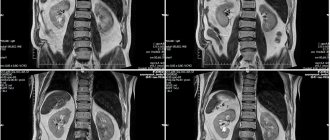
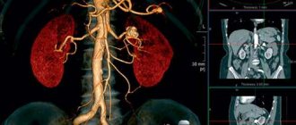
An excretory urogram shows the functional and structural features of the kidneys, ureters and bladder, renal pelvis and calyces, and anomalies of their development.
Intravenous urography is one of the main tools in the urologist’s arsenal.
Intravenous urography is prescribed when the following symptoms occur:
- pain in the lumbar region;
- hematuria (blood in the urine);
- high blood pressure;
- frequent urinary tract infections;
- urinary dysfunction.
The study is indicated for diagnosing the following conditions:
- urolithiasis disease;
- polycystic kidney disease;
- pyelonephritis;
- hydronephrosis;
- malignant neoplasms of the kidneys, ureters, bladder;
- complications after surgery on the urinary tract;
- congenital anomalies of the urinary tract.
In what cases is a study prescribed?
The procedure is prescribed in the following cases:
- lower back injuries;
- presence of blood in the urine;
- suspected stones in the kidneys, ureter or bladder;
- postoperative control;
- suspicion of a tumor disease;
- unknown cause of hypertension;
- suspicion of kidney development abnormalities;
- impaired urine output, presence of edema.
Restrictions and contraindications
The procedure involves ionizing radiation, so it has some contraindications. In addition, the use of a contrast agent has its own additional limitations.
The study is not carried out in the following cases:
- pregnancy and breastfeeding
- stroke and heart attack;
- iodine intolerance;
- a positive test for allergies to a substance used as a contrast;
- renal failure;
- state of shock;
- high degree of bleeding.
What types of research are there and what is the difference?
There are three main types of renal urography: survey, excretory and intravenous:
- A survey is essentially a regular x-ray of the kidney area. With the help of such a study, the presence of stones, parasites and tumors is determined. The study is prescribed if necessary to further study the location of the kidneys, their shadows, contours.
- Excretory is a radiopaque study based on the excretory function of the kidneys. The patient is injected with a contrast agent and a picture is taken while they begin to remove it.
- Intravenous also requires the administration of a radiocontrast agent, but several photographs are taken. The study is used to evaluate the rate of contrast removal. It can detect the following diseases: hydronephrosis and hydroureter, wrinkling and stretching of the kidneys, cysts, neoplasms and others.
FAQ
What can you see on x-rays of the kidneys?
X-ray of the kidneys reveals a number of abnormalities:
- Congenital kidney anomalies;
- Pathological increase or decrease in organ size;
- Kidney prolapse;
- Presence of neoplasms;
- Tissue rupture;
- Presence of stones.
They are signs of conditions that require medical treatment because they are dangerous to the health and life of the patient and can lead to serious complications.
Is there an alternative to kidney x-rays?
Alternative research methods are MRI and MSCT. They allow you to obtain more detailed information and clarify the diagnosis. It is important to understand that the doctor chooses the method of conduction based on the indications and contraindications, as well as the purposes of the diagnosis.
Answers questions:
Kochetov Sergey Anatolievich
Urologist, Candidate of Medical Sciences, doctor of the highest category
33 years of experience
doctor
Preparing for the study
There are several points that affect the quality of the x-ray image when performing urography, namely:
- amount of urine in the bladder;
- the amount of feces in the intestines;
- increased gas formation.
In order to maximize the information content of the procedure and protect yourself from possible side effects, when preparing for kidney urography using a contrast agent, you should follow some rules and recommendations from your doctor:
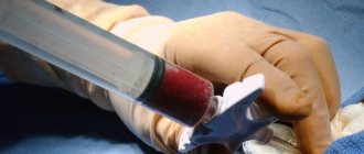
- To exclude renal failure, it is necessary to donate blood for biochemistry .
- Conduct an allergy test ; some time before the test, you need to inject a small amount of contrast agent subcutaneously and monitor the reaction. Sometimes an iodine mesh is applied.
- For 48 hours, exclude fiber-rich foods that increase gas formation: brown bread, legumes, sweets, fresh fruits and vegetables. It is also recommended to exclude foods that are poorly tolerated by individuals, such as dairy products.
- your last meal no later than the night before.
- Do an enema in the morning , or twice - the evening before and the morning before the procedure.
- In some cases, the doctor prescribes laxatives .
- Drinking less fluid the day before will increase the contrast of the images by increasing the concentration of urine.
- Sometimes the patient is asked to take pre-absorbing drugs , in particular activated carbon.
How long does the procedure take?
The sequence of performing intravenous urography is usually as follows:
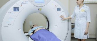
- After changing into a gown, the patient is placed on a special table, which is located under the X-ray machine. Hands are placed behind the head, the patient lies on his back.
- Before the administration of a contrast agent, 1-2 survey photographs of the abdominal cavity are taken in direct and oblique projections. The images are assessed by a radiologist: they must capture all structures of the collecting system (from the upper poles of the kidneys to the level below the pubic symphysis). Sometimes calcifications are detected on a survey image.
- The nurse inserts a catheter into a peripheral vein.
- The drug (for example, Omnipaque) is taken into two 50 ml syringes and quickly injected into a vein. The contrast begins to be distributed with the blood flow throughout the body. After a few minutes, its excretion by the kidneys begins.
- Attention! After administration of the drug, the patient may feel a fever and a metallic taste in the mouth, which normally passes quickly.
- The first photo is taken after 1 minute.
After intravenous administration of a contrast agent, you need to wait 5 minutes, after which the first image is taken. Subsequent X-ray images are taken after 14, 23 minutes. If kidney function is reduced, the image is repeated after 50 minutes.
Preparation for excretory urography involves cleansing the intestines on the eve of the study. You should cleanse the intestines with an enema, laxatives from stool, and also prevent increased gas formation, since air reduces image clarity.
To reduce flatulence, it is recommended to take Espumisan and follow a diet excluding legumes, flour products, cabbage, carbonated drinks, and sweets.
Despite the rather simple mechanism, this procedure can warn and indicate many urological diseases and injuries, such as:
- damage to the lumbar muscles;
- diseases of the spine and organs close to it (scoliosis, spondylitis, etc.);
- in some cases, shadows of the kidneys can be seen. Incorrect position of the kidney shadow may indicate its pathological diseases - abnormal organ mobility or nephroptosis. (It is worth mentioning that according to statistics, the right kidney is subject to mobility several times more than the left);
- stones and other foreign bodies in the area of the kidneys, bladder and ureters.
One of the popular reasons for performing excretory urography is the detection of blood in the urine. The study allows us to roughly determine the source of the appearance of red blood cells in urine. Indications may also include:
- pain in the urinary organs (in the lower back, radiating to the groin) and/or diuresis disturbances;
- differentiation of the source of edema or “causeless” hypertension;
- urinary tract infections characterized by a chronic recurrent course in order to exclude pathologies with similar manifestations;
- identification of areas in the tissues of organs with a disturbed structure or changes in the size of organs (dystrophy, hypertrophy);
- the presence of symptoms that may indicate the presence of stones in the urinary organs;
- suspected blockage of the ureter (obstruction);
- the likelihood of complications due to surgical interventions;
- detection of neoplasms;
- diagnosis of genetically determined anomalies in the structure of organs;
- traumatic injuries.
The advantages of urography over ultrasound are especially obvious when it is necessary to examine hollow organs - the bladder and ureters.
Like any x-ray examination, urography is unacceptable during pregnancy and lactation. You should also refrain from it if you have such diagnoses as:
- intolerance or severe allergy to iodine (contained in contrast);
- infectious diseases during exacerbation;
- diabetes;
- disorders of the blood clotting process;
- circulatory disorders (heart attack, stroke);
- acute glomerulonephritis;
- tuberculosis (open form);
- pheochromocytoma;
- chronic and acute renal failure;
- endocrine disorders (hyperthyroidism);
- sepsis (blood poisoning);
- state of organ failure of various body systems.
If there are indications for emergency urography (for example, in case of trauma), the doctor does not always have access to the patient’s medical history. But even in these cases, the study cannot be performed on a person who is in a state of shock or has lost a lot of blood.
In general, excretory urography is a safe and informative study, which in most cases leaves patients with a positive impression.
How is the research conducted?
The examination itself lasts about 45 minutes. But the time may vary depending on individual indications.
Before a diagnostic study, the doctor must explain to the patient in detail how it will be carried out, what medications can be used, warn about side effects, and obtain written consent.
Possible use of analgesics or sedatives if the patient is very worried.
It is also necessary to tell your doctor if you are taking any medications.
Before urography begins, all metal objects are removed from the patient’s body. Often urography with the use of a contrast agent is done in combination with a survey.
Thus, first of all, an x-ray of the ureter, kidneys and urinary tract is taken. The doctor evaluates the result.
The drug Urografin, Ultravist, or another drug used for fluoroscopy is administered intravenously.
A preliminary allergy test is required if it has not been done previously. The contrast agent is administered in a dose of 20-40 ml.
If intravenous urography is performed, then pictures are taken, most often, at 3, 7, and 15 minutes. In some cases, later images and images in a standing position are added to assess kidney function over time.
Our doctors
Perepechay Dmitry Leonidovich
Urologist, Candidate of Medical Sciences, doctor of the highest category
Experience 39 years
Make an appointment
Mukhin Vitaly Borisovich
Urologist, Head of the Department of Urology, Candidate of Medical Sciences
33 years of experience
Make an appointment
Kochetov Sergey Anatolievich
Urologist, Candidate of Medical Sciences, doctor of the highest category
33 years of experience
Make an appointment
Khromov Danil Vladimirovich
Urologist, Candidate of Medical Sciences, doctor of the highest category
34 years of experience
Make an appointment
Patients say
Reviews from patients who have experienced kidney urography with the use of a contrast agent in their personal experience.
They performed a procedure on a child in order to determine the quality of the kidney, the functionality of which was in question. Before the procedure, the doctor asked if I was pregnant, since it turned out that pregnant mothers were not allowed to attend the urography.
We had to urgently call the grandmother, who found it quite difficult to hold the child for such a long time, and the child was very nervous. But everything turned out well.
Katerina, 29
He was taken by ambulance to the hospital with a high fever. The doctor immediately determined that there was a problem with the kidneys. They brought an ultrasound machine and performed diagnostics. According to the ultrasound, everything is clear.
Then the doctor suggested that perhaps an x-ray should be tried. They performed an enema and injected a contrast agent. Then, while lying down, we took several pictures at short intervals.
The pictures revealed that there was a very small stone in one of the kidneys, but not in a very good place. Then it was removed. It is good that the hospital had the opportunity to conduct this study.
Larisa
I developed severe pain in the lumbar region, went to the hospital and got tested, and also visited a urologist. After performing an ultrasound, they discovered some strange inclusion. The doctor suggested a tumor. And the tests were not very good. Then the doctor suggested undergoing urography.
An examination and an intravenous test were prescribed. I followed a diet for several days and did an enema. There was a rather unpleasant moment when the medicine was administered, plus then a rash appeared, and the doctor prescribed me to take antihistamines. A cyst was discovered.
Valery
Where to do urography
We invite you to do urography in one of the branches of SM-Clinic.
The first one is located near the Ladozhskaya
and
Bolshevikov Avenue
.
The second is located near the Danubskaya
,
Kupchino
and
Mezhdunarodnaya
.
In the Vyborg region, urography can be done in close proximity to the Prospekt Prosveshcheniya
.
In the Krasnoselsky district - not far from the Leninsky Prospekt
.
Experienced specialists of our clinic guarantee high-quality examination and accuracy of results.
Come, we will take care of your health!
| Name of service (price list incomplete) | Price, rub.) | In installments* (rub.) |
| Urography intravenous | 5150 | — |
* You can read more about the conditions here - Treatment on credit or in installments.