Kidneys are organs that are responsible for the chemical balance of the blood, the balance of macro- and microelements and the excretion of various substances from the body. Disorders of the vascular apparatus of the kidneys cause their failure and can lead to a number of serious complications - from thrombosis or arterial hypertension to acute renal failure. Early diagnosis is a key link in maintaining the functional health of the urinary system. Most often, it is performed using ultrasound of the renal vessels.
Indications
Most often, specialists prescribe a simple ultrasound examination of the kidneys, without paying attention to the health of the arteries and blood vessels. The purpose of their examination occurs in the following cases:
- for colic in the kidneys, in addition, OAM, intravenous urography and chromocystoscopy are additionally prescribed;
- with difficulty urinating, which may be caused by insufficient blood supply, which can cause the development of an inflammatory process;
- with severe swelling of the face or limbs, which arises due to malfunctions of the excretory function;
- with high blood pressure, because dysfunction of the organ is very often a factor in increasing blood pressure;
- with a pathological analysis of urine, in which red blood cells, leukocytes, protein in increased concentration were diagnosed, or there was a violation of urine density;
- with late toxicosis in pregnant women - gestosis. In this case, it is necessary to conduct an examination as soon as possible, because emergency delivery may be required;
- in case of tissue damage in the lumbar region, to exclude kidney damage;
- in case of chronic or acute kidney diseases, thanks to the study, it is possible to assess the state of urine outflow and the state of blood supply;
- for diabetes, vasculitis or other systemic diseases that require complete control. If you do not regularly monitor the patient’s condition in these pathologies, the development of oxygen starvation and inflammation may occur;
- for various neoplasms that can disrupt the blood supply to the organ.
In this case, pinching or deformation of the blood vessels may occur. This examination is also recommended before kidney surgery for preventive purposes.
Detection of pathologies
Doppler ultrasound can detect the following diseases:
- Arterial stenosis;
- Failure in the blood supply to the kidneys;
- The speed of blood circulation in the artery;
- Vascular failures at an early stage, due to which atherosclerotic plaques appeared.
This examination is used not only to identify any pathologies, but also to analyze the effectiveness of the completed treatment .
Indicators
After an ultrasound scan of the renal vessels, information about the condition is obtained:
- speed and volume of blood flow, which must correspond to age limits; the presence or absence of blood clots or plaques;
- the health of the vascular wall, which may be damaged;
- lumen of blood vessels;
- the presence of stenosis or spasms;
- the effectiveness of therapy that was previously prescribed by a specialist.
In addition to ultrasound examination, the doctor may prescribe additional methods for examining blood vessels:
- Color Doppler – using this technique, you can combine a black-and-white image of an organ and Doppler assessment of blood flow.
- Ultrasound ultrasound – allows you to evaluate the speed of the bloodstream and the anatomical characteristics of the vessels.
- Doppler ultrasound allows you to assess the patency of the entire vascular bed using special charts.
Color Doppler mapping
Color Doppler, as a type of ultrasound based on the Doppler effect, allows you to assess vascular blood flow. The basis of color flow is a combination of black-and-white ultrasound images and Doppler assessment of blood flow. When the device is set to Color Domain Mode, the doctor can see a normal ultrasound image on the monitor. In the part that is being studied, the blood flow rates are given in color. The methods for color coding a cartogram are as follows:
- shades of red color code the speed of blood flow moving towards the sensor;
- shades of blue - the speed of blood flow moving from the sensor.
Color Doppler mapping gives the doctor the opportunity to most accurately examine the characteristics of blood flow in the renal vessels, using color.
The less saturated the color, the correspondingly lower the speed. In addition, the monitor displays the tint scale and its interpretation (description of the hue-speed correspondence). The CDC visually displays and analyzes: direction, speed and nature of blood flow, patency, resistance and diameter of the vessel being studied. CDC allows you to diagnose: thickening of the vessel wall, indicating it, the presence of blood clots and atherosclerotic plaques in the parietal space, allows you to distinguish them from each other; aneurysm and excessive tortuosity of blood vessels.
Preparation
This examination requires special preparation in order to obtain the most reliable result. First of all, it is necessary to reduce the amount of intestinal gases. That is why the patient should:
- a few days before the ultrasound, exclude from the diet foods that can cause increased gas formation, for example, cabbage, grapes, legumes and carbonated drinks;
- take enterosorbents or simethicone-containing medications, which must be selected in advance by a doctor;
- undergo the study strictly on an empty stomach. You should also not drink or take medications;
- Avoid taking dairy products that contain lactic acid bacteria;
- do not drink oxygen cocktails containing large amounts of air, which may remain in the body.
If the examination is scheduled for the afternoon, then the last meal should be at least 6-8 hours before. For seriously ill patients, this indicator can be reduced to 3 hours.
In addition, if, in the opinion of the doctor, the patient needs to take medications, he must take them. In this case we are talking about patients with heart disease or diabetes.
It is also best to conduct an ultrasound examination after gastroscopy and colonoscopy, because these diagnostic methods contribute to excessive accumulation of gases, which can negatively affect the result.
What does an ultrasound show?
The blood supply to the kidneys is carried out due to the coordinated work of blood vessels (arteries and veins). The arteries arise from the aorta and then divide into smaller branches. The processed blood leaves the kidneys through the venous pathways.
If these processes are disrupted, the function of the organ will suffer. And since the kidneys are responsible for electrolyte and water metabolism, this threatens serious systemic disorders. Therefore, it is important to undergo timely diagnosis if any changes are suspected.
During the examination, most attention is paid to the condition of the arteries.
There are three main types of diagnostics of the renal vascular system:
- CDC of renal veins and arteries. The essence of color Doppler mapping is that the doctor receives a color image from which he evaluates blood flow. The brighter the color, the better the blood supply.
- Doppler ultrasound is a study of the renal vessels using an ultrasound machine with a Doppler effect, obtaining blood flow graphs. This method allows you to find out the features of blood flow in graphical form.
- Ultrasound duplex scanning (ultrasound duplex scanning) - an image of the vessel is displayed on the screen, so you can study its characteristics.
The principle of Dopplerography of renal vessels is that ultrasound waves that are sent through the sensor are able to be reflected from red blood cells. By scanning arteries and veins, a converted image is projected onto the screen, from which you can judge:
- diameter of the lumen of blood vessels (normal or there are narrowings and dilations);
- the presence of pathological changes (thrombi, atherosclerotic plaques, protrusions of the vascular wall);
- characteristics of blood flow (volume, speed, signs of lack of blood flow);
- effectiveness of previously prescribed treatment.
Additionally, read the article about ultrasound of the kidneys, indications and procedure.
Physics of the method and types of ultrasound
The essence of ultrasound examination of blood vessels is to reflect the ultrasonic waves emitted by the sensor from moving red blood cells. Information based on the type of electrical impulses is transmitted to an ultrasonic device and converted into a graphical form. The graph is displayed on the monitor.
Ultrasound of the renal vessels is performed in three options:
- Ultrasound Dopplerography. Determines the patency of veins and arteries;
- Ultrasonic scanning - scanning using the duplex method (duplex). Duplex scanning of renal vessels, in addition to blood flow speed, shows anatomical deviations in the structure of the vascular network;
- Color flow Doppler analysis of blood flow with color image. Red color on the monitor screen indicates blood moving towards the sensor. Blue – blood flow directed in the opposite direction. The intensity of the color depends on the speed of blood flow.
A comprehensive ultrasound scan determines:
- Speed, total volume of blood flow in accordance with age standards;
- Presence of plaques, blood clots;
- Dimensions of the lumen of blood vessels;
- Presence of stenoses;
- Pathological changes in the vascular walls.
Ultrasound diagnostics of the state of the vascular network is a painless technique for identifying changes at a very early stage. Interpreting the results of an ultrasound scan allows the doctor to accurately diagnose and prescribe appropriate treatment. Doppler ultrasound and ultrasonography are safe procedures for health and can therefore be carried out without restrictions.
Ultrasound of the kidneys and blood vessels is a widely used diagnostic method. The examination can be done at the clinic for free. As a rule, you will have to sign up for the procedure several weeks in advance. In private medical centers, Doppler sonography is done on the day of treatment, and its cost is within one and a half thousand rubles.
The only disadvantage of ultrasound of renal vessels is the inability to obtain accurate data on the blood flow of the smallest vessels. In this case, ultrasound examination is supplemented with CT, MRI or angiography.
Doppler sonography is also used to determine the effectiveness of treatment. With the help of vascular ultrasound, drug therapy is adjusted.
Indications
Ultrasound scanning of the renal veins and arteries is carried out when a person complains of problems with the urinary system (pain, changes in urine), as well as in case of certain diseases or suspicion of them.
Examination of renal vessels using ultrasound is carried out for the following indications:
- lower back pain;
- swelling of the limbs, face or whole body;
- changes in the volume and characteristics of urine excreted;
- changes in blood or urine test results;
- arterial hypertension;
- renal colic;
- lower back injury;
- kidney diseases;
- toxicosis of pregnant women;
- diseases that cause vascular damage (diabetes mellitus, systemic connective tissue diseases);
- before kidney surgery;
- suspicion of cancer;
- after transplantation to assess how effectively the donor kidney has taken root.
Who should be tested?
Ultrasound of the renal vessels is not always accompanied by a diagnosis of vascular blood flow, but there are certain indications for which an assessment of the characteristics of the organ’s blood supply is necessary:
Increased pressure in the arteries – arterial hypertension. The causes of this disease may lie in various pathologies of the kidneys, both with impaired excretory function and with involvement of the renal arteries in the process.- Suspicion of anomalies of the organ's blood vessels.
- Separately, it should be said about such pathologies as atherosclerosis and thrombosis. If a patient has ascending thrombophlebitis of the inferior vena cava or atherosclerotic lesions of any vessels, a study of the renal blood supply is necessary - if it is disrupted, serious consequences can occur.
- The last stage of prolapse (displacement) of the kidneys, the so-called nephroptosis. With this disease, deformation of blood vessels can be observed - their stretching, expansion and even compression.
- Tumors of close localization can put strong pressure on surrounding tissues, including arteries and veins, and impede the outflow of lymph.
- To confirm the diagnosis of stenosis (narrowing), aneurysm (expansion), vascular ruptures.
- Screening for the treatment of a variety of chronic kidney diseases, for example, systemic autoimmune diseases or nephropathy in diabetes.
- Various operations on the organ, as well as monitoring it after surgery or transplantation.
- Injuries in the lumbar region.
In addition to these direct indications, in certain cases, ultrasound examination of the renal vessels may be prescribed in the presence of conditions such as:
colic;- endocrine disorders;
- pain in the lumbar region;
- swelling, especially of the face and eyelids in the morning;
- toxicosis during pregnancy;
- diseases of the genitourinary system;
- cardiovascular pathologies;
- abnormalities in urine analysis.
Due to the fact that the negative impact of ultrasound on the body has not been established, it is prescribed even to pregnant women; there are no direct contraindications to the use of the method in the generally accepted regulations.
Time restrictions include:
- Serious damage to the skin in the area where the sensor is applied, for example burns.
- After endoscopic examinations of the gastrointestinal tract, the intestines may become excessively filled with air bubbles, which makes ultrasound diagnosis difficult.
- Most urgent conditions, especially those requiring urgent surgical intervention.
Rules for preparing for the examination
Preparation for ultrasound of renal vessels in women and men is carried out in the same way. Preparation for ultrasound of the renal veins and arteries includes:
You need to start the diet 3 days before the test date. You should not eat foods that increase the formation of intestinal gases (milk, fiber, carbonated drinks, baked goods, fatty, smoked, fried, alcohol). Only easily digestible foods are allowed.
You cannot eat on the day of the ultrasound; the examination is carried out on an empty stomach. If the ultrasound is scheduled after lunch, then a light snack is allowed 6 hours before.
To reduce the amount of gases directly on the day of the examination, you need to take adsorbents: “Activated carbon”, “Smecta”, “Espumizan”.
How to check the condition of the kidney vessels
With duplex of the renal arteries and veins, the person takes a sitting or lying position on his side. The doctor applies the gel to the skin and moves the sensor in the projection of the organs being examined.
The specialist analyzes the image that comes to the monitor, describes the characteristics and makes a conclusion. It will indicate whether there are deviations from the norm. Ultrasound of the kidney vessels is done quickly - no more than half an hour.
The final diagnosis after ultrasound is determined by the attending physician based on the totality of research results.
How is the procedure done?
Modern examination methods allow you to look inside the vessel and see disturbances in its operation in real time.
Ultrasonic waves are reflected from red blood cells, which are constantly in motion.
Thanks to this, diagnostics make it possible to accurately assess the functioning of blood vessels and measure the speed of blood movement in them.
The diagnostician applies a special gel to the skin, which improves contact between the skin and the sensor. Next, the doctor moves the sensor over the organ, studying the image on the device’s monitor.
If the doctor cannot examine the organ properly, he advises the patient to hold his breath as much as possible.
After the procedure, the patient can lead his usual lifestyle; no rules are required. If the doctor does not prescribe a special diet, the diet can also be left as usual.
An ultrasound examination usually lasts from 3 to 5 minutes for a conventional ultrasound examination and 15 to 20 minutes for a Doppler examination. The doctor gives out the results of the procedure in 10 – 15 minutes.
In his conclusion, the doctor deciphers all the examination data. In normal condition, a healthy kidney should have a bean-shaped shape, the contours should be smooth and even.
Both kidneys should not differ by more than 2 cm, the right one is slightly lower than the left one. The system of cups and pelvis is not visible.
The anterior-posterior size of the organ is 15 mm, the mobility of the organ during breathing is 2.5 - 3 cm.
This method allows you to quickly determine the presence of stenosis. This technique simultaneously provides images in black and white and color, thanks to which the specialist can monitor blood flows and study their speed.
With a black-and-white study, the physician cannot always visualize the renal artery, but color scanning does an excellent job of this task.
Color duplex scanning also helps to see the renal veins and diagnose arteriovenous fistula, vein thrombosis, damage to certain areas of blood vessels, and aneurysms.
This study is increasingly used by doctors to diagnose various kidney pathologies. For patients with pyelonephritis, this diagnosis is mandatory.
Decoding the results
The results of a duplex scan of the renal arteries indicate the data that the specialist identified during the study.
Normal indicators
Sample normal ultrasound report:
- Characteristics of the vascular wall: smooth, not thickened, integrity is not broken, there are no deposits.
- Resistance index (parameter that determines the ratio of blood flow velocities): basilar artery 0.69–0.71; interlobar arteries 0.36–0.74.
Approximate blood flow speed values:
- Main trunk: systolic 47-99 cm/sec; diastolic 36-38 cm/sec.
- Interlobar arteries: systolic 29-35 cm/sec; diastolic 9-17 cm/sec.
The video clearly shows what the kidney vessels look like during duplex scanning:
Options for changes
Ultrasound changes can reveal:
- reduction in the thickness of the vascular wall due to thinning of the layers;
- increase in thickness due to parietal thrombus, cholesterol plaques, calcifications or tissue proliferation;
- protrusion of a section of the vascular wall;
- spasm;
- block in the path of blood flow.
The specialist determines these changes by changes in size and visual detection of obstacles or changes on the screen.
An example of renal vessel deviation in the video:
The essence of the method
The method of ultrasound scanning of renal vessels is based on ultrasonic waves that are reflected from red blood cells present in human blood. After their reflection, the waves are recorded by a special sensor of the main apparatus and converted into electrical impulses. Only after this can the doctor examine the overall picture of the body’s condition.
All converted pulses are displayed on the device monitor in graphic format and color photographs. They give a detailed idea of the state of the blood flow. The main feature of this research method is the ability to monitor the activity of the organ and its surrounding vessels in real time.
What does a duplex scan of the renal arteries show?
Doppler ultrasound of the renal arteries guarantees the ability to monitor blood flow during spasm, any narrowing or even thrombosis. A correctly conducted analysis helps to compile a picture of possible pathological processes during the onset of the disease. The study will be especially useful in cases where the disease is asymptomatic due to its early stage or after a series of therapeutic measures to confirm the emerging dynamics of the patient’s recovery. Painless duplex scanning of the renal arteries also helps in assessing:
- architectonics of the problem area (structure, type, location and volume of the affected area);
- functionality (indicator of blood resistance).
Advantages and disadvantages
The main positive quality of ultrasound of the renal arteries using a Doppler sensor is the ability to immediately receive the result along with an initial transcript from the specialist conducting the examination immediately upon completion of the examination. At the same time, the final diagnosis of the patient must still be made by the attending physician after studying the results obtained and based on incoming complaints from the person who applied.
The method based on the Doppler effect does not involve any injection interventions, which makes it comfortable for the patient, as safe and painless as possible. In addition, this option for studying the problem is considered a relatively budget version of execution compared to other variations in the context of visualizing a possible disease.
From the point of view of its effect on the body, kidney Doppler has no contraindications, since it does not use ionizing radiation. According to experts, it is unlikely that it will be possible to cope with the examination of soft tissues more effectively, because even the most advanced X-ray machine is not capable of producing a clear image of the vascular network. It is much more convenient to use Doppler angiography of the organ, which will confirm the preliminary diagnosis or refute the presence of problems in the kidney.
Service cost
Moscow clinics offer ultrasound of kidney vessels for 1000–5100 rubles, St. Petersburg clinics for 800–4000 rubles, clinics in other regions for 1300–2300 rubles.
The average price varies between 1500–2000 rubles.
You can contact specialists from both private and public clinics for this service. Consult your doctor, he will recommend a good clinic with affordable prices.
Are you familiar with ultrasound of renal vessels? Have you or your loved ones had this examination done? What was revealed? Share your experience. Repost on social networks. All the best.
The vital activity of each organ depends on the supply of nutrients and oxygen, that is, blood supply. The kidney parenchyma is very sensitive to a lack of oxygen (hypoxia), so blood circulation in it is supported by several regulatory mechanisms.
Pathologies of the blood supply can be detected using ultrasound of the vessels of the kidneys, and its variety Doppler ultrasound.
Anatomical features and essence of the procedure
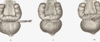
Blood enters the kidneys through the paired renal arteries, which arise directly from the aorta. This anatomy allows them to constantly maintain good blood flow and pressure. The renal artery is divided into small lobar vessels, capillaries, which envelop the renal glomeruli.
A decrease in blood pressure in the kidneys stimulates the renin-angiotensin system, which produces and releases substances into the bloodstream that cause vasospasm and increased blood pressure. Disruption of this regulatory mechanism often leads to persistent hypertension, which cannot be treated with conventional drugs for hypertension.
Indications for use
Ultrasound of the renal arteries is an informative and safe study for the body, which is prescribed if pathological processes in the parenchyma, vessels or surrounding tissues are suspected. The study is prescribed if the patient has the following complaints:
- lower back pain;
- violation of urine separation;
- high blood pressure;
- phenomena of late toxicosis in pregnant women, etc.
Indications for examination are also:
- cardiovascular diseases;
- endocrine pathologies;
- suspicion of a malignant or benign tumor of the kidneys or adrenal glands;
- abnormalities in urine analysis;
- persistent hypertension;
- symptoms of acute, chronic diseases of the excretory system;
- injuries;
- control over organ engraftment during transplantation.
In childhood, ultrasound is performed to determine abnormalities in vascular development.
Why is an ultrasound scan of the kidneys done?
Ultrasound is a common instrumental examination method in case of problems with the pelvic organs and kidneys. The procedure is safe and therefore suitable even for pregnant women. Unlike computed tomography, ultrasound is performed without the use of a contrast agent and allows you to examine stones and sand in the kidneys, cysts, neoplasms and the general condition of the internal organs. The procedure has no contraindications; there is no need to take a long break between examinations.
Ultrasound echography will show changes in the renal parenchyma and help determine the presence of stones, neoplasms, and organ displacement. The device forms an image due to the peculiarities of the reflection of sound waves from tissues of different densities. Sound easily passes through liquid, encountering difficulty only in the form of gases. Doppler ultrasound helps monitor the movement of blood in the vessels of the kidneys. The Doppler effect used in this method implies different frequencies of ultrasonic waves reflected by blood components.
An ultrasound machine has a transducer that emits sound waves that are too high in frequency for the human ear to hear. Sound waves penetrate through the skin to the internal organs, are reflected from them and again end up on the transducer, which transmits them to processing devices and then to the device’s display. The gel that is applied to the transducer not only makes it easier to glide over the skin, but also prevents air from being an obstacle.
Air creates interference and distortion, being the medium through which ultrasonic waves travel the slowest. They pass most quickly through bones. During an ultrasound examination of the kidneys, it is possible to see the blood flow inside the organs. A weak signal or its complete absence may be a sign of low patency or blockage of the vessel.
Kidney ultrasound is often prescribed for regularly high blood pressure, which cannot be reduced. Also on the list of common indications for ultrasound examination:
- pain in the lumbar region;
- changes in urine tests;
- renal colic;
- failure of the kidney;
- enuresis;
- loss of strength, weakness;
- traumatic injuries;
- diseases of the endocrine system;
- diagnosis of neoplasms;
- infectious diseases, inflammatory processes;
- kidney transplantation (monitoring the rehabilitation process after surgery);
- medical examinations.
Preparation for ultrasound of renal vessels
Preparation for ultrasound of the renal arteries is aimed at reducing gas formation in the intestines. This is achieved through diet and medication. For 2-3 days, the patient is recommended to exclude from the diet foods that cause increased gas formation:
- black bread;
- fresh vegetables and fruits;
- cabbage in any form;
- legumes;
- milk;
- sweets, baked goods and carbonated drinks.
Heat-treated vegetables, cereals, and fermented milk products are allowed. But it is necessary to reduce the intake of spices, salt, and avoid eating smoked meats and marinades, as this can contribute to irritation of the kidney tissue and distort the results of the study.
Technique for performing ultrasound of renal vessels
The study is prescribed in the morning, on an empty stomach. Last meal 8-12 hours before the procedure. If for some reason it is postponed to the daytime, a light breakfast is allowed, which consists of liquid porridge, tea, 6 hours before the ultrasound.
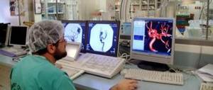
During the procedure, the patient lies down on the couch, the doctor applies a sensor lubricated with gel to the skin of the abdomen and turns it at different angles, receiving an image of the kidneys and blood vessels in different projections, which is displayed on the monitor.
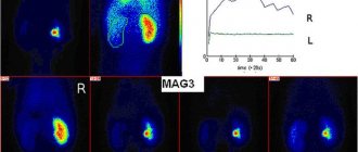
Then, using Doppler ultrasound, blood flow is studied. A special computer program processes the information received from the sensor and displays it on the screen in the form of color pictures and graphs. The higher the blood speed, the brighter the image. Therefore, it is possible not only to judge the speed of blood flow, but also to determine obstacles to blood flow, their location, and size.
The patient may be asked to turn to one side, stand up, and take a breath to get a good look at the structure of the kidney tissue and blood vessels. The examination is painless and lasts 20-30 minutes. Upon completion, a conclusion is issued describing the results obtained.
Interpretation of results: norm and pathology
Vascular examination is carried out in several modes. The doctor examines the kidneys and the vessels suitable for them in different projections. The normal indicators that are taken into account are the following:
- the shape of the organ is bean-shaped;
- thickness and echogenicity of the capsule - hyperechogenic, no more than 1.5 mm;
- contours are smooth, slight hypertrophy in the cortex is considered normal (Bertin's columns);
- mobility during inhalation and exhalation 25-30 mm;
- structure - medium echogenicity, less dense in the area of the cups;
- the walls of the vessels are smooth, without thickening or ruptures;
- The diameter of the vessels at the gate is about 7 mm, the size of the interlobar ones is from 3 mm and above.
In the color flow mode, the speed of blood flow, the presence of turbulences, which indicate damage to the intima, the presence of atherosclerotic plaques, and blood clots are determined. In such cases, mapping reveals a narrowing of the lumen of the vessel. An area of increased blood flow may indicate a malignancy.
Based on the results obtained, the doctor issues a conclusion. In some cases, additional more precise studies may be needed to clarify. For example, tomography (CT, PET CT, MRI). The diagnosis is made taking into account the clinical picture, laboratory results and examination of the patient.