When performing an ultrasound of the kidneys, adrenal glands and retroperitoneal space, preparation for the procedure is of particular importance. The ultrasound diagnostic method is used to obtain more detailed and informative data about the urinary system. Such a study is carried out simultaneously for all organs, since they are located nearby, connected and function together. Moreover, pathologies, inflammations, infections, affecting one organ, negatively affect the condition of another. Therefore, when checking the kidneys, the doctor simultaneously sees diseases that affect the adrenal glands and peritoneum. The results obtained during ultrasound are complex. The procedure is considered safe, painless, effective, it evaluates the condition of organs and their functions. This makes it possible to detect pathologies and formations, even if they have just arisen and begun to develop.
Indications for ultrasound of the kidneys and retroperitoneum:
- Painful symptoms localized in the lumbar region, not caused by physical activity;
- As part of a preventive examination;
- After suffering back injuries;
- For urolithiasis (UCD);
- For any disturbances in urine excretion;
- In case of renal failure;
- In pathological conditions that precede renal failure;
- Suspicion of neoplasms of a benign or malignant nature;
- In the process of preparing for the operation to determine the tactics and volume of intervention;
- In the postoperative period to assess the effectiveness of treatment;
- To monitor the kidney after transplantation.
Indications for use
An ultrasound is performed only in cases where the patient experiences certain symptoms that suggest the presence of disorders in the functioning of the kidneys and adrenal glands.
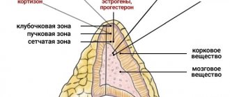
The adrenal glands produce hormones, so disturbances in the hormonal cycle may be a reason for an ultrasound.
Frequent increases in blood pressure
In particular, if pigmentation or stretch marks on the skin appear on the patient’s body, this is already a reason for an ultrasound examination.
In addition, it is very important to undergo an ultrasound scan of the adrenal glands when blood pressure rises and falls for no apparent reason, the patient is constantly accompanied by weakness and loss of strength.
Excessive excess weight, which appears in a patient even if the basics of proper nutrition and an active lifestyle are observed, are also an important reason for conducting an ultrasound.
Women who cannot become pregnant are referred for such examination.
Of course, the most serious reason for an immediate ultrasound examination is the suspicion of the presence of a tumor of the kidneys or adrenal glands.
Headache
The adrenal glands and kidneys belong to the urinary system, so any pathology associated with urine excretion must be examined in order to find out the causes of the pathology and then prescribe effective treatment.
Reasons for undergoing an ultrasound may also be:
- systematic severe pain in the lumbar region;
- increased swelling of different parts of the body;
- headache;
- sudden jumps in blood pressure;
- abnormalities during urination.
All of these symptoms may characterize malfunctions of the kidneys and adrenal glands. Additionally, patients are recommended to undergo a urine test for laboratory testing.
Such an integrated approach to studying the circumstances of the pathology allows you to correctly determine the cause, establish a diagnosis and prescribe the most effective treatment for both the kidneys and adrenal glands.
Preparation for ultrasound of the retroperitoneum, kidneys and adrenal glands
In order to achieve the desired accuracy of a diagnostic test, the patient needs to prepare for it. All measures in this case should be aimed at minimizing the processes of gas formation, since the gas environment makes it difficult for ultrasonic waves to pass through, which does not allow achieving the desired visualization. To do this you need:
- Avoid consuming foods that cause gas three to four days before the test;
- On the recommendation of a doctor, start taking enterosorbents;
- Take your last meal no later than nine hours before diagnosis;
- An hour and a half before the procedure, drink one to one and a half liters of water or fruit drink and do not empty your bladder.
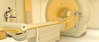
Ultrasound examination (ultrasound)
Ultrasound examination (ultrasound) plays an important role in diagnosing diseases of internal organs. Due to its relatively low cost and high availability, ultrasound is a widely used method for examining a patient and allows diagnosing a fairly large number of diseases. Due to physical characteristics, not all organs can be reliably examined by ultrasound; for example, the hollow organs of the gastrointestinal tract are difficult to access due to the gas content in them. For better information, it is advisable to prepare for the ultrasound examination the day before.
Liver
Ultrasound of the liver is quite highly informative. The doctor evaluates the size of the liver, its structure and homogeneity, the presence of focal changes, as well as the state of blood flow. Ultrasound allows one to detect both diffuse changes in the liver (fatty hepatosis, chronic hepatitis and cirrhosis) and focal ones (liquid and tumor formations) with fairly high sensitivity and specificity.
Gallbladder and bile ducts
In addition to the liver itself, the condition of the gallbladder and bile ducts is assessed - their size, wall thickness, patency, the presence of stones, and the condition of surrounding tissues are examined. Ultrasound allows in most cases to determine the presence of stones in the cavity of the gallbladder.
Pancreas
When examining the pancreas, its size, shape, contours, homogeneity of the parenchyma, and the presence of formations are assessed. High-quality ultrasound of the pancreas is often quite difficult, since it can be partially or completely blocked by gases in the stomach, small and large intestines.
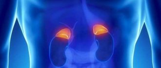
Kidneys and adrenal glands, retroperitoneum
The study of the retroperitoneal space, kidneys and adrenal glands is quite difficult for a doctor due to the peculiarities of their location, the complexity of their structure and the versatility and ambiguity of interpretation of the ultrasound picture of these organs. When examining the kidneys, their number, location, size, shape, contours, structure of the parenchyma and pyelocaliceal system are assessed. Ultrasound allows you to identify kidney abnormalities, the presence of stones, fluid and tumor formations, as well as changes due to chronic and acute pathological processes of the kidneys.
Thyroid
In the study of the thyroid gland, ultrasound is the leading one and allows you to determine the presence of nodes, cysts, changes in the size and structure of the gland. Obstetrics, gynecology and prenatal diagnostics Ultrasound examination is used to study the internal genital organs of a woman, the condition of the pregnant uterus, anatomy and monitoring of intrauterine development of the fetus.
Preparing for an ultrasound
- Preparation for ultrasound of the abdominal organs: 3 days before the examination, it is necessary to exclude brown bread, whole milk, raw fruits and vegetables from the diet, take 2-4 tablets of activated charcoal or “Espumizan”, “Filtrum”, etc. during these days. P. according to the diagram attached to the drug package. The last meal is at 19.00 the previous day - if the study is in the morning, the study is carried out strictly on an empty stomach. If the examination is carried out in the afternoon, do not eat before the examination for at least 6 hours, preferably 10 hours.
- Preparation for an ultrasound of the abdominal organs with determination of gallbladder function: 3 days before the study, exclude brown bread, whole milk, raw fruits and vegetables from the diet, take 2-4 tablets of activated charcoal or “Espumizan”, “Filtrum” during these days " and so on. according to the diagram attached to the drug packaging. The last meal is at 19.00 the previous day - if the study is in the morning, the study is carried out strictly on an empty stomach. If the study is carried out in the afternoon, then before the study do not eat for at least 6 hours, preferably 10 hours. Take with you two raw yolks, or 0.5 liters of kefir 3.2% or cream at least 10% - 100-200 ml.
- Preparation for ultrasound examination of the kidneys: If the patient is overweight and has increased gas formation in the intestines, 3 days before the examination, exclude brown bread, whole milk, raw fruits and vegetables from the diet, take 2-4 tablets of activated carbon or "Espumizan", "Filtrum", etc. according to the diagram attached to the drug package.
- Preparation for ultrasound of the pelvic organs (for men transrectally, for women transvaginally): The night before, do a cleansing enema of 1.5 liters. In the morning, if necessary, empty your bowels. Empty your bladder before the test. You can eat and drink on the day of the test; the test is not carried out on an empty stomach.
- Ultrasound diagnosis of the uterus and ovaries: preparation for transabdominal echography - tight filling of the bladder, i.e. 1 hour before the test, drink 1 liter of water, do not go to the toilet. A bladder filling is not required for a transvaginal scan.
- Ultrasound diagnosis of the thyroid gland: no preparation is required for this study.
- Ultrasound diagnostics of the mammary glands: as planned - on days 5-7 of the menstrual cycle; in the presence of pain or formation - any day.
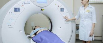
How is ultrasound diagnostics of the kidneys and adrenal glands performed?
Ultrasound of the kidneys and adrenal glands is a painless procedure that does not require the use of painkillers. The patient lies down on a medical couch and frees the anatomical area of the abdomen from clothing. A special medical gel is applied to it. Its use ensures close contact with the ultrasound scanner sensor and improves the passage of ultrasonic waves.
Having applied the sensor to the treated area, the diagnostician begins to move it and rotate it at different angles, achieving maximum clarity of the image that is displayed on the screen. Penetrating into the soft tissues of the body, ultrasonic waves are reflected from them, which allows the formation of visual images. Based on them, the specialist makes a conclusion about the condition of the organs and existing pathologies.
Methodology
The standard examination protocol includes the study of internal organs, large vessels and surrounding tissues. During the diagnosis, the doctor asks the patient to lie down on the couch, exposing the stomach and lower back. A contact gel is applied to the skin, which facilitates the passage of ultrasonic waves. As the sensor moves across the body, a picture is displayed on the screen, based on which conclusions can be drawn about the state of the area being examined.
During the procedure, the patient primarily lies on his back. The doctor may ask you to roll over on your stomach, side, or stand up. The average procedure time is 20-30 minutes.
If necessary, in addition to the standard examination of organs, vascular diagnostics is carried out - Doppler sonography (CDC). With its help, the doctor evaluates blood flow, which allows him to make a conclusion about the quality of blood supply to tissues.
How is an adrenal ultrasound performed?
During an ultrasound examination of the adrenal glands, the patient can lie on his back or stomach, lying on his back, or even standing. The area of the lower back and lower abdomen being examined should be freed from clothing. A medical gel is applied to them, which ensures tight contact with the ultrasound scanner sensor and improves the passage of ultrasonic waves.
It is important to know that during the examination, healthy adrenal glands are not visible due to the fact that the structure of their tissue coincides with the structure of the retroperitoneal tissue. As a consequence, diagnostic capabilities are limited to determining the area in which they are located... But even here, the success rate is not always 100%. Thus, it is possible to determine the desired area on the right in 90% of cases, on the left - only in half of the cases.
When starting a diagnosis, the doctor first of all finds the right kidney, determines the right hepatic lobe and the inferior vena cava. The right adrenal gland is located in the area formed by the extreme points of the above organs. After visualization and examination of the right adrenal gland, the patient is asked to lie on the right side and begins to identify the left one.
How is ultrasound performed?
Conduct and contraindications. To undergo the procedure, the patient must lie on a couch. The doctor will ask you to take a lying position (or on your back, stomach, side). If the organs are not visualized, then the patient will have to stand. The kidneys are examined in several different positions. To do the examination, you need to remove your clothes, exposing an area of your body. It is lubricated with gel, and then the diagnostician begins to move the sensor over the skin. This is how the kidneys, adrenal glands, and retroperitoneal space are studied. Even with a comprehensive diagnosis, the procedure takes no more than half an hour.
Ultrasound usually does not cause injury to patients and is therefore considered safe. There are no contraindications to the study, but it all depends on the patient’s condition. An ultrasound examination of organs cannot be performed if the patient has severe purulent skin lesions, inflammation in the organs begins, which can make visualization very difficult. Ultrasound can be performed on pregnant women, young children, and nursing mothers.
Diagnostics does not cause side effects, which makes it a universal method for studying the disease. Doctors use this to establish the correct diagnosis, prescribe the necessary medications, and find formations: cysts, cancer, stones, salt deposits.
Ultrasound of the kidneys, adrenal glands and retroperitoneal space is prescribed for various conditions, including when the patient has complaints or alarming symptoms are detected. Diagnostics is also necessary for preventive purposes, before and after surgery, during pregnancy, in order to find pathologies in the development of the child. Separately, ultrasound is necessary to monitor the progress of the operation and its results.
An ultrasound helps check why the patient complains of severe pain in the upper abdomen or in the back, lower back, or right hypochondrium. The procedure is prescribed if a person has symptoms of flatulence, a feeling of heaviness in the stomach, which intensifies after eating. Typically, diagnostic results show that such symptoms indicate kidney pathology, neoplasms, dysplasia, hyperplasia in the adrenal glands. The results for vascular diseases and lymph nodes, including the appearance of metastases, are considered separately.
Thanks to the study, the doctor is able to localize the pathology, reduce the degree of tissue damage to neighboring organs, and determine the connection with other processes of inflammation and infection.
Interpretation of the study of ultrasound results of the kidneys and adrenal glands
After performing an ultrasound of the kidneys and retroperitoneal space, the diagnostician begins to decipher the results. To do this, he compares the results obtained with the results that determine the norm according to the following parameters:
- The shape and size of organs - for each of them there are average indicators;
- The location of the internal organs relative to each other - due to the density of placement, if one of the organs is pathological, it can exert compression on the others;
- Tissue homogeneity, which makes it possible to detect inflammatory processes, malignant neoplasms and even microscopic injuries.
The sizes of healthy kidneys and adrenal glands are presented in our table below:
| Options | Kidneys | Right adrenal gland | Left adrenal gland |
| Length | About 11 cm (13 cm for pregnant women) | From 1 to 1.5 cm | From 0.8 to 1.5 cm |
| Width | About 6 cm | From 0.3 to 1.6 cm | From 0.8 to 1.5 cm |
| Height | From 4 to 5 cm. | From 1 to 2 cm | From 0.8 to 1.5 cm |
How should you prepare for the procedure?
How to prepare for diagnosis? Before undergoing the examination, patients must carefully prepare for the procedure. It all depends on the purpose, the patient’s complaints, and the need for a comprehensive or single diagnosis. These factors are taken into account by the doctor in order to correctly prescribe preparatory measures.
First of all, preparation for diagnosis should rid a person of flatulence. An ultrasound of the kidneys, adrenal glands and retroperitoneal space is performed when there are no gases in the body that would interfere with the procedure.
Gas formation must be completely eliminated, as it interferes with echolocation, visualization of the kidneys or adrenal glands. In order to get rid of flatulence, you need to reduce the amount of foods that cause fermentation processes. These are dairy foods, legumes and peas, lentils. It is worthwhile to completely remove fatty and spicy foods, fresh bread, pastries, vegetables and fruits, coffee, carbonated water and drinks from your diet for a while.
If a patient is diagnosed with symptoms of flatulence, then the doctor prescribes a carminative, as well as an enterosorbent substance. They are taken 24 hours before diagnosis.
Before the procedure itself, most often in the morning, you should not eat food. The meal should take place in the evening, but no later than 8 hours before the ultrasound examination. In the case where the bladder needs to be tested, separate preparation is prescribed. It is worth following the above requirements, but before the procedure itself you need to drink half a liter of water. You cannot go to the toilet because the procedure requires the patient to have a full bladder.
For the procedure itself, you need to bring napkins to remove the gel, and a diaper to cover the couch.
What can be seen with an ultrasound of the adrenal glands
If your adrenal glands are healthy, they may not be visible at all, since the structure of these glands and the retroperitoneal space is the same.
In this case, the examination can only show the size of the adrenal glands, their shape and outline. A description of the ultrasound diagnostic results will show where there are deviations. Ultrasound of the adrenal glands will help identify various pathologies in the early stages, they can be:
- presence of hematomas. This is possible in the event of a strong blow to the lumbar region,
- inflammatory processes in the thickness of the adrenal gland. Most often occurs with sepsis, syphilis and autoimmune diseases. This disease is characterized by changes in the structure of the gland,
- the tumor is benign. An adenoma is a neoplasm that can develop in the body for a long time without any symptoms. It can be detected on ultrasound only after its size has reached 2 cm,
- pathologies of the adrenal glands. This is an increase in gland size due to thickening of the adrenal cortex. In the future, this can cause hormonal imbalances,
- endocrine disorders. These diseases are characterized by the appearance of a large amount of hair on the body, a late menstrual cycle, and growth arrest. In addition to ultrasound of the adrenal glands, additional diagnostics are also necessary.
The doctor first of all analyzes the size of the adrenal glands, because it is these readings that can signal the first failures in the vital system of the human body. Any discrepancies with the norm are recorded and transferred to the attending physician, who will decipher the data and make a diagnosis.
Do not forget that you should not try to palpate the adrenal glands on your own or analyze the diagnostic results obtained. The adrenal glands should only be examined by specialists; any amateur activity can lead to a deterioration in your well-being and health.
Carrying out diagnostics
Ultrasound of the kidneys is much easier to perform than the adrenal glands, since they are located in a difficult-to-reach place for diagnostics.
Ultrasound diagnostics
Most often, the examination begins with the right kidney, the right part of the liver and the inferior vena cava. It is in this zone that the adrenal glands are located.
A special gel is applied to the skin in the projection area and a special ultrasonic sensor is passed over it. To facilitate the search for the adrenal glands, the patient is asked to take a deep breath.
During normal functioning of the adrenal glands, they are practically invisible during examination. And since the right one is much easier to detect, the doctor initially examines it, after which he proceeds to search for the left one.
In 90% of cases, the right adrenal gland can still be detected, while the left one has to be looked for with special effort. The patient is asked to lie on the side which adrenal gland is currently being examined.
Instrumental diagnostics
Although the patient does not have to lie down to perform an ultrasound. If the adrenal gland cannot be detected, the patient’s position is changed, asking him to lie on his stomach or stand up.
Statistics say that the success rate of detecting the left adrenal gland is only 50%.
Before the ultrasound, the patient takes a laxative to cleanse the intestines, and an enema is not recommended, because filling the intestines with air prevents normal ultrasound.
The duration of an ultrasound is generally about half an hour. After it, the patient is necessarily given either a video recording or photographs so that the attending physician can personally examine the results obtained.
What does ultrasound of the kidneys, adrenal glands, and retroperitoneal organs show?
Normally, the sonologist determines the usual size, shape, location and structure of organs. There are no neoplasms or inflammatory foci.
For various types of pathologies the following can be diagnosed:
- displacement of organs, their enlargement or reduction;
- pathological inclusions;
- cysts;
- tumors;
- vascular changes, etc.
If necessary, to clarify the results of ultrasound, the patient is additionally referred to CT, MRI, biopsy, and radioisotope study.
At the Spectra clinic, ultrasound diagnostics are performed by sonologists of the highest category. Highly qualified specialists and the latest technical equipment guarantee our patients obtaining reliable results. A detailed conclusion is issued a few minutes after the examination.
Decoding the results
During the study, the density, structure, size, and contours of the lymph nodes are assessed. Normally, they are small in size, oval in shape, and not fused to each other. The contours are clear and even. The structure is homogeneous, without areas of decreased or increased echogenicity. The sizes are small, do not exceed 1-3 cm. An increase in the size of the lymph nodes can be a sign of infection, autoimmune pathology, inflammation, cancer or hematological disease.
Conclusion Ultrasound of the retroperitoneal lymph nodes is not a final diagnosis. It can only be determined by a doctor based on all the results of tests and studies!