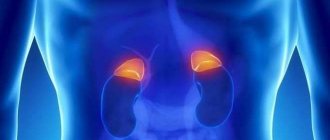
The adrenal glands are organs of the human endocrine system that are located above the kidneys, in the retroperitoneal space. These are endocrine glands that produce many hormones (adrenaline, norepinephrine, steroids, female sex hormones, cortisol, etc.).
Diseases of the adrenal glands of endocrine etiology are much less common than similar pathologies of other organs.
Various methods are used for differential diagnosis of the pathology of these organs. The safest and most informative is MRI (magnetic resonance imaging). The earlier any tumor or other pathology is diagnosed, the more effective its therapy will be.
Detection of pathological changes in the early stages makes it possible to avoid various consequences for human health.
CT or MRI of the adrenal glands. what's better?
A more budget-friendly option for MRI of the adrenal glands is a computed tomography (CT) procedure.
The diagnostic results are not as detailed and accurate as with MRI, but in general an assessment of the causes of the disease can be provided. To conduct a computed tomography (CT) scan of the adrenal glands, you need to specially prepare and undergo a series of tests. The results of computed tomography can also identify benign and malignant adrenal tumors, neoplasms and various mechanical damage and injuries.
The cost of computed tomography is significantly lower than that of magnetic resonance imaging (MRI) of the adrenal glands.
The difference in price can be explained by the price of the equipment and the cost of the resources used. It should also be noted that computed tomography of the adrenal glands uses X-rays, while MRI uses only a magnetic field, which is absolutely safe for the body. Preparation is required for a CT scan, but not for an MRI.
CT or MRI of the adrenal glands, which will be better? It can be said that computed tomography of the adrenal glands is a diagnostic method for little money, but it should not be used often. If repeated studies are necessary, it is better to resort to MRI of the adrenal glands with contrast. An MRI can provide the most complete picture of your disease.
general information
MRI allows you to examine the structure of tissues; the more fluid they contain, the better the image. For this reason, bones are less visible on MRI than parenchymal organs. Objects that do not consist of soft tissue, for example, kidney stones, cannot be examined; they do not change their structure during manipulation. Against the backdrop of this state of affairs, urolithiasis is not diagnosed using MRI; kidney CT is used to identify pathology.
The patient does not feel any discomfort during the procedure; all changes are completely reversible. Magnetic and radio radiation are not dangerous to humans. All organs and systems in the patient’s body are safe and sound. Therefore, MRI can be used for women carrying a child and small children.
MRI scan results: what is normal and what is abnormal
Normally, the adrenal glands are presented in the form of homogeneous formations, up to 10 mm thick. Thanks to the contrast agent, the cortex and medulla are distinguished.
Nodular hyperplasia - thickenings up to 1.5 cm with uneven, bumpy outer contours.
Myelolipomas are round formations with unclear contours, homogeneous structure, and large sizes.
In adrenal tuberculosis, the cortex and medulla are separated, and the glands are affected by calcifications.
In malignant tumors (pheochromocytomas), heterogeneous, clear formations with a high degree of intensity are visualized.
Benign tumors (aldosteromas) are smaller in size, round in shape, and have clear contours. They affect one adrenal gland and are prone to accumulation of contrast media.
Metastases on MR images have a heterogeneous structure, uneven contours, and tend to accumulate contrast agents. Both adrenal glands are affected.
Pheochromoblastoma of the left adrenal gland
Malignant corticosteromas appear as unclear contours, a heterogeneous structure with cystic or fatty deposits. They accumulate contrast and are large in size. In a benign course of the disease, an even contour and homogeneous structure are visualized.
What are the main diseases of the adrenal glands called?
- Cushing's disease or syndrome. Main symptoms: high blood pressure, round face, increased body hair growth, menstrual irregularities, brittle bones, impaired glucose metabolism, muscle weakness and incapacity, excess weight, depression, headaches, fragility of blood vessels, impotence, the appearance of boils, poor circulation.
- Addison's disease or hypercortisolism. In this case, the following is observed: decreased appetite, decreased immunity, frequent colds, weight loss, increased fatigue, weakness, increased pigmentation, excessive tanning from ultraviolet rays, hyperpigmentation of the cheeks, lips, nipples, loss of pubic and axillary hair, depression, decreased libido, night urination, nausea and vomiting, low blood pressure, impaired concentration and memory; alternating constipation and diarrhea, tachycardia, decreased blood glucose levels. This adrenal disease manifests itself from 30 to 40 years of age.
- hyperaldosteronism. This is a disorder in which aldosterone is produced in excess. Main symptoms: headaches (migraines), rapid heartbeat (tachycardia), muscle weakness, swelling, constipation, convulsions, increased volume of circulating blood and plasma, convulsions, fatigue, increased urine production.
- adrenal tumors. These include: aldosteroma, pheochromacetoma, glucocorticosteroma, corticoestroma, glucoandrosteroma. Symptoms: night urination, muscle weakness, tachycardia, high blood pressure, disorders of sexual development, convulsions, nausea and vomiting, pallor, redness or bluishness of the skin, migraine, shortness of breath, chills, pain in the chest and abdomen, sweating, panic attacks. Adrenal adenoma on MRI is a benign tumor of the adrenal gland that develops in the cortex.
In what cases is it prescribed?
The job of the adrenal glands is to produce hormones. The latter are necessary for the functioning of the entire body. The adrenal glands are an important link in the functioning of the entire human body. If there is a malfunction in their work, it affects the entire body. Disruption of the normal functioning of the adrenal glands can seriously affect a person’s health and become a threat to his life.
In connection with the above, an important point is the early detection of any malfunctions in the functioning of the adrenal glands.
There are statistics that the cortex of these organs is most often susceptible to pathological changes. There are a number of changes in the human body, if they occur, you need to immediately contact a medical institution for examination and diagnosis. These include:
- High blood pressure.
- Weight gain in a short period of time.
- The appearance of swelling on the face.
- The appearance of stretch marks on the skin. This is associated with an increase in body weight.
- Diabetes.
- Disruption of the immune system of the human body.
- Osteoporosis.
If a person comes to a medical facility with the above symptoms, the doctor prescribes tests and an MRI or CT scan.
Which doctor should I contact?
In case of endocrine disorders, contact an endocrinologist, who issues an appointment for examination.
The doctor conducts an oral interview, an in-person examination, and prescribes laboratory and instrumental research methods.
Sometimes consultation with doctors of related specialties is required: oncologist, therapist, surgeon.
No specific preparation is required. The patient is diagnosed on an empty stomach; if contrast is used, the patient is refrained from taking potent substances the day before.
This minimizes the risk of nausea, vomiting, and vomit entering the respiratory tract. The doctor is informed in advance which drugs have been used previously.
Avoid soda, coarse fiber, fermented milk products and foods that cause increased gas formation.
People with chronic constipation and flatulence use laxatives and carminatives.
The patient will be asked to remove metal jewelry, hearing aids, and dentures.
Communication means and mobile phones are left away from the tomograph. Loose clothing made from natural fabrics is preferred.
The patient lies down on the table, the limbs are secured with belts for complete immobility.
The table slides into the tomograph machine, and communication with the doctor and medical staff remains throughout the scan.
The patient can stop the procedure at any time by pressing the panic button.
The duration of the study is up to 40-50 minutes. After completion, they wait for the results to be deciphered and return to their usual way of life.
How to prepare for an MRI?
The doctor explains the essence of the MRI procedure
If you plan to use contrast during MRI of the adrenal glands, you should not eat food 5-6 hours before the start of the study. Otherwise, no special training is required.
- clothing should not contain metallic inclusions, this may affect the results of the study or damage the device;
- magnetic and plastic keys should not fall into the field of operation of the device; it is better to remove them during the preparation process;
- It is also better not to use decorative cosmetics; some cosmetics contain metal particles;
- Tattoo inks contain metal particles, so irritation may occur in the area after an MRI.
MRI is contraindicated for owners of pacemakers and metal endoprostheses. In doubtful cases, preparation for an MRI includes checking the patient's body with a hand-held metal detector.
How are they diagnosed?
MRI of the kidneys and adrenal glands is performed as follows:
- Before entering the room where the diagnosis will take place, the patient must put away his phone, watch and keys, and also remove all metal objects. If your clothing has metal buttons, these will also have to be removed. This is due to the fact that metal distorts the magnetic field and can affect the information content of the study.
- The patient lies down on the tomograph conveyor table. If necessary, a contrast agent is injected at this time. The patient's limbs are secured using special straps.
- The table moves into the tomograph together with the patient. This is where the research takes place. Scanning is done in two planes: frontal and transverse.
- The patient should lie still. It is very advisable to synchronize breathing with taking pictures.
- During the procedure, the doctor will sit in the next office at the computer. You can contact him at any time through the public address system.
Typically, the patient is alone during the procedure. However, if the diagnosis is being carried out on a child, one of the adults may be with him (before entering the office, he must also remove all metal objects from himself).
The duration of an MRI is usually no more than 50 minutes.
Computed tomography of the kidneys
CT diagnostics of the kidneys and adrenal glands also has another name - CT urography, since it also examines the organs of the retroperitoneal space. This method is very important for identifying various diseases, neoplasms, urolithiasis, vascular abnormalities in the kidneys and adrenal glands.
When is kidney diagnostics necessary?
The kidneys and adrenal glands are different organs, each performing its own function, so the indications for computed tomography of the kidneys and adrenal glands can be completely different. It is necessary to resort to the CT method for diagnosing the kidneys and adrenal glands in case of hormonal changes, severe causeless changes in weight, back injuries, suspected urolithiasis, tumor, or injury to internal organs. You should also undergo an examination in case of chronic infectious pathology of the kidneys and dysfunction of their function, to determine the causes of renal colic, diagnose developmental abnormalities, in inflammatory processes, pathology of blood vessels and lymph nodes.
How is diagnostics carried out?
Before carrying out diagnostics, it is necessary to get rid of all metal objects, remove the smartphone, as well as those devices that can affect magnetic waves.
After this, you need to take the correct position, a specialist will help with this, and will also give an injection if a contrast MRI is prescribed. If necessary, the patient's arms and legs are secured with belts, after which the table begins to move inward.
There is no need to worry if you suddenly feel unwell during the examination; the device is equipped with a connection through which the subject will be able to contact a specialist and receive the necessary help.
MRI is performed lying with your stomach up, motionless; according to the doctor’s requirements, you must stop breathing. The time depends on the health of the person being examined, but does not take more than 50 minutes.
If the procedure is performed on a child, then one of the adults remains with him in the room with the device, who must also first leave metal objects and all kinds of equipment outside the door. A relative of the child can report anything to the doctor via speakerphone to a specialist.
MRI of the adrenal glands. How is it done and what preparation is needed?
It is worth saying that there is no need to prepare in any special way for the examination. The only requirement that should be adhered to is to refrain from eating for at least 6 hours before the procedure.
Also the day before, you should refrain from drinking alcohol, drinks that contain gases, and foods that contain coarse fiber. It is also not recommended to consume dairy products.
How does the procedure work?
During the study, the patient is placed in a separate room on a horizontal table moving along a magnetic coil in a horizontal direction. Before the procedure, he must first remove metal objects from himself and leave mobile devices in another place.
The medical technician explains the rules of conduct during the scanning process, puts on headphones to reduce noise, and provides a panic button to alert medical staff in alarming situations. During the MRI scan, the patient is left alone; the study lasts about 40 minutes.
MRI is performed according to standard techniques, in the frontal and axial planes, obtaining T1-T2 weighted images with a slice thickness of 4-5 mm. Their quality improves when using short sequences with breath holding.
The cortex and medulla are distinguished using a surface coil and high resolution with 2mm slice thickness. MRI of the adrenal glands with contrast is used for differential diagnosis of tumors. Healthy tissues of this organ do not accumulate contrast; the structure of tumors is clearly visualized with intravenous administration of a paramagnetic substance - Gadolinium. Malignant and benign formations accumulate and wash it away in different ways.
Research methodology
Before entering the room with a tomograph, the patient takes off all items that contain metal (earrings, chains, rings, piercings, clothes with metal buttons, buttons, cufflinks), and he also leaves his watch and keys outside. This is done due to the fact that the metal distorts the magnetic field emitted by the tomograph, which affects the results of the study.
The patient is then placed on the tomograph conveyor table in a supine position. At the same time, the doctor's assistant injects a contrast agent into his vein, after which the diagnostic process immediately begins.
The table with the patient on it enters the tunnel where the scanning itself takes place, stopping at the level of the kidneys and adrenal glands. Scanning is carried out in two planes: transverse and frontal. The optimal cutting thickness is 5 mm. To obtain the best possible images, the patient must lie still and synchronize their breathing with the taking of the images.
During the procedure, the person is alone in the room with the tomograph (if the study is performed on a child, one of the relatives stays with him, who also leaves all metal objects outside the door), but he has the opportunity to communicate with the radiologist who is in the next room behind him. computer monitor, speakerphone. On average, the procedure lasts from half an hour to 45 minutes.
What does a CT scan of the kidneys and adrenal glands show?
CT diagnostics of the kidneys and adrenal glands is ideal for determining the shape of organs, since changes in structure or size can indicate diseases and pathologies. Also, using computed tomography it is easy to detect signs of kidney cancer, diagnose the development of a neoplasm, size, character, and also see hemorrhages or necrosis. An examination helps to see diseases associated with the vascular system of organs and urinary canals.
#!KTseredina!#
MRI of the adrenal glands is performed as follows:
- conversation with health workers at the diagnostic center. You should inform about chronic diseases, drug intolerance and allergic reactions, implanted implants, foreign bodies in the body (bullets, metal foreign bodies, etc.), possible pregnancy. The center staff will tell you in detail how to prepare for an MRI of the adrenal glands. Do not hesitate to ask questions that concern you;
- changing into suitable clothing (without metal elements - zippers, buttons, hooks, etc.), removing dentures, jewelry (rings, chains, bracelets, etc.), hairpins, belts with buckles, suspenders, removing them from pockets keys, magnetic cards, etc.;
- moving to the room with the tomograph. Laying on the tomograph table, explaining the rules of behavior during the examination of the adrenal glands. If the person cannot lie still, then anesthesia is administered at this stage. If necessary, intravenous administration of a contrast agent is performed;
- the patient being examined is given a device that should be pressed in case of deterioration of health inside the device. The device makes slightly unpleasant sounds - you should not be afraid of this and be prepared to hear them;
- the device is activated and the patient moves inside the device. There you need to remain calm and not move. The doctor is in the next office and controls from there.
- process and communicates via speakerphone with the patient; The obtained MRI images of the adrenal glands are transferred to a computer screen and 3D photo processing is performed using a special application;
- The adrenal gland examination lasts about half an hour, less than the preparation for it. The more accurate and new the device, the longer the study will take, since the interval between photographs will be longer.
- After the doctor allows you to get up, you can change clothes and wait a little or pick up the MRI transcript of the adrenal glands at the agreed time.
Preparing for an MRI
Magnetic resonance imaging of the adrenal glands does not require any special preparation or use of special medications. There is only one condition. Before the procedure, the intestines must be cleared of contents. If necessary, the attending doctor will recommend taking a course of a special remedy a few days before the examination, which will allow you to get rid of the problem as quickly as possible.
Also, before an MRI, you should limit your intake of food that can cause gas formation in the body or other problems with the digestive tract. Typically, such food includes fast food, fatty, spicy foods, soda and alcohol, as well as bakery and confectionery products.
Compliance with these few rules will allow you to obtain undistorted images and most accurately determine the disease. When an MRI using a contrast agent is indicated, the patient should refrain from eating for several hours before the diagnosis.
By adhering to these simple recommendations, the MRI process will not leave negative consequences, and the data obtained with a deliberate approach to therapy will be the most accurate.
Kidney MRI with contrast
MRI: metastases to the adrenal glands in lung carcinoma (indicated by arrows)
To improve visualization capabilities according to indications, MRI of the kidneys and adrenal glands is performed with contrast. The use of the indicator is especially important for identifying a tumor at an early stage of development, sometimes before the appearance of full-blown clinical symptoms. To enhance magnetic resonance scanning, a drug based on soluble gadolinium salts is used.
Significant side effects after it enters the body develop in less than 1% of people. Gadolinium is better tolerated compared to iodinated contrast, so there are fewer absolute contraindications to its administration:
- a history of aggravated allergic reactions to the substance in previous studies;
- acute or chronic renal failure with a decrease in glomerular filtration rate of less than 30 ml/min, receiving replacement therapy.
Relative restrictions include:
- childhood;
- pregnancy;
- lactation.
Magnetic resonance imaging, regardless of the amplifier used, is not available if:
- the patient has metal in his body - shavings, shrapnel, bullets, vascular clips, etc.;
- implanted devices are functioning - pacemaker, defibrillator, insulin pump, cochlear implant, orthopedic structures, neuromodulator, etc.
- the subject's body weight exceeds 120 kg (for closed-type devices);
- there is severe pain or the condition requires resuscitation;
- a pathology has been diagnosed in which the patient cannot lie quietly on his back, for example, Parkinson's disease, Alzheimer's disease, dementia, claustrophobia, etc. The study is possible within the walls of a hospital under sedation.
MRI of the kidneys and adrenal glands with contrast for children under 12 years of age is performed in a hospital setting. During lactation, you should make a reserve of milk for 16-18 hours, express the excess and pour it out. During pregnancy, enhanced magnetic resonance imaging is performed if the benefits of the examination outweigh the potential risks. There is no convincing evidence that gadolinium chelates are harmful to the human body, including if contrasting was performed during embryogenesis at any stage or lactation.
Indications for referral for diagnostics
Dysfunction of the endocrine organs is expressed by various symptoms; endocrinologists suspect diseases of the adrenal glands if there are complaints:
- Changes in body weight, blood pressure, appearance.
- Disorders of menstrual function, sexual development, sexual desire, erection.
- Muscle weakness, frequent bone fractures.
- Interruptions in the functioning of the heart.
- Hair loss, increased sweating, changes in mental health, appetite.
- Skin hyperpigmentation, hirsutism.
- Infertility.
- Dehydration, mainly night urination, thirst.
- Frequent hypertensive crises, paresthesia, headache, blurred vision, respiratory arrhythmia, pain of various localizations.
Hormonal examinations confirm pathology of the adrenal glands - dysfunction, cortical hyperplasia, benign and malignant tumors, which may be hormonally inactive. To clarify the cause of the lesion, the endocrinologist chooses an instrumental examination method.
Due to the small size of the organ and the need for a large number of thin sections, CT and MRI are preferred. If a tumor is suspected, it is recommended to determine the problem using MRI.
Indications for MRI of the adrenal glands are suspicions:
- For Cushing's syndrome.
- Congenital dysfunction of the adrenal cortex.
- Hyperaldosteronism.
- Pheochromocytoma.
- Incidentaloma.
- Metastases of tumors from other organs.
- Secondary hypertension.
- Adenoma.
- Hyperplasia.
To diagnose pathology of the adrenal glands, computed tomography is used, with its help the size, contours, and density of the formation are determined. The endocrinologist and the radiologist decide together which method to choose.
Pros and cons of MRI diagnostics
Examination of the body using MRI means the accuracy of the results and safety. The magnetic radiation of the device does not harm the body at all and does not disrupt its functioning. The kidney image results are produced without distortion, and the image has natural contrast. Disadvantages of the procedure:
- duration (depending on the severity of the examination), which often affects the purity of the results;
- expensive equipment and costs associated with its repair;
- special requirements for the room where the device is installed.
Indications for the study
Magnetic resonance imaging of the adrenal glands is recommended for patients who show signs of endocrine pathology, probably caused by a pathological process in these organs.
In particular, these are:
- Itsenko-Cushing syndrome (characteristic redistribution of fatty tissue in combination with muscle atrophy of the upper and lower extremities);
- disorders in the psychoemotional sphere;
- high blood pressure, combined with severe muscle weakness and symptoms of kidney dysfunction;
- tachycardia (rapid heartbeat) in combination with the above symptoms;
- changes in appearance that do not correspond to the patient’s gender;
- premature sexual development;
- changes found in the blood (decreased glucose and sodium levels, increased potassium levels);
- laboratory-recorded changes in the level of adrenal hormones in the blood.
MRI is also performed when a tumor of this organ is accidentally discovered (incidentaloma) in order to verify its structure and after surgical treatment in order to check whether the tumor has arisen again.
Order of conduct
The scanning procedure itself is carried out in several stages:
- Preliminary consultation. The doctor examines the patient and finds out whether an MRI scan can be performed.
- The administration of a contrast agent is carried out immediately before diagnosis. It is done in cases where it is necessary, for example, when vascular and tumor pathologies are suspected.
- The scanning procedure itself. The person lies down on a movable couch, on which he is fixed with special belts. During the 15-20 minutes that the procedure lasts, the patient must remain completely still in order to obtain clear images.
- After the diagnosis is completed, the couch moves out of the tomograph tube, and the person is released from the belts. Now you need to wait a little to make sure that the scan was successful and all the data was recorded.
- Processing the results. The specialist processes the received data, on the basis of which he prints out or records images on an electronic medium, and, if necessary, builds a three-dimensional image of the problem area, which allows for a clearer visualization of the pathology.
After these manipulations, the person can lead a normal lifestyle, undergo further examination and treatment. The data obtained from MRI scanning in most cases makes it possible to make an accurate diagnosis even in the early stages of the disease, so you can begin treatment of the disease in a timely manner and avoid many negative consequences.
This article contains general information only, is not scientific material and should not be construed as a substitute for medical advice.
Benefits of the study
Why is MRI considered a safe, effective method for diagnosing and monitoring kidney treatment? The procedure is considered the most informative diagnostic measure, which helps to identify cancer formations in the early stages of development. Using this technique, doctors are able to track the slightest changes in tissue metabolism at the cellular level. Early diagnosis increases the likelihood of a successful outcome for the patient.
The advantages of MRI include:
- Preparation for manipulations takes a little time;
- the possibility of carrying out the procedure in cases where other studies are prohibited;
- MRI shows the physician the existing problem and allows you to track the effectiveness of the prescribed therapy;
- the reliability of the results is not affected by distracting signals: the patient’s breathing, arterial pulsation, heartbeat, the presence of adipose tissue;
- doctors do not need to take tissue for cytological examination, minimizing the risk of infection;
- the method allows for early diagnosis of cancer;
- erroneous results are minimized. The images are processed by a computer, as a result the doctor receives a reliable picture.
Given the advantages of this diagnostic method, it is often used in all areas of medical practice. The only disadvantage of the study is its inaccessibility in some localities. A tomograph is an expensive device; the cost of the procedure itself can be about 6,000 rubles, which today is a large amount for some citizens. An additional obstacle is that the device is not available in all medical institutions, which forces the patient to look for a clinic equipped with a tomograph.
Advantages of magnetic resonance imaging over computed tomography
- MRI does not use harmful x-rays during scanning;
- visualizes soft tissues and blood vessels better than CT;
- allows you to differentiate benign tumors from malignant ones;
- allowed during pregnancy (except the first trimester);
However, MRI of the adrenal glands is usually performed after obtaining CT or ultrasound data in cases where more detailed or clear images are required to make an accurate diagnosis. MRI is slightly more expensive than CT and ultrasound, so doctors try to make do with lower costs for the patient. If a tumor is suspected, MRI is usually used, as with vascular pathologies.
At the same time, magnetic resonance imaging also has disadvantages:
- it is necessary to remain motionless in the tomograph for a long time;
- high noise level produced by the tomograph;
- Contraindicated for persons with implanted electronic devices that may be affected by the magnetic field of the tomograph...
Only a specialist can determine which diagnostics will be best in your case.
Contraindications for the procedure
Diagnosis using computed tomography is not suitable for all people. This procedure is prohibited for pregnant women and those who are breastfeeding. Also, the method is not suitable for people suffering from claustrophobia, mental disorders, or acute pain that will not allow them to remain motionless for a long time. CT scanning is not recommended for patients with diabetes and children under 14 years of age. Kidney CT with contrast is prohibited for patients with an allergic reaction to a contrast agent, as well as in the presence of renal failure.
Structure and functions of the adrenal glands
The adrenal glands (gl. Suprarenalis) are a paired organ of the endocrine system; they produce vital hormones. They are laid in utero in the third week of pregnancy, located above the upper edge of the kidney, have the shape of a triangle or leaf, weighing up to 20 grams. These glands of the endocrine system are covered with a capsule and consist of two different layers - the cortex and the medulla.
Both layers have different structures, origins, and produce different hormones. The cortical layer occupies up to 90% of the organ's area and is divided into three zones. The medulla has a common origin with the nervous system; the substances it produces adapt the body to acute stress and inflammatory reactions.
| Layer | Zone | Hormone | Function |
| Cortical | Glomerular | Aldosterone | Controls the absorption of electrolytes |
| Beam | Glucocorticoids | Regulate metabolic processes | |
| Mesh | Androgens | Participate in the production of sex hormones | |
| Cerebral | Adrenaline, Norepinephrine | Transmitters of the sympathetic nervous system |
In addition to these substances, the cortex produces about fifty steroids, has complex hypothalamic-pituitary regulation, and is an important link in the renin-angiotensin-aldosterone system, which controls blood pressure levels.
The essence of MRI, advantages
MRI of the adrenal glands is a diagnostic method that is based on recording NMR (nuclear magnetic resonance or response) of organ tissues in response to an induced magnetic field acting on them. The structure of the gland is scanned layer by layer, the information is sent to the computer, and a three-dimensional image of the examined structure is projected onto the monitor.
In MRI there is such a thing as T1 and T2-weighted images. T1 and T2 are the relaxation times during which the protons of the biological tissues under study return to an equilibrium state.
We will not delve into the jungle of the physical foundations of these processes - this is not what the article is about, and such information is not important for the reader.
Let's just say that these indicators are different in healthy and diseased tissues.
- The study allows the radiologist to detect space-occupying formations in the area of the adrenal glands, determine their exact location, structure, clarity or blurring of contours, and relationships with tissues located nearby.
- The obvious advantages of this diagnostic method include non-invasiveness (it is not associated with the penetration of foreign bodies into the body’s structures), the absence of exposure to harmful ionizing radiation on the body, the possibility of detailed examination of even the smallest formations and, of course, high information content and reliability of the information received.
- MRI can reveal the following adrenal tumors: adenoma, aldosteroma, corticoestroma, glucoaldosteroma, androsteroma, neuroblastoma, pheochromocytoma.
Adrenal glands. Symptoms of the disease in women
A woman's body is prone to hormonal changes. The main function of the adrenal glands is to produce hormones. The main disturbances in the functioning of organs are associated with insufficient production of these substances.
How do you know when you should have your adrenal glands checked? Symptoms of the disease in women may vary. Since their nature depends on the disease that is present in the body. For example, the signs may be the following:
- bone fragility;
- lack of libido;
- disruptions of the menstrual cycle;
- tendency to high blood pressure;
- obesity;
- drowsiness;
- increased weakness;
- panic attacks;
- increased irritability;
- headache;
- frequent muscle cramps.
- appetite decreases;
- bluish lips;
- unnatural thinness appears;
- tachycardia;
- severe fatigue;
- tremor of the limbs;
- increased gas formation;
- depressive states.
In these and some other cases, the doctor may prescribe an MRI of the adrenal glands.