Kuznetsov Alexander Viktorovich
Head of the Urology Department
Urologist of the highest category
The urology department is equipped with modern endourological, laparoscopic, ultrasound and R-scopic equipment, allowing to diagnose and treat patients with urological, oncourological and urogynecological pathologies.
How to deal with a kidney stone?
A 5 mm kidney stone, what to do in this situation is a frequently asked question among patients. In the modern world, there are many different methods of treating urolithiasis, both conservative and surgical. The choice of method depends on the size of the stones, also called stones. What types of kidney stones are there, and how are they diagnosed?
Types of formations
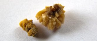
Stones formed in the kidneys are diverse and vary in their structure, chemical composition, size and shape.
Both one and both kidneys are affected. The sizes of the stones vary from a few millimeters to tens of centimeters. If we are talking about stones with a diameter of up to 3 mm, then they are defined not as stones, but as sand in the kidneys.
Classification of renal solid foreign bodies according to various criteria is the main criterion for choosing further tactics for treating the disease. Stones are formed from a mixture of minerals and organic substances. There are the following main groups of foreign bodies formed in the kidneys:
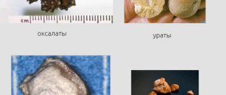
- Oxalate deposits belong to the most common category of formations. They are dense, small bodies with sharp edges, spines and processes.
- Phosphates are smooth formations that can grow to significant sizes. Consist of calcium salts of phosphoric acid.
- Urate formations are formed from uric acid salts against the background of an increase in their concentration. They have a round shape and a smooth surface.
- Xanthine stones are rare. They are the result of a complex genetic defect in which amino acid metabolism is disrupted.
- Cystine stones are rare types of formations. Often formed as a result of impaired absorption of amino acids during digestion.
Renal coral-shaped foreign bodies are identified as a separate group. These deposits occupy the entire space of the pelvis, unlike other types of formations, and then they move to the area of the renal calyx.
Diagnostic method
Hardware research methods are considered the most informative and accurate methods for diagnosing the presence of kidney stones, since this cannot be done by other means. Similar visualization methods include:
- MRI;
- CT with contrast;
- Ultrasound of a characteristic organ, ureter and bladder;
- X-ray examination.
Using ultrasound, it is possible to establish the features of the physiological renal structure, position and size of the formation. This method allows you to exclude diseases that have similar symptoms. However, when a stone moves into the ureter, ultrasound will provide little information, since this area is poorly visible by the mentioned device.
A more informative method is x-ray examination. In controversial situations, computed tomography (CT) is performed, which allows you to get a clearer picture - not only to determine the location, size, shape, boundaries of the stone, but also to assess the general condition and performance of the kidney.
MRI is used to assess damage to a characteristic organ, neighboring tissues, and clarify the chemical composition of deposits. This method is considered the most accurate.
Treatment of urolithiasis is carried out in three ways:
- conservative therapy;
- surgical removal;
- stone crushing.
Conservative treatment
In most cases, a 5 mm kidney stone is not accompanied by any symptoms. Only sometimes do you experience discomfort when urinating: itching, burning or pain. Often, urolithiasis is discovered accidentally during a routine medical examination.
This kind of formation, the size of which ranges from 4 mm to 6 mm, can independently exit the pelvis, pass through the ureter and be excreted in the urine. For these purposes, diuretic and antispasmodic drugs are prescribed to promote the release of stones.
If a 5 mm stone does not pass on its own, medications are used to help reduce the size of the foreign body and its subsequent removal from the body.
The choice of medicine depends on the identified chemical composition of the formation, since individual salt compounds are poorly broken down. Do not forget that stone-dissolving agents are used exclusively as prescribed by the treating specialist.
Self-medication can cause deterioration in health and the need for surgery.
Surgery
Surgery is indicated if the patient experiences intense pain during stone removal that cannot be relieved. If a stone in the ureter has blocked the passage, a decision is also made to promptly remove the foreign body from the urinary tract.
Therapy is performed endoscopically or laparoscopically. After this kind of manipulation, the patient recovers quickly, and all kinds of complications are minimized. The operation is performed without cutting the kidney or damaging the integrity of the epidermis. The instrument is introduced through natural routes:
- ureter;
- bladder cavity;
- lumen of the urethra.
Abdominal surgery is performed if the doctor detects a staghorn calculus, a blockage of a characteristic organ, a foreign body of significant size, or severe bleeding. In modern medicine, this technique is used less and less due to the development of minimally invasive surgery.
Stone crushing
Kidney stone crushing is one of the modern methods of stone removal. It allows you to crush and remove these formations from the body. To avoid violations of tissue integrity, the doctor may recommend that the patient undergo stone crushing instead of surgery. There are two types of this procedure: shock wave and laser lithotripsy.
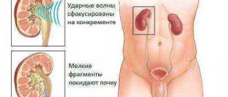
Shock wave lithotripsy
With the help of shock wave lithotripsy, you can get rid of kidney stones up to 2 cm in size. Most often, such manipulations are prescribed by a doctor if the solid formation exceeds 1.5 cm in size. Multiple sonic shocks, without harming the soft tissues, affect the stones and destroy their structure to the state of sand. It is subsequently eliminated from the body.
Ultrasound waves may cause pain in the patient as there is minimal impact on soft tissue, so the procedure is performed under local anesthetic. There is a risk that during the procedure the foreign body will break into sharp fragments that can provoke renal colic. Therefore, shock-wave destruction of stones of solid formations is carried out several times.
Laser lithotripsy
Laser lithotripsy treatment is designed to destroy small stones. The laser is directed pointwise to the area where the stone is localized. This manipulation is performed using an endoscope, which is inserted under the skin through a small incision. The solid body is crushed under the influence of a laser to the state of sand, and then excreted along with urine. Experts recommend laser crushing, since such a procedure allows you to get rid of any stones in one session.
If a patient has formed 5 mm kidney stones, only a qualified specialist can decide what to do and how to treat this disease. Therefore, at the first symptoms of urolithiasis, you should not delay a visit to the doctor. Self-medication is strictly prohibited.
Chop or delete?
I suffer from urolithiasis. And I am faced with a choice: should I crush my stones or remove them? Which is better to choose?
Olga, Nizhny Novgorod
–?Traditional surgical methods for removing kidney stones (open surgery) are now used in rare cases when the patient already has a violation of the outflow of urine, caused, for example, by a narrowing of the ureter (congenital or acquired), and plastic surgery of the upper urinary tract is required.
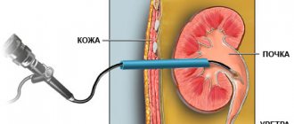
In other cases, doctors resort to three methods: extracorporeal lithotripsy (ESLT), transurethral (through the urethra) contact and percutaneous nephrolitholapaxy.
In the first case, crushing of a stone (usually medium in size - from 1 to 1.5 cm) is carried out using a special remote lithotripsy apparatus, which, by creating a special field, fragments kidney stones without internal intervention in the human body.
The other two methods involve tissue penetration and are more often used for larger and more multiple stones. The essence of these methods is as follows: under conduction or general anesthesia, the patient is punctured in the skin above the kidney and, under ultrasound and X-ray control, an optical system is inserted through a special tube or an instrument is brought into the stone through the urethra along the ureter.
Then, using special devices (ultrasonic, laser, pneumatic, electric pulse), the stone is crushed. Thanks to these techniques, you can immediately destroy all the stones and remove their fragments. In this case, the patient returns to active life much faster than if traditional open surgery was used.
However, the choice of stone removal method is a matter for the doctor.
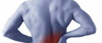
Urolithiasis can be recognized by the following signs:
>>?pain and feeling of heaviness in the lower back, just above and to the side of the sacrum;
>>?pain in the lower abdomen, as well as in the groin, in the genital area;
>>?blood in the urine;
>>?pain when urinating, frequent urination, cloudy urine;
Source: https://www.aif.ru/health/life/kamni_v_pochkah_drobit_udalit_ili_podozhdat
Urolithiasis rightfully occupies a leading place among the most common urological diseases in Voronezh. The MedStar clinic provides non-surgical removal of stones from the kidneys and ureters. We are for crushing stones.
Kommuna.ru - Main news from Voronezh and the Voronezh region. Print version: At the end of last year, the hospital purchased the latest lithotripter, designed for contact crushing of stones in the bladder and ureters, as well as in the kidneys.
Related About: “crushing kidney stones Voronezh”. cone crusher. how to crush a kidney stone. Fibroscopy - MINIMALLY INVASIVE. kidney stones, crushing kidney stones clinic, methods of removing kidney stones with a laser.
Our center is already known far beyond the Lipetsk region; patients of the center are also residents of Tambov (Tambov region), Voronezh (Voronezh region), Tula (Tula region), Ryazan (Ryazan) Fast painless crushing of stones in the kidneys.
If you make the necessary adjustments, kidney stones will stop forming. Tell me, is it necessary to crush the stone? What are the gentlest crushing methods that are best for me?
Remote crushing of kidney stones with ultrasound or laser is recommended for sizes from 5 to 20 mm. This procedure is performed, of course, only in the kidney or ureter, but not in the bladder, where stones are never crushed remotely.
Another question is how to crush kidney stones using traditional medicine. I found this method of crushing kidney stones in one of the health magazines. Irina Igorevna wrote about him.
Federal AIF St. Petersburg Arkhangelsk Barnaul Belgorod Vladimir Volgograd Voronezh Yekaterinburg Izhevsk Irkutsk Kazan Kamchatka Tell me, does the composition of kidney stones matter? Have you heard that carbonate stones crumble best when crushed?
The safest crushing of stones using the Lazurit complex is only in our clinic. Laser crushing of stones in the kidneys, ureter, bladder without incisions in Kyiv.
Flax seeds have a good effect in crushing kidney stones. 1 tbsp. crush the seeds, mix with 3 tbsp. fresh milk and boil until 1 tbsp remains. strain.
First of all, these are methods of crushing kidney stones, such as ultrasonic and laser waves. Each method has its pros and cons. Ultrasound crushing of stones.
Home kidney stones recipes for herbal crushing of kidney and bladder stones. And one more interesting remedy: “Ash of cabbage root crushes kidney stones.”
Specialists from the Federal State Budgetary Institution "KDC with a polyclinic" crush kidney stones using a unique modern device called Lithoskop, principle of operation. The advantage of remote crushing of kidney stones is that the preparation of the patient and the procedure itself last about one hour.
External lithotripsy is a safe method of crushing kidney stones
One of the common urological diseases is urolithiasis. It is manifested by the formation of stones in the kidneys, bladder and ureters.
Stones can appear for many reasons, the main ones being metabolic disorders, abnormalities and kidney diseases, which impair the flow of urine.
Mostly stones are calcium, but mixed types, uric acid and cystine, are also found.
External lithotripsy is a progressive method of treating urolithiasis. The therapy has virtually no contraindications, and the patient experiences a minimum of negative sensations throughout the procedure.
What is the essence of the method
The method is based on hardware crushing of stones using a shock wave (short-time pulse with high pressure) aimed at the formations.
After this, they pass out on their own in the urine. The fragments may go away for several days or months.
Sometimes patients' stones are very hard and require several procedures to crush them.
Conditions in which the procedure is most effective
External shock wave lithotripsy (ESWL) will be more effective in patients with kidney stones up to 2.5 mm in size.
Treatment is prescribed when there is free urinary outflow below the area where the stone is located. It has been established that the effect of stone crushing is related to its size and physicochemical properties.
Stones composed of calcium oxalate and uric acid break down better than stones made of cystine and calcium oxalate monohydrate.
Before the operation the following is taken into account:
- size, chemical composition of the stone, volume and density;
- patency in the urinary tract;
- kidney condition;
- the nature of organ dysfunction and urinary tract infection;
- level of patency in the urinary tract.
Restrictions for the procedure
The main contraindications include:
- body weight more than 130 kg;
- person's height is more than 2 m;
- dangerous arrhythmias;
- cardiac dysfunction;
- decrease in kidney functionality over 50%;
- disorders in the blood clotting system;
- pregnancy, menstruation;
- acute inflammatory processes in the genitourinary tract (pyelonephritis, cystitis and urethritis);
- renal failure.
Patient preparation
Before prescribing a surgical intervention, a detailed examination of the patient is carried out in accordance with the data of computed tomography, x-ray and ultrasound examination. There are no contraindications to treatment.
The patient is also prescribed blood tests for group, Rh factor, general and biochemical analysis. A general urinalysis and an electrocardiogram are performed.
At least 5 days before surgery, medications that thin the blood and stop blood clotting (Warfarin, Aspirin, etc.) are discontinued. If there are infections in the urine culture, antibacterial agents are taken first.
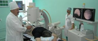
Progress of the procedure
External lithotripsy of kidney stones can be performed on an outpatient basis, after which the patient is sent home. To increase the effectiveness and comfort of the patient, the operation is performed with anesthesia (with intravenous anesthesia or slight sedation).
It is possible to operate without anesthesia using painkillers.
The patient is placed on the lithotripter table, the skin in the area where the focuser is placed is covered with a special gel, after which shock waves are focused onto the stone using ultrasound or X-rays.
After this, waves are generated with a certain strength and frequency, destroying the stone. The duration of the procedure usually does not exceed 60 minutes.
The impact of impulses is carried out under constant supervision. After the operation, a dull pain may appear in the lumbar region, but gradually its intensity decreases.
In some cases, the pain is colicky and is characterized by the passage of a stone.
You may also experience blood in your urine for several weeks after surgery.
Sometimes the effectiveness of the treatment used is proven immediately after the operation to change the stone shadow on x-rays. But in general, the final result becomes known only after a few weeks.
It is impossible to talk about shock waves being completely non-traumatic, since the kidney experiences bruise and stress, which can be eliminated in 5-7 days.
Possible complications
In some cases, distance lithotripsy is associated with some complications. They are divided into the following types:
- complications due to the passage of stone fragments;
- complications due to the action of the shock wave on nearby tissues.
The first complication includes blockage (obstruction) of the ureter by the remains of a damaged stone.
If the stone is large, large fragments may accumulate in the main part of the ureter, causing obstruction in the urinary tract.
Basically, the resulting obstruction in the form of pain in the lumbar region is similar to renal colic and requires instrumental intervention.
Stones are removed from the urinary tract using a ureteroscope or secondary crushing is done.
Complications after obstruction of the urinary canals by stone fragments include pyelonephritis before the appearance of urosepsis, bruising, and hemorrhage into the perinephric tissue.
But such complications rarely appear and do not reduce the benefits of the technology.
They've been through it
Reviews from patients who have successfully undergone extracorporeal lithotripsy.