Today, extracorporeal lithotripsy of kidney stones is a very effective procedure. It causes virtually no complications, and also does not last long, unlike techniques that were done to remove kidney stones in the past.
All these advantages were obtained through the use of technologically advanced lithotripters. Nowadays, extracorporeal lithotripsy is very common.
In addition, the risk that a repeat procedure will be required in this case is reduced to a minimum. Everything will depend on how well the specialist performs the work.
Symptoms of urolithiasis
Urolithiasis can occur as a result of various factors. Heredity plays a big role in its occurrence. Another reason is metabolic disorders.
The following conditions can also cause urolithiasis:
- pyelonephritis;
- various infectious diseases;
- injuries;
- environment – working and living conditions, etc.
The location of the stones is the urinary tract, the patient will experience the following symptoms:
- pain in the lumbar region;
- bloating;
- renal colic;
- nausea, vomiting, etc.
If you notice symptoms of urolithiasis, you should immediately consult a doctor.
What is required for a successful procedure?
The process of removing kidney stones will be effective when their size does not exceed 2.5 mm.
Lithotripsy is performed only if the outflow of urine below the location of the stones is not impaired.
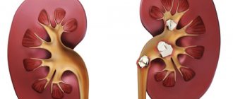
The effectiveness of removing kidney stones will depend not only on their size, but also on their properties.
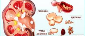
Kidney stones can be made up of different substances. Thus, it was found that those stones that consist of uric acid and calcium oxalate are best crushed, which cannot be said about stones that contain calcium oxalate monohydrate and cystine.
Before performing the operation, the following indicators must be taken into account:
- what are the stones made of?
- their size, as well as density and volume;
- the condition of the kidneys themselves;
- presence of kidney problems;
- presence of infection;
- whether passage through the urinary tract is impaired.
Like every medical procedure, lithotripsy has both indications and contraindications.
Indications for extracorporeal lithotripsy are:
- the presence of stones whose size does not reach 2.5 cm;
- The chemical composition of the stone makes it possible to break it with a laser.
When the procedure is prohibited:
- the presence of inflammatory processes in the body;
- the presence of diseases such as, for example, prostatitis and pneumonia;
- blood clotting disorder;
- pregnancy period;
- bleeding and menstruation;
- obesity stage 3-4;
- tuberculosis;
- oncology;
- the occurrence of complications after previous lipotripsy;
- increased gas formation;
- scoliosis and other problems associated with the spine;
- pathologies in the functioning of the cardiovascular system.
There are many contraindications to lithotripsy. Only a doctor, after a thorough examination, during which no contraindications are identified, will have to prescribe it.
Contraindications
The method is characterized by fewer complications when compared with surgery or minimally invasive treatment. The recovery period is also due to the short period of time. Patients return to normal activities much faster. A number of contraindications:
- inability to accurately target (diseases of the musculoskeletal system, obesity);
- decreased blood clotting;
- gastrointestinal diseases, as there is a risk of intestinal bleeding;
- renal tumor;
- cavernous tuberculosis of the kidney;
- chronic renal failure.
Insufficiency of the heart and lungs is a limitation for this procedure. The presence of inflammatory processes of different localization requires preliminary treatment. A course of antibiotics, vitamins and infusion therapy are prescribed.
Methods of carrying out
There are several options for carrying out the procedure, the differences between them lie in the generation of shock waves, which are:
- electromagnetic;
- piezoelectric;
- electrohydraulic.
The goal of each of the listed exposure options is to destroy kidney stones to such a size that they can then be eliminated from the body on their own.
The doctor performing the procedure will need to use x-rays or ultrasound to target the kidney stones and break them up. Depending on the distance of the shock wave, there are several methods of carrying out the procedure: contact and remote. What are they?
The remote method of removing kidney stones began to be used in medicine about 30 years ago. Its main advantage is that it effectively removes kidney stones and does not cause injury.
A machine located outside directs shock waves to where the kidney stones are located. This method is very often used in medical practice. It is prescribed not only to adults, but also to children, which is also an advantage.
However, the remote method has its contraindications, these include:
- inability to direct the shock wave to the location of the stones;
- bleeding disorders;
- pathologies associated with the gastrointestinal tract, there is a risk of internal bleeding during the procedure.
Extracorporeal shock wave lithotripsy is another option for removing kidney stones. The procedure is carried out using a special device - a lithotripter. This device can collect a shock wave into a beam, and then, using guidance, redirects it directly to the place where the stones are located.
During the course, the integrity of the muscles and skin is not compromised. Another advantage is that lithotripsy is completely painless and therefore does not require anesthesia. Its duration is about 30 minutes.
Indications for use
Extracorporeal shock wave lithotripsy is not recommended for all categories of patients (maximum 15-20% of the total flow). Certain conditions must be met:
- Inadmissibility of inflammatory processes inside the affected organ.
- Normal patency of the ducts. If they are impassable, exit will be impossible.
- Efficient bile organ. If its contractions are weakened, emptying the gallbladder is difficult, and the fragments will not be able to leave it.
Wave crushing is recommended only for those patients who have formed only one stone no larger than 3 cm, or several stones no larger than 1.5 cm.
The extracorporeal crushing procedure is performed in a small number of medical institutions. Most experts consider this method ineffective and life-threatening for the patient. But, in Germany, for several decades now, doctors have been doing and trying to improve this operation to remove gallstones, make it more effective and safe, and reduce the cost. The exact price of a non-contact operation can only be announced after a personal conversation with the doctor, a comprehensive examination, the size and number of stones.
Lithotripsy and laser for gallstone disease.
Even 20-30 years ago, the only alternative for treating gallstone disease was surgery to remove the gallbladder, cholecystectomy. Crushing gallstones is a modern, minimally invasive method that allows you to remove stones from the body without surgery. This article presents the techniques that are most in demand at present.
The crushing of gallstones using various physical methods is called lithotripsy. This treatment method is becoming increasingly popular because it does not require long-term anesthesia and is suitable for people with many chronic diseases, in particular the elderly.
Minimally invasive methods of treating calculous cholecystitis can be divided into two groups: contact and remote.
- Contact methods are performed during endoscopic retrograde cholangiopancreatography (ERCP) or fistulography. Thus, the diagnostic method simultaneously becomes a therapeutic one.
- Remote methods are based on the physical properties of ultrasonic waves to penetrate deep into the body and destroy stones at the required depth.
The indications for this treatment method are expanding every year. In any case, the decision whether lithodestruction can be used in a particular patient is made by the attending physician, who knows all the features of the disease. There are the following general indications for this manipulation:
- Single stone with a diameter of more than 1 cm;
- Multiple stones occupying less than half the volume of the bladder;
- Combination of cholecystitis with cholangitis;
- Preserved contractile function of the bladder and large duodenal papilla.
How might the patient feel?
Often after the procedure, the patient may experience some symptoms. There is no need to worry about this, since such a manifestation is absolutely normal.
Symptoms that a patient may feel after lithotripsy:
- abdominal pain;
- the presence of blood in the urine;
- urinary retention;
- nausea and vomiting;
- increased blood pressure;
- pain when urinating;
- constipation;
- increased body temperature;
- bloating.
Symptoms may continue until the stones are passed from the body. The duration of this period should not exceed a month.
The following factors will play a role in the duration of such symptoms:
- properties and size of the pebble;
- patient activity;
- kidney condition;
- amount of fluid consumed.
If a patient develops renal colic, then in this case he needs to undergo an emergency procedure.
If urine output is impaired, the patient should be prescribed an anesthetic.
There is another way to remove stones - contact laser lithotripsy of the kidneys. Recently, this procedure has become more widespread and the necessary equipment for its implementation is available in many city hospitals. Its main difference is that during the procedure the device is introduced into the human body.
Remote stone crushing
Extracorporeal shock wave lithotripsy (ESWL) has become a breakthrough in the treatment of gallstone disease. Over the past 20 years, the technique has been improved, and the number of complications has been reduced to a minimum. Unlike crushing gallstones with a laser, the removal of crushed stones in this way occurs naturally through the intestines. But the dangerous effects of X-rays remain only at the diagnostic stage.
The patient does not require anesthesia during the procedure, and the manipulation itself lasts no more than an hour. A course of extracorporeal lithotripsy usually consists of 1-7 procedures, depending on the size and number of stones. After undergoing ESWL, the risk of developing obstructive jaundice and re-formation of stones is significantly reduced.
Description of the method
Currently, it is possible to use three different variations of ultrasonic stone crushing:
- Underwater spark discharge. During the procedure, the patient lies down in a bath filled with water. Lithotripters are placed next to it, each of which transmits ultrasonic waves of a certain frequency through a liquid medium.
- Generating waves using an electromagnet. Vibrations of electromagnetic waves are transmitted to a special membrane, which creates ultrasound. A focusing screen is installed along its path, with the help of which all the energy is directed to the stone.
- Ultrasonic crushing using piezocrystal. The operating principle is similar to sensors installed on ultrasound machines. Vibrations of the piezocrystal under the influence of electric current produce ultrasound, which is directed to the stone through a system of focusing lenses.
Advantages
The advantages of the method are:
- Remote methods crush gallstones without anesthesia;
- The patient experiences a minimum of discomfort;
- Quite large stones with a diameter of more than 3 cm can be crushed.
After completing a course of extracorporeal shock wave lithotripsy, a positive result is achieved in almost 95% of patients. However, the method does not remove the cause of stone formation. Therefore, there is no guarantee that stones will not form again. Within 7-10 years, relapse occurs in those patients who did not take ursodeoxycholic acid to dissolve gallstones.
Flaws
Like any method of treatment, distance therapy is not without its disadvantages:
- Good results will only occur if there are cholesterol or pigment stones;
- Efficiency sharply decreases in the presence of calcifications (X-ray positive stones);
- The danger of obstruction of the bile ducts by stone fragments with the development of obstructive jaundice.
Ultrasonic crushing is prohibited if the patient has signs of purulent cholangitis, purulent cholecystitis and adhesions of the bladder to surrounding tissues.
Performing shock wave lithotripsy
Efficiency of the procedure
Many people are interested in how effective extracorporeal lithotripsy is, reviews of which can be read on the Internet.
Some who have already gone through this procedure claim that the method did not help them. This can be explained by the fact that in each specific case, kidney stones break down differently and are also excreted through the urinary tract differently.
In some cases, it is not possible to crush the stones the first time, so in this case a repeat procedure will be required.
Remote lithotripsy, the price of which in some places is about 8-10 thousand rubles, is high for some. The cost of the procedure consists of a number of different factors, which include the method of its implementation, the size of the stones, etc.
But unlike other methods of removing kidney stones, extracorporeal lithotripsy causes virtually no complications, it is practically painless and its cost is much lower than other procedures. Another advantage is that the body recovers quite quickly after lithotripsy.
External lithotripsy does not guarantee that kidney stones will not recur. Therefore, after the procedure, a person must carefully monitor his health, eat right, and lead an active lifestyle.
mkb.guru
Treatment of the gallbladder with lithotripsy
This is a collective concept that implies methods for removing stones. Currently, there are two main methods - remote procedure and contact. The first includes shock wave and ultrasonic techniques.
Contact crushing methods include:
- chemical;
- mechanical;
- laser lithotripsy.
Contact interventions are carried out using chemicals or lithotripters (devices designed for crushing), as well as precisely directed laser beams. The procedure requires general anesthesia or local anesthesia. Most often, the latter is used for elderly patients.
External lithotripsy of formed gallstones is the most common technique. This is largely due to the lack of intervention in the body and the need to stay in a hospital. Unlike contact methods, manipulation is possible only with small formations.
general information
The essence of remote lithotripsy is that stones are crushed without direct contact with them. In this case, the stones can be located in the bladder, kidney or ureter. A special apparatus is used, with the help of which a large wave is produced, under the influence of which the stones are crushed to a sandy state. After this procedure, the sand can come out along with the urine on its own.
The device creates impacts of a certain depth and strength. The waves created by a special device recede in a cone and are directed to one point. Thanks to the maximum amount of energy, the stone is crushed.
Procedure process
Preparation
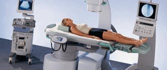
In the case of extracorporeal lithotripsy, the patient does not need complex special preparation. An important step before this procedure is an X-ray examination of the kidneys, ureter and bladder or ultrasound. This helps determine the focal point to which the waves of the device will need to be directed. In addition, doctors must ensure that the patient's intestines are completely cleansed. This can be done with a special drug or an enema.
The process of performing extracorporeal lithotripsy
This procedure consists of several stages:
- The patient is given general anesthesia or intravenous anesthesia.
- Depending on where the stones are located, a special crushing device is applied to the side of the abdomen or lumbar region. The duration of extracorporeal lithotripsy ranges from 40 minutes to 2 hours and depends on the patient’s condition, the size of the stone, its structure and other factors.
- The device creates shock waves that can reach up to 5 thousand waves in one session. If this amount of wave radiation is not enough, then the procedure may need to be repeated.
- The doctor regulates the intensity of the waves and gradually reduces the time between shocks so that the patient can adapt to what is happening.
Rehabilitation period
After the extracorporeal lithotripsy procedure, the patient needs regular examination. If he has pain in the lumbar region and renal colic occurs, this indicates the need for repeated surgery.
The patient may experience the following unpleasant symptoms for several days after the procedure:
- pain at the site where the device is applied;
- nausea with vomiting;
- pain during urination, which occurs due to the fact that sand comes out through the urinary tract;
- There are blood impurities in the urine.
During the postoperative period, the patient must consume a sufficient amount of fluid so that parts of the crushed stone and sand can freely exit the body.
Two weeks after extracorporeal lithotripsy, the patient undergoes a control examination of the organs using ultrasound or x-ray.
Lithotripsy of kidney stones
External lithotripsy of kidney stones is a method of crushing kidney stones without damaging the skin. This procedure allows them to exit the urinary tract to the outside. Let's consider the features of this procedure, its indications, contraindications.
What is lithotripsy?
External lithotripsy (ESLT) is one of the non-surgical methods of treating urolithiasis in humans. The essence of this crushing of kidney stones is that they are crushed without direct contact.
The required effect is achieved by using a shock wave. In turn, contact lithotripsy involves direct crushing of stones using a laser beam, compressed air, and ultrasound.
A special shock wave generator is used for grinding or crushing. It creates shock waves that repeat at a certain frequency.
During DLT, waves are collected in one place, which makes it possible to crush stones into sand. Then it comes out on its own with the blood flow.
To determine the point where the shock waves should converge (focus), the ultrasound diagnostic method is used. This makes it possible to significantly increase the efficiency of ESWL.
This operation is a closed type intervention, with the exception of crushing the so-called coral stones. The patient must be observed by a specialist and, if necessary, can stay in the hospital for several days.
Types of lithotripsy
In modern conditions, the following types of lithotripsy are performed:
- Extracorporeal.
- Remote.
Extracorporeal lithotripsy - crushing kidney stones in this way is carried out using a high-energy shock wave. This causes the stone to become deformed.
To achieve the desired effect, crushing is carried out for about 1 hour. Extracorporeal lithotripsy is not done during pregnancy, if the patient's weight deviates, especially if there are stones in the pelvis.
In contact lithotripsy, the stone is destroyed using a laser beam or ultrasound. Using a urethroscope, fragments are removed from the genitourinary tract. After lithotripsy, the patient is placed a stent in the ureter area.
After a few days it is removed. This procedure is performed only with the use of anesthesia (most often general). The patient can get rid of the stone in just a few sessions.
Remote lithotripsy is carried out, as already indicated, using ultrasound, which is focused into the area of the kidney stone. Typically, crushing is used in cases where they are small in size (that is, do not exceed 25 mm in diameter).
It can sometimes be used as an emergency treatment option for renal colic. For stone sizes up to 5 mm, the lithotripsy procedure is usually not prescribed.
This method of fragmentation must be carried out under the condition that frequent and severe inflammatory phenomena develop in the urinary tract. Lithotripsy is safe and can be prescribed to children.
Features of the event
To perform ESWL, the patient is given anesthesia (most often intravenous). Sometimes the doctor may use general anesthesia (it all depends on the patient’s health, age and other factors).
Next, the device is applied to the abdomen (usually it is installed in the lumbar region, where a renal calculus is most likely to be located). The duration of the wave effect on stones can be different: it depends on the number and their size.
In one wave session, the number of blows reaches 5000. During the procedure, the doctor changes the intensity of the wave radiation, which makes it possible to effectively control the process of stone removal.
During this operation, an endoscope is inserted through the urethral canal, bladder and ureteral cavity directly to the stone. After this, a laser beam is directed towards it.
This crushing method allows the stone to be crushed into dust. This means that it disappears in the kidneys without a trace.
After laser lithotripsy, there are no fragments left in the kidneys and urinary tract. Then such dust is completely removed from the kidney and urinary tract naturally.
The advantages of such crushing are as follows:
- laser removal is most suitable if there are stones of a complex chemical nature;
- in just 1 procedure you can achieve complete expulsion of a kidney stone;
- the patient does not feel pain during destruction;
- in the specified localization, it is possible to completely achieve the expulsion of calculi;
- laser removal is almost completely suitable for small stones;
- Due to the fact that the holmium laser is used during the procedure, it does not affect the surrounding tissue.
In most cases, this procedure is effective. Often, you can say that it was successful immediately after the end of the procedure (when radiography shows that the impact method of removal reduces the shadow). However, it may take some time to assess the condition of the body after such a procedure.
In some cases, the doctor prescribes repeat lithotripsy to his patient. If several sessions are ineffective or ineffective, then percutaneous lithotripsy is used.
How to prepare?
As with any procedure, you need to prepare for the destruction of a kidney stone. An initial consultation is recommended prior to this.
During the initial consultation, the doctor collects and carefully examines the medical history. Before fragmentation, it is necessary to take an x-ray, and in some cases an MRI.
Before crushing it is necessary to pass the following tests:
- general clinical urine test (it is necessary to determine the platelet count);
- general examination of urine, necessarily with bacterial culture;
- biochemical blood test (performed according to a standard procedure);
- blood coagulogram;
- Chest fluorography should be done no more than a year before removing stones in this way;
- All patients who have reached the age of forty should have an electrocardiogram.
A week before the wave destruction, you need to stop using all drugs containing acetylsalicylic acid.
Are there complications after lithotripsy?
Despite the fact that lithotripsy has a high treatment effect, it, like any other procedure, can lead to complications. First of all, during the intensive passage of stones, acute pyelonephritis, blockage of the upper urinary tract and even renal colic can develop.
All patients need to be aware of the possibility of developing such dangerous complications in order to respond in a timely manner to their occurrence. If renal colic occurs, you should immediately call an ambulance.
Blockage of the ureter is a rather dangerous condition. It is otherwise called a stone path.” Manifests itself in the form of lower back pain similar to renal colic.
In some cases, urgent removal of the stone using a urotroscope is necessary. A consequence of pyelonephritis is urosepsis with hemorrhage into the renal tissue.
Other complications of the removal procedure:
- hematuria (usually should go away within a few days);
- severe spasmodic pain (relieved by analgesics);
- internal bleeding (refers to emergency conditions);
- urethral damage.
If you experience very severe pain, blood in the urine, or a strong urge to urinate, even if your bladder has recently been emptied, you should immediately call an ambulance.
When is lithotripsy not necessary?
This procedure is not carried out in the presence of the following diseases and phenomena:
- prostatitis;
- pneumonia;
- menses;
- complications after lithotripsy;
- stones of urate origin;
- poor blood clotting;
- aneurysms;
- obesity, increased formation of gases;
- spinal deformities;
- tuberculous process of renal cavities;
- various exacerbations and disorders in the gastrointestinal tract;
- the presence of an artificial pacemaker in the heart, atrial fibrillation;
- heart and lung failure;
- renal dysfunction;
- sharp narrowing of the ureter (if it is narrowed below the location of the stone, in such cases there is a risk of developing blockage of the urinary tract and other unfavorable complications);
- pregnancy (regardless of duration).
So, stone lithotripsy is an effective and safe procedure to get rid of stones. This is one of the most effective ways to treat urolithiasis and its complications.
Patients note an improvement in their condition after lithotripsy. Complications after this method of crushing are rare. If you still feel unpleasant symptoms after removal, you should urgently call a doctor to provide emergency care.
Indications
External lithotripsy has a number of the following indications:
- the presence of renal colic, which is caused by the presence of stones;
- frequent inflammatory reactions to the presence of stones in the urinary tract;
- the presence of stones in the ureter, bladder or kidneys that reach more than 0.5 cm.
Contraindications
It is strictly prohibited to perform extracorporeal lithotripsy in the following patient conditions:
- frequent hemorrhages or bleeding, which indicate a blood clotting disorder;
- pregnancy;
- the presence of an inflammatory process in the urinary system or infection in these organs (cystitis, pyelonephritis, urethritis);
- narrowing of the ureter in the lower area of the stone;
- taking medications that thin the blood (these medications must be stopped at least 7 days before the procedure).
How to prepare for lithotripsy
It is important to tell your doctor about all the medications you are currently prescribed and any treatment you are undergoing. Certain drugs, such as aspirin, ibuprofen, warfarin, and their blood thinners, can negatively affect blood clotting during lithotripsy. Because of this, your doctor will likely ask you to stop taking these medications for a certain period of time before surgery. But under no circumstances stop taking any medications unless prescribed by your doctor.
Some patients receive local anesthesia for pain relief during lithotripsy. However, most people undergo surgery under general anesthesia. In this case, you will be advised to refrain from eating and drinking 6 hours before the procedure.
Also ask one of your relatives or friends to take you home after the operation under general anesthesia, because This method of anesthesia has a fairly long duration and may make you sleepy for several hours after leaving the hospital, so you should not risk driving yourself.
External shock wave lithotripsy removes kidney stones
External shock wave lithotripsy is currently one of the most promising methods for treating urolithiasis. The essence of extracorporeal lithotripsy is the non-invasive crushing of stones in the urinary tract, that is, crushing occurs without direct contact with the stone. A calculus that has undergone lithotripsy is subsequently passed out along with the urine without any problems, without causing pain to the patient and preventing the occurrence of complications, the development of which is possible when using other methods of treating urolithiasis.
External shock wave lithotripsy:
— main types of shock wave lithotripters;
— indications for shock wave lithotripsy;
— contraindications to shock wave lithotripsy;
— technique of shock wave lithotripsy.
Contact crushing of stones
Before the actual procedure of contact lithotripsy, the patient undergoes ERCP. This is an endoscopic operation in which the surgeon inserts a probe into the duodenum through the esophagus and stomach. The endoscope is brought to the large duodenal papilla. A papilotomy is performed with a special scalpel, that is, the sphincter that closes the ampulla of the nipple of Vater is cut.
Then the doctor inserts a special tube into the formed mouth, through which a contrast agent will be supplied. The bile ducts and the location of the stones are visualized on the screen. After this, the probe is passed through the common bile duct directly to the stone. The entire operation is performed in fluoroscopy mode.
Contact crushing methods are divided into two types:
- Mechanical biliary lithotripsy;
- Laser contact lithotripsy.
Mechanical biliary lithotripsy
This crushing method is one of the first. It is carried out using a special reinforced Dormia basket (lithotripter), which captures the calculus and crushes it. After this, stones are removed from the gallbladder, followed by washing it. At the end of the manipulation, cholangiography is performed again. If large fragments of stones are seen on the image, the procedure begins again.
Advantages of the method:
- Manipulation is possible with the development of obstructive jaundice;
- You can crush any gallstones;
- Relatively cheap.
Despite the simplicity and accessibility of mechanical lithotripsy, its implementation is associated with certain difficulties. The disadvantages of this method of treatment are:
- Technically difficult to carry out;
- High risk of perforation of the gallbladder wall and ducts;
- Possibility of bleeding;
- There are often wedgings of the remains of crushed stones into the inflamed mucous membrane of the biliary tract;
- The lithotripter may break, which will require open surgery.
Laser contact lithotripsy
Crushing gallstones with a laser is as follows. During ERCP, a light guide with a laser radiation source is connected to the stone. Upon contact with the stone, the energy supply is turned on and it breaks into several parts. The fragments are removed using special forceps, and the bile ducts are washed with saline solution.
Benefits of laser removal of gallstones:
- The method is suitable for the destruction of any large stones of the extrahepatic bile ducts;
- Used for the development of obstructive jaundice;
- Can be used without general anesthesia by percutaneous transhepatic puncture.
Removing gallstones with a laser has its disadvantages:
- High risk of perforation of the walls of the biliary tract;
- Possibility of bleeding;
- The most common complication is a burn of the mucous membrane.
Performing laser contact lithotripsy
Main types of shock wave lithotripters
Today, more than 40 types of lithotripters are used in the world, the most widespread of which are three main methods of generating shock waves:
- electrohydraulic lithotriptors generate shock waves as a result of a high-voltage discharge in water between pairs of electrodes located on the discharger. Shock wave guidance is performed under X-ray or ultrasound guidance. Such lithotripters make it possible to obtain a powerful shock wave of over 1300 bar;
- electromagnetic lithotriptors make it possible to generate a shock wave in a special shaper and focus it on the calculus using a system of acoustic lenses. The targeting of the wave to the stone is also controlled by X-ray or ultrasonic devices;
- Piezoceramic lithotriptors are the newest to date, have a complex structural mechanism and allow absolutely painless crushing of stones in the patient’s body. At the same time, piezoceramic lithotriptors are not effective against very large and strong stones.
«>
Indications for shock wave lithotripsy
External shock wave lithotripsy is possible if a patient with urolithiasis has indications for this procedure. The following are the main indications for shock wave lithotripsy:
- single kidney stones up to 25 mm in size, provided there are no disturbances in the outflow of urine below the stone;
- multiple kidney stones with a total size of up to 30 mm;
- one or two stones in the ureter up to 10 mm in size. provided there is no pronounced dilatation of the upper urinary tract and an active form of chronic pyelonephritis;
- coral kidney stones with a total size of up to 40 mm.
How is treatment performed using extracorporeal lithotripsy at the Central Clinical Hospital of the Russian Academy of Sciences?
- Painless. During the procedure, anesthesia is performed - local or intravenous. The choice of anesthesia is discussed in consultation with a doctor.
- Under visual control. Using ultrasound guidance, the localization of the stone in the kidney or urinary tract is determined. Throughout the entire process, the doctor monitors the kidney stone using a machine monitor.
- Safely. The device creates shock waves that gradually destroy the stone. The procedure is stopped when the stone reaches an acceptable size to pass through the ureter and urethra. During the procedure, anesthesia is performed - local or intravenous. The choice of anesthesia is discussed in consultation with a doctor.
Contraindications to shock wave lithotripsy
There are also contraindications to performing extracorporeal shock wave lithotripsy, the main of which are:
- the inability to visualize and accurately guide the stone under shock waves;
- obstruction of the urinary tract below the level of stone localization;
- severe spinal deformity;
- disorders of the blood coagulation system;
- pregnancy;
- gross disturbances of cardiovascular activity;
- splenomegaly;
- oncological pathology;
- acute inflammatory processes in the body;
- active tuberculosis;
- sepsis;
- acute obstructive pyelonephritis;
- disturbances in the secretory function of the kidney as a result of its shrinkage.
Benefits and effectiveness
The main advantages of the procedure are:
- harmlessness;
- the ability to avoid surgical intervention;
- short rehabilitation period;
- relatively low cost;
- Rare occurrence of negative consequences.
The effectiveness of lithotripsy depends on a number of factors. Sufficient experience of the specialist and excellent equipment of the clinic come first. It is very important that the doctor correctly assess the patient’s condition and prescribe the appropriate method for the procedure.
Its results also depend on the volume and chemical structure of the stone, as well as on the condition of the patient’s ureters and biliary tract.
Equally important is the localization of the formation and the ability of a specialist to penetrate exactly into the affected area without affecting neighboring ones.
Shock wave lithotripsy technique
Preparing the patient for shock wave lithotripsy involves cleansing and reducing intestinal pneumatization, which allows for the best possible visualization of stones. During the procedure, the patient must be on his back or stomach. Crushing begins with low-energy modes - 500-600 pulses; if ineffective, the number of pulses is increased. Crushing of stones of the renal pelvis and ureters is performed on a pre-installed stent. As a result of the impact of the shock wave on the calculus, it splits into small fragments, which subsequently, along with the urine, painlessly exit the patient’s body. To create favorable conditions for the passage of stone fragments, the patient is recommended to take antispasmodic drugs during the first 7-10 days after the procedure.
estet-portal.com
Crushing stones in organs without breaking the skin is called lithotripsy . It is carried out when stones cannot leave the body on their own. This method has been used in medicine for decades. Its meaning is that a shock wave aimed at a stone destroys it with the help of short pulses, this happens gradually and lasts until only dust remains from the stone. The Germans were the first to use this procedure; they quickly realized that this method was one of the safest. There are two types: contact and extracorporeal lithotripsy of ureteral kidney stones. As with any intervention in the body, it has both pros and cons.
The method of external shock wave lithotripsy (ESWL) was discovered and put into practice in the 80s of the twentieth century. This method has actually revolutionized the treatment of urolithiasis, as it is non-invasive, low-traumatic and highly effective.
The principle of this method of treating urolithiasis is to use shock waves to break up the stones. This allows the stone to be broken into smaller fragments, which can more easily pass through the ureter or dissolve.
Currently, there are various devices for lithotripsy with different sources of shock wave generation (electrohydraulic, electromagnetic and piezoceramic).
The lithotripsy method is characterized by a lower number of complications compared to other surgical and minimally invasive methods of treating urolithiasis. In addition, ESWL is characterized by a shorter recovery period, as well as a half-shorter period of pain. Patients after ESWL are discharged faster and return to work.
It should be understood that lithotripsy does not cure urolithiasis, but only destroys existing stones. That is, the patient in this case is not insured against the fact that he will develop stones again. Therefore, after a course of lithotripsy, he needs to constantly take measures to prevent stone formation.
Indications for lithotripsy
Among the main parameters that were previously taken into account as indications for lithotripsy is the size of the stone. In the early 80s, the main indication for lithotripsy was stones up to 1.5 cm in size. However, modern ESWL devices make it possible to crush stones ranging in size from 0.5 to 2.5 cm. Recently, it is believed that the effectiveness of stone crushing is influenced by not so much the size of the stone, but its physical and chemical properties. We also note that the lithotripsy method is effective in both adults and children.
Contraindications to lithotripsy
Contraindications to lithotripsy include the inability to accurately position the stone in the shock wave zone (urate stones, deformation of the spine, musculoskeletal system, obesity, etc.). Absolute contraindications to lithotripsy include bleeding disorders, as well as conditions in which the patient is taking anticoagulants, as well as menstruation.
Another contraindication to ESWL is pathology of the gastrointestinal tract during its exacerbation, since a shock wave hitting the area of the inflamed intestinal wall can lead not only to aggravation of the disease, but even to intestinal bleeding and hemorrhages into the intestinal wall.
Lithotripsy is also contraindicated for any purulent and inflammatory processes (pneumonia, prostatitis, etc.). Urological contraindications to lithotripsy include kidney tumor and cavernous tuberculosis of the kidney.
In addition, lithotripsy is contraindicated in cases of cardiac dysfunction (atrial fibrillation, artificial pacemaker, presence of cardiopulmonary failure), a decrease in kidney function by more than 50%, or pregnancy.
Preparation for lithotripsy
Since inflammatory processes are a contraindication to ESWL, before this procedure a complex of antibiotic therapy, infusion therapy, drugs that improve microcirculation and vitamins is usually carried out. This allows you to prepare the kidney for shock waves and reduce the likelihood of complications, as well as shorten the recovery time.
Anesthesia for lithotripsy
Many modern lithotripsy machines allow this procedure to be performed without anesthesia. Despite this, most patients experience moderate pain. As a rule, it is associated with the passage of shock waves through the skin and irritation of nerve endings.
How does the procedure work?
The lithotripsy procedure usually lasts an average of 1 hour, during which the patient can receive up to 8,000 shock waves. Typically, a lithotripsy procedure begins with low-energy shock waves and long intervals between pulses. This allows the patient to get used to the procedure. Gradually, the strength and frequency of the shock waves gradually increases. If there is bone (such as a rib) in the path of the shock wave near the stone, the procedure becomes more painful as the wave causes some resonance in the bone.
In order to facilitate the removal of stone fragments, a ureteral stent can be used, which expands the lumen of the ureter.
Complications with lithotripsy
Despite the fact that lithotripsy is a non-invasive method of destroying stones, it also has some complications, since this method is associated with a mechanical effect on the kidney tissue.
Complications may arise due to the characteristics of the patient himself, or due to incorrect technique for performing the lithotripsy procedure, or incorrect selection of indications for the procedure. Complications of lithotripsy include:
- Kidney hematomas are hemorrhages into the kidney tissue. The reasons may be that the patient has factors that contribute to its formation (for example, bleeding disorders, taking anticoagulants, menstruation, pyelonephritis).
- Hematuria is the presence of blood in the urine. Hematuria occurs in almost all (100%) patients after ESWL. However, with the correct technique for performing lithotripsy, it passes through 1–2 acts of urination.
- Obstruction of the urinary tract. This is one of the current problems of lithotripsy. The fact is that a large stone, when it is in one place, usually does not interfere with the outflow of urine, while its fragments already have greater mobility and there is always a risk of blockage (obstruction) of the ureter.
www.klinika29.ru
How stones are crushed
The essence of extracorporeal shock wave lithotripsy is to generate shock waves into the lumen of the gallbladder. Typically, repeated shocks are required that focus on the stones.
Extracorporeal shock wave lithotripsy is suitable for patients with a small number of cholesterol stones without calcification with a diameter of up to 30 mm. There should not be more than 4 stones.
The stones are destroyed into fragments no larger than 3 mm when exposed to a high-power shock wave, which occurs when a piezoelectric element is discharged. These fragments spontaneously exit with bile into the duodenum. 1-7 such “crushing” sessions are carried out
Complications of extracorporeal shock wave lithotripsy.
- blockage of the bile duct with the development of obstructive jaundice if the stones are too large;
- damage to the gallbladder wall with further perforation;
- inflammation of the pancreas.
Contraindications to crushing gallstones
- Heart disease (especially heart rhythm disturbances)
- Hypertension
- Acute cholecystitis
- Blood clotting diseases
- Calcified and/or bilirubin stones.
- Stones more than 3 cm in diameter
- A large number of stones
Removal of gallstones by percutaneous transhepatic cholelitholysis
Another method of crushing gallstones allows you to dissolve not only cholesterol, but also other types of stones. Moreover, their number does not matter.
Through a catheter inserted through the skin and liver into the gallbladder, special drugs are injected that will dissolve the stones over several weeks. The disadvantage of this method is that systematically inserting a catheter into the liver tissue can cause complications. Many authors believe that it is easier to perform an operation with guaranteed success.
Endoscopic removal of stones from the gallbladder.
When using this method to remove stones, the patient swallows the endoscopic tube. The operating instrument thus travels through the digestive tract (mouth, esophagus, stomach and duodenum) to the mouth, often blocked by stones, of the bile duct in the duodenum.
Why is bile duct endoscopy needed?
If severe pain occurs, for example, in the back and immediately after this jaundice appears, this may mean that a stone is stuck in the bile duct and it is interfering with the outflow of bile. At the same time, older people suffering from concomitant diseases can also be quickly helped without laparotomy. Using an endoscope (a flexible tube with a camera), the bile duct is reached through the stomach with the duodenum and the stone is removed.
These methods of accessing the gallbladder and bile duct can also be used during the dissolution or crushing of stones. The goal is always to ensure free flow of bile into the intestines. If this goal cannot be achieved or it is possible to achieve it only partially, this threatens with severe, sometimes fatal complications due to stagnation of bile with severe inflammatory complications and breakthroughs (perforations).
Any method that deals only with removing the stones and leaving the gallbladder behind encourages new stones to form since the site of gallstone formation, the gallbladder, is still present.
Not everyone is familiar with the concept of lithotripsy, but many people know about the possibility of foreign bodies forming in the bladder. Oddly enough, this concept, like the process of getting rid of the disease itself, has existed for more than 100 years.