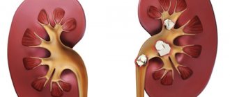
A breakthrough in urology, in operations to remove kidney stones, was a method that is least painful and at the same time quickly copes with this disease. The modern medical society suggests removing kidney stones using a laser. This technology expanded the horizons of urology, and made it possible not to resort to complex operations, after which the patient was left with unpleasant scars and the functioning of the internal organs themselves was disrupted. Laser crushing of kidney stones minimizes the risk associated with the development of complications of urolithiasis.
General information about urolithiasis
Before delving into the stages of lithotripsy, you should understand in what cases it should be performed. First of all, stone removal is prescribed for urolithiasis, or in other words, “urolithiasis.”
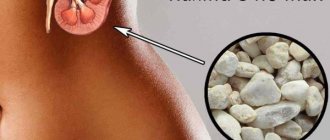
The main sign of the pathology is the formation of kidney stones, which provoke severe pain during movement and discomfort at rest.
Urologists around the world note that urolithiasis is still difficult to respond to conservative treatment methods, and often occurs in severe form with complications.
Often, patients with urolithiasis experience renal colic so severe that they cannot move and are forced to constantly take painkillers.
However, it is important to remember that medications only block pain, but do not solve the root cause of the problem or speed up the healing process.
Important. You should not ignore the symptoms of urolithiasis and delay visiting a doctor until the last minute. The sooner a patient seeks medical help, the sooner his condition will improve, and the risks of complications will be minimized.
Removal methods
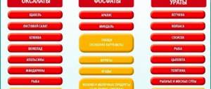
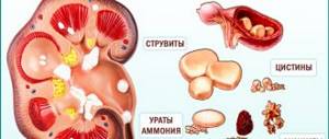
In the initial stages of stone formation, you can do without surgery. The gentle method of removing kidney stones involves drug therapy. This does not apply to oxalates, phosphates and urates, which are difficult to respond to drug therapy.
The possibilities of modern surgery are expanding every year, and today traumatic abdominal operations are being replaced by minimally invasive methods that allow minimal trauma to tissues and removal of stones. Non-traumatic stone removal operations have a significant disadvantage - high cost. Judging by patient reviews, the price varies from 20,000 to 30,000 rubles.
At the same time, there is a risk that stones will form again with any type of intervention. The patient will have to change his lifestyle and diet in many ways after the operation, so as not to fall under the surgeon’s knife again.
To give you an idea of the differences between methods for removing kidney stones, let’s take a closer look at each of them.
Open abdominal surgery
This method of removing kidney stones has been used for a long time, and sometimes it cannot be replaced with minimally invasive methods due to the significant size of the stones, purulent inflammation of the diseased kidney and frequent relapses.
During this operation, all layers of tissue are excised so that the doctor can easily access the kidneys. If the stones are located directly in the renal pelvis, then they are removed, the edges of the organ are sutured with self-absorbing threads, leaving a drainage tube there to remove possible purulent discharge. The fabrics are also sewn together layer by layer. On days 7-10, the tube is removed.
Complications after open surgery are common: suppuration, scars that do not heal for a long time, the appearance of postoperative hernias, and others.
When urolithiasis has reached its extreme stage, necrotic areas have appeared, and one of the kidneys is constantly being injured by huge stones, the doctor decides to perform a nephrectomy, removing the affected kidney or part of it.
Important! Such an operation is possible only if the second kidney is healthy and functioning normally, and also if the age and condition of the patient allow such a radical intervention.
Laparoscopy
This method of stone removal is most in demand today by both doctors and patients. It allows you to remove stones with minimal blood loss and the risk of complications. Through microincisions (no more than 10 cm), a nephroscope and a camera are inserted into the kidney cavity, through which the surgeon will monitor the progress of the operation. Large formations are crushed with a special tool and removed in small parts. This intervention is quite reliable and does not require a repeat procedure. Cosmetic defects are almost invisible afterwards, and the risk of bleeding is minimized.
Lithotripsy
This method of endoscopic intervention came to Russia in the 90s of the last century. Removal of a kidney stone during such an operation occurs without a single skin incision, which means that the patient is not at risk of infection, suppuration, or prolonged healing of the sutures. The doctor, carefully monitoring the location of the formations using an ultrasound machine, directs an ultrasound shock wave at them through the skin. The stones cannot withstand its pressure and begin to crumble into pieces. Their fragments are then excreted along with urine completely painlessly. This applies to small stones, up to 1 cm in diameter. For larger ones, a microtubule is inserted into the renal pelvis to extract particles of the formations.
This method also has some contraindications:
- pregnancy;
- impaired blood clotting;
- pathologies of the musculoskeletal system, in which the patient cannot position himself correctly on the couch;
- heavy weight (over 120 kg), height above 2 meters or below 1 meter.
In general, reviews from doctors and patients about lithotripsy are not bad, but the latter note that they experienced pain during the intervention due to local anesthesia.
Laser lithotripsy: general characteristics
Contact laser lithotripsy is an effective modern method of treating urolithiasis, in which stones are removed from the kidneys and other organs of the urinary system (see illustration below).
The big advantage of manipulation is the way of removing salts. As can be seen in the schematic image, the removal of crushed stones is carried out naturally - through the urethra.
In addition, since a pneumatic lithotriptor is not used during lithotripsy, the mucous membranes and tissues of the urinary organs are not injured, so the likelihood of infection is leveled.
According to numerous clinical studies, laser stone removal is an absolutely safe technique for patients of all ages.
Moreover, this procedure can be performed even during pregnancy, since a laser with a power of 80 W is used to destroy mineral formations.
Research directions
For the last decade, the clinic has been working on the following main scientific areas:
- the problem of urolithiasis;
- problems of uro-oncology;
- surgical and conservative treatment of benign prostatic hyperplasia;
- treatment of strictures and obliterations of the urethra in men;
- anomalies of the genitourinary system in children and adults;
- minimally invasive methods for diagnosing and treating urological complications after kidney transplantation;
- new technologies in the treatment of urological diseases.
Currently, the clinic has 60 beds, including 10 beds for children.
Indications for laser lithotripsy
Indications for the procedure may include the following points:
- Unsuccessful conservative treatment, which did not improve the patient’s condition over 2 months of therapy.
- The diameter of stones in the kidneys or other organs of the urinary system exceeds 5-6 millimeters and the likelihood of their independent removal through natural channels is almost impossible.
- Due to its specific location in the organs of the urinary system, kidney function, which is necessary for human life, is impaired.
- Stones were found not only in the kidneys, but also in the bladder or ureter.
- The patient has concomitant urological inflammation leading to urosepsis.
Indications and contraindications
Indications and contraindications for surgery to remove kidney stones are taken into account in each individual situation. Removing kidney stones is important if:
- the diameter of the stones is greater than the diameter of the ureters and urethra;
- stones do not come out on their own;
- drug therapy did not bring results;
- ureteral obstruction was detected;
- there is renal failure;
- carbuncle detected;
- there is purulent inflammation;
- the normal flow of urine is disrupted;
- there is severe pain.
Surgery is contraindicated if:
- pregnancy; excess weight (over 120 kilograms);
- blood clotting disorders;
- obstruction;
- atrial fibrillation;
- cardiovascular diseases.
Description of laser lithotripsy
If the patient has passed the necessary examinations and his diagnosis has been confirmed, if necessary, the doctor will prescribe laser stone crushing.
At the first stage, the specialist will administer an anesthetic drug, after which he will wait a few minutes until the anesthesia takes effect.
Then, through the urethral opening (see illustration above), he will insert a very thin, plastic endoscope, similar to a regular cord, with a laser attached to the end.
The probe with the laser will move to the desired location in the kidney or other organs of the urinary system along the urethra and urinary tract.
Due to the fact that the doctor uses an endoscope - a camera, he can observe the entire process of movement of the probe and correct its trajectory during manipulation.
After the probe is positioned in the right place - near the stone, the doctor will turn on a special device and the laser will begin crushing. When the formation is broken into small particles, a special solution will be injected into the kidney to flush out the sediment.
Most often, at the end of the procedure, the patient can independently relieve himself of minor need, during which all remaining salts will be eliminated naturally.
In the following days, from one to two weeks, there may be a frequent urge to urinate with blood. This process is considered normal, since due to the crushing and removal of stone fragments, the tissues of the urinary organs were injured.
If a person experiences severe pain during the period after lithotripsy, the attending physician may prescribe painkillers.
In addition, during the recovery period the patient must undergo drug therapy, which consists of:
- Plant-based preparations that dissolve mineral deposits, salts and remains of calculi in the organs of the urinary system - “Cyston” or “Urocholum”.
- Antispasmodics (painkillers) – “No-shpa”, “Drotraverine”, “Spasmomen”, etc.
- Antibacterial drugs (antibiotics) - Ceftriaxone or its third generation alternatives.
- Non-steroidal (non-hormonal) anti-inflammatory drugs - Ibuprofen or its alternatives.
As is known, according to clinical practice around the world, laser stone removal is effective in more than 95% of cases.
Possible complications
Any surgery has a potential risk of complications. With the development of medicine and the improvement of instruments, these risks are now minimized. However, you should be aware of possible problems before deciding to choose a surgical treatment method.
- Bleeding
. Since the kidney is very well supplied with blood, some blood loss is present with any operation, but it rarely requires a blood transfusion or any active intervention. Before surgery, your consent is required for a blood transfusion in the event of an emergency. - Infectious complications
. Despite the fact that before the operation and for some time after you will receive antibiotics to prevent infectious complications, in some cases you may experience fever, pain in the kidney area, and frequent urination. Treatment usually requires a change in antibiotic or removal of the drain. - Serious damage to the kidney or adjacent organs
is a very rare complication. Damage to the intestines, liver, and spleen is possible during puncture of the renal cavity system. It is also possible for scars to form around the kidney and ureter, which may impair their function and require additional surgical treatment. - Transition to open surgery
. If difficulties or serious complications arise during endoscopic surgery, to eliminate them, it may be necessary to switch to open surgery, in which a 15-20 cm incision is made in the lumbar region. - Incomplete stone removal
.
The goal of treatment is to remove all
stone fragments. But in some cases, due to the structural features of the abdominal system or other reasons, some fragments may remain in the kidney. To remove them the next day or a few days later, you can resort to a repeat procedure (pyeloscopy) through an existing passage into the abdominal cavity system of the kidney. Pyeloscopy is low-traumatic, is performed using a flexible instrument and requires only slight anesthesia. Another possible option is to perform extracorporeal lithotripsy after some time.
Recently, technological progress has led to the miniaturization of instrumentation, allowing this technique to be performed using a smaller access (<18F), thereby reducing trauma to the renal tissue and blood loss to a minimum. Such operations are called mini-PCNL and ultra-mini-PCNL.
.
What is possible after the procedure
For some time after the operation, you will remain in the recovery room under the supervision of an anesthesiologist. After you fully awaken, you will be transferred to a room.
Pain in the surgical area
is a normal reaction of the body. To reduce pain, you will be prescribed painkillers, the frequency of administration or frequency of administration of which you can control depending on how you feel.
Vomiting and headache
possible on the first day after surgery and are associated with anesthesia. Treatment is medicinal.
Nephrostomy
. The presence of a drain in the lumbar region may cause you discomfort, but you need to carefully monitor the integrity and patency of the drain, especially when you are lying or sleeping in bed. Usually the nephrostomy is removed 2-3 days after surgery, but in some cases you may be discharged home if it is necessary to remove the drainage after a longer period of time.
Stent
. In some cases, you may have a ureteral stent placed. To remove it, you will need to return to the clinic in 1–2 weeks. Stent removal does not require hospitalization or general anesthesia.
A urethral catheter (bladder drainage)
is installed during surgery and removed within 24 hours.
After removal, a slight admixture of blood in the urine may be observed for several weeks. Diet
. Immediately after the operation, in agreement with the attending physician, you can drink and take liquid food; the next day there are no restrictions. Try to drink more.
Weakness
may occur for several weeks after surgery.
Physical activity
. The next day after surgery, there is no need to lie in bed or limit your activity in any way unless bed rest is prescribed by your doctor. After discharge, you can return to your normal amount of physical activity; the exception is heavy physical labor or active sports in the first two weeks.
You can shower the day after surgery.
After discharge
Pain
. It is possible that a dull pain in the lumbar region will persist, but gradually the intensity of the pain will decrease. If necessary, your doctor will prescribe you medications to relieve pain.
Taking antibacterial drugs
. During surgery, stone fragments will be sent for testing for sterility. In most cases, if the result is positive and the presence of microorganisms in the stone, long-term use of antibiotics may be required to achieve sterility of the urine. In the case of infectious stones (struvite), avoiding their re-formation is possible only by completely removing the fragments and achieving complete sterility of the urine.
Hygiene
. At home you can shower without restrictions. If you have a nephrostomy tube, remove any surrounding bandages before showering. Wash the skin around the tube or wound with warm water and soap and dry with a clean cloth. After a shower, apply the bandage yourself as shown in the picture. Try not to take a bath for two weeks after surgery until the surgical wound has completely healed.
Physical activity
. After discharge, avoid heavy physical labor and active sports for 1–2 weeks, then you can lead your normal lifestyle.
Stent removal
.
In some cases, a ureteral stent may be placed during surgery. Because the stent is inside the body, you may not feel it. The time after which the stent must be removed depends on the complexity of the procedure and individual characteristics. Usually this is 1–4 weeks. Your doctor will set a date for your next visit. Stent removal does not require hospitalization or general anesthesia. It is important to remember that prolonged use of a stent is dangerous due to the development of encrustation and obstruction, which can lead to kidney death.
Nephrostomy care
.
The purpose of installing a nephrostomy tube is to ensure the outflow of urine from the kidney, so ensure the integrity of the tube and prevent it from being kinked or stretched. The nephrostomy is fixed to the skin with a thread, so make sure that the thread is intact when dressing. The urine collection bag should always be below lumbar level. Monitor the flow of urine into the bag and its color (there may be a slight admixture of blood or isolated small clots) - if there is a sharp decrease in the amount of urine or a change in its color, notify the doctor. Try to shower every day or simply wash the skin around the nephrostomy with warm water and soap. Afterwards, dry the skin with a clean, dry cloth and apply a sterile sticker. Post Views: 824
Laser crushing methods
Patients are often interested in how many methods of crushing stones using a laser exist and which of them are the best and most painless.
However, doctors assure that lithotripsy is carried out exclusively according to one standard, and its effectiveness and efficiency does not depend on specific equipment or instruments.
The use of laser in the treatment of patients suffering from the disease.
Unlike the outdated method of stone removal, the use of laser has significantly increased the effectiveness of treatment. The main difference is considered to be its minimal pain, and this is perhaps one of the important criteria in evaluating the treatment of any disease. The process itself is the crushing of stones. A thin tube called an endoscope is passed to the area where the unwanted particle is located, the laser is turned on, and treatment begins. The laser is capable of crushing a stone of any size, thereby turning it into dust, which in the future will easily come out of the patient along with the liquid.
It is worth noting that before starting treatment, antibacterial and vitamin therapy is carried out and at the same time, special medications are taken that improve the patient’s microcirculation.
Advantages and disadvantages of stone removal manipulation
This type of therapy, such as laser lithotripsy, is one of the most modern and painless, and therefore has a number of undeniable advantages.
- After the medical intervention, the patient is left with no scars or other noticeable defects.
- Laser equipment allows you to destroy stones of any diameter and density, regardless of location.
- The laser on a thin and plastic probe practically does not injure the walls of the urinary organs.
- There is no possibility of stone fragments forming.\
- In order to get rid of tumors, it is enough to perform one lithotripsy procedure.
- High rate of efficiency and effectiveness.
- There is no pain during the procedure.
However, the main disadvantage of this method of contact removal of stones is the high cost, despite the fact that the price is fully justified by its effectiveness.
At the same time, there are some contraindications to laser fragmentation of tumors, which may include:
- Acute stage of prostatitis.
- Malignant formations (oncology).
- Kidney hematoma.
- Coral stones.
- Complex form of arrhythmia.
- Cysts in the genitourinary system.
- Incoagulability of blood (or poor clotting - hematocrit).
- Infectious or purulent processes in the body.
- The patient's serious condition and other complex conditions.
Important. Women in the last months of pregnancy should be especially careful, therefore it is necessary to listen to the recommendations of the attending physician, both during lithotripsy and during the recovery period.
Reviews
Most patients who have undergone laser lithotripsy note that the operation is painless and minimally invasive. In one session you can forget about the problems associated with kidney stones. But there is also a negative assessment of such treatment, which is associated with the high cost of the procedure.
Doctors clearly confirm the advantage of laser lithotripsy over traditional treatment methods. After all, this method of treating renal stone pathology is characterized by minimal trauma and postoperative consequences.
Duration of recovery after laser lithotripsy manipulation
The average recovery period lasts from eight to ten days.
But each patient can speed up this process by strictly following all the doctor’s prescriptions, namely: drinking at least two and a half liters of clean water without gases every day and adhering to the prescribed diet.
Water and diet are essential to ensure that the crushed remains of stones are quickly released naturally.
As for the menu, for the first 14 days the patient needs to eat small meals and often vegetables, fruits, and foods rich in calcium.
In addition, it is important to remember that for several days after the procedure, any physical activity on the body is prohibited.
What patients say
Some people who have undergone laser stone removal have shared their personal experiences. “I had kidney stones removed by laser about two years ago. Initially, I doubted this procedure for a long time, since the method is considered new and modern, but it still scared me. However, after a couple of months I decided to undergo lithotripsy, which I am very happy about, since the terrible pain no longer bothers me” (Ivan, 26 years old).
“I suffered from urolithiasis for several years. According to my doctor, the problem arose due to poor quality water, which contained a large number of various sediments. For a long time I was treated with medications, but when the pain became unbearable and the medications did not help at all, I agreed to lithotripsy. The procedure was absolutely painless, and the main thing is that now I am not bothered by pain at all, and my health has improved significantly” (Anna, 34 years old).
“I was diagnosed with urolithiasis more than 6 years ago. At the beginning, I experienced virtually no pain and simply forgot that I was suffering from such a disease. But just a few years later, the colic became so severe that I agreed to any procedure, just to get rid of the stones as quickly as possible. Despite the fact that laser crushing seemed too expensive to me, my wife persuaded me to have this procedure, which I am sincerely glad about. The intervention itself took place very quickly and, most importantly, the stones were crushed in just one procedure. For the first three days I was in the hospital and followed all the nutritional recommendations under the supervision of doctors. As a result, after just seven days I felt like a new person, as if I had been born again!” (Oleg, 52 years old).
As can be seen from the reviews of real patients, laser therapy really lives up to expectations for the treatment of urolithiasis.
Scientific, organizational, methodological and educational work
The discovery of large stones in the kidneys almost always results in surgery. Surgical treatment of urolithiasis (UKD) can lead to complications and deterioration of health. It is much better to use preventive measures or apply conservative therapy at the first signs of nephrolithiasis than to undergo kidney surgery.
It is much easier to take medications to dissolve stones than to experience pain from the release of small fragments after lithotripsy. Drug therapy for urolithiasis under the supervision of a physician is an effective and safe way to get rid of kidney stones. However, when indicated or if drug therapy is ineffective, stone crushing or surgery is used.
Scientific, organizational, methodological work in the Moscow region is one of the priority areas of our institute and clinic as well.
To date, 12 medical districts have been deployed in the Moscow region. There are 1,187 urological beds in 24 urological and surgical departments, including 30 for children. Urological specialized care to the population is provided by regional institutions - the Moscow Regional Research Clinical Institute (MONIKI), the Moscow Regional Hospital of War Veterans (MOGVV), the Moscow Regional Oncology Dispensary (MOOD), the Moscow Regional Anti-TB Dispensary.
Urology departments in the Moscow region are equipped with new equipment that makes it possible to use new technologies. Thus, in MOGVV, Serpukhov, Podolsk, Zhukovsky, Kolomna, Odintsovo, Krasnogorsk, Noginsk, Solnechnogorsk, domestic and foreign lithotripters have been installed. Many departments (Korolev, Kolomna, MOGVV, Zhukovsky, Krasnogorsk and others) are equipped with surgical endoscopic resectoscopes.
This ensures the performance of minimally invasive high-tech operations in the urological departments of medical institutions in the Moscow region. The Moscow Regional Society of Urologists has existed for more than 35 years. Since 1999, it has the status of a branch of the Russian Society of Urologists. Every month, urologists of the Moscow region under the leadership of the Chairman of the International Educational Organization of Urologists, Doctor of Medical Sciences, Prof. Dutova V.V.
On-site and joint conferences with doctors of other specialties are being widely introduced. They discuss border issues in medicine. Thus, in 2004-2005, together with gynecologists of the Moscow region, conferences were held on the problems of urinary tract infections in women (Sergiev Posad), overactive bladder and urinary incontinence in women (Kolomna).
In 1990 On the basis of the Order of the Ministry of Health of the Russian Federation, a faculty of advanced training for doctors was created at MONIKI, which currently includes 27 departments and courses in which more than 4.5 thousand medical workers annually improve their qualifications. Thus, the function of postgraduate training of doctors has again returned to MONIKI.
Every year, 150 doctors study in clinical residency in 19 specialties, and up to 600 doctors in the Moscow region are trained in specialization cycles (28 titles). Over the 15 years of its existence, the faculty has trained more than 35 thousand medical workers in the Moscow region, including about one and a half thousand in clinical residency, more than 400 in internship, and 80 doctors in graduate school. Among all doctors in the Moscow region, 75% completed postgraduate training within the walls of MONIKA.
Since 1991, a course in urology has been created at the Faculty of Advanced Medical Training. In 2005, the course of urology was reorganized into the Department of Urology of the Faculty of Internal Medicine of MONIKI. The head of the Department of Urology at the Faculty of Internal Medicine of MONIKI for a long time was the head of the urological clinic of MONIKI, Honored Scientist of the Russian Federation, Academician of the Russian Academy of Medical Sciences, Doctor of Medicine.
Sciences, Professor M.F. Trapeznikova. After her death, the department was headed by Doctor of Medical Sciences, Prof. Dutov V.V. The department has two professors (Doctors of Medical Sciences V.V. Dutov and V.V. Bazaev), three associate professors (Doctor of Medical Sciences Urenkov S.B., Candidate of Medical Sciences Bychkova N.V., Ph.D. M.Sc. Pozdnyakov K.V.), three assistants (Ph.D. Romanov D.V., Ph.D. Kolobova L.M., Ph.D. Podoynitsyn A.A.
) and senior laboratory assistant (Vinogradov A.V.). Postgraduate education programs are created for residents and medical students undergoing professional retraining, general or thematic improvement in urology. In recent years, more than 450 urologists have been trained and certified, who successfully work in the Moscow region, Moscow and other regions of Russia.
A significant part of the work is occupied by the training of scientific and medical personnel of foreign specialists. The students of the clinic honorably develop the traditions of the school of academician M.F. Trapeznikova in the regions of Russia, near (Ukraine, Moldova, Georgia, Armenia, Azerbaijan) and abroad (Guinea, Palestine, Syria, Lebanon, Jordan, Egypt).
Currently, the clinic staff continues the glorious traditions of their teachers. This is a doctor of medical sciences. Professor Bazaev V.V., Doctor of Medical Sciences, Professor Dutov V.V., Doctor of Medical Sciences Urenkov S.B., Doctor of Medical Sciences Morozov A.P., Ph.D. Sobolevsky A.B., Ph.D. Bychkova N.V., Ph.D. Romanov D.V., Ph.D. Kolobova L.M., Ph.D. Podoynitsyn A.A., Ph.D. Pozdnyakov K.V., Ph.D. Abrahamyan N.S., Ph.D. Ivanov A.E., Ph.D. Shibaev A.N., Beizerov I.M., A.A. Morozov., A.V. Vinogradov, Ovcharova Yu.O.
DETAILS: Tablets for cystitis - 15 popular drugs
In this they are helped by a friendly and close-knit team of nurses and junior medical staff under the leadership of the head nurse of the department Sazhnova S.Yu.: Bekreneva I.V., Berezina G.V., Vasina N.A., Dolgopolova T.A., Doskich E. N., Drozdova M.M., Ionova I.S., Kazakova E.V., Konchits T.S., Koroleva T.A., Morozova E.V., Morozova M.M., Moiseeva O.V. ., Noskova E.V., Urvacheva O.V., Yakushkova E.S., Andreeva L.V., Borisova L.I., Divnova N.A., Zakharova E.A., Ivanova T.M., Pustoshkina N.I., Siparkina O.V., Stenkina G.A., Turkina M.V., Fadeeva T.P.
Laser lithotripsy price
The cost of lithotripsy may vary depending on the city, the specific hospital and the package of services included in the price.
For example, in private clinics in the capital, the cost of laser therapy also includes: an initial consultation with a urologist, anesthesia, hospitalization in the postoperative period and dietary nutrition.
Therefore, taking into account the factors described above, the price range in large cities ranges from 8 to 50 thousand rubles, and in smaller cities - from 4.5 to 39 thousand.
As medical practice shows, laser removal is the most effective, therefore, a one-time investment in lithotripsy, for many patients, is actually a better option than suffering from pain for many years.
Percutaneous stone removal option
Lithotripsy is a medical procedure during which stones are removed while the patient's skin remains intact.
This method of treatment has been practiced for more than forty years, earning special gratitude from patients, since it is highly effective and less traumatic.
Lithotripsy is based on the use of shock and ultrasonic waves, the actions of which are aimed at the area where stones are concentrated. Under the influence of such waves they are crushed to the smallest state.
The percutaneous option is used for stones filling the renal pelvis. In this case, contact and remote options may be ineffective and even dangerous. This method involves a puncture in the lumbar region, the introduction of endoscopic devices and the removal of stone formations. For coral stones, this method is considered the most optimal and effective.
To perform this type of lithotripsy, general anesthesia, antibiotics, and postoperative medical supervision are required. The same cases as with the remote method become contraindications.
Concomitant diseases and intolerance to anesthesia become an obstacle to percutaneous lithotripsy. For the intervention to be effective, experts recommend combining a set of measures: drinking regimen, physical exercise, hot baths.
Reviews about this method of lithotripsy are mixed. Some note severe pain, some deny the fact of suffering.
The advice of everyone who has undergone lithotripsy is the same: you must choose a qualified specialist and follow all the doctor’s recommendations.
