Personal data
Name: Sergey Age: 32 years Address: Voronezh Profession: programmer Date and time of admission to the hospital: 10 00 Diagnosis on admission: Urolithiasis. Renal colic on the right. Associated pathologies: chronic gastritis, remission stage.
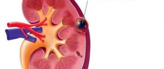
Diagnosis of kidney cyst
Timely detection of a neoplasm is seriously difficult due to the lack of symptoms. If it is diagnosed in the early stages, it is by chance, during preventive studies or diagnosis for another reason. Signs that the attending physician must pay attention to include changes in urine levels and a rise in blood pressure for unknown reasons. Diagnostic tests are as follows:
- Ultrasound scanning, including Dopplerography of blood vessels, allows one to visualize the neoplasm and determine what effect it has on the blood supply to the kidneys;
- Computed tomography makes it possible to distinguish cystic formations from neoplasms of a malignant nature;
- Renal function studies - allow you to determine the activity of the MVS, malfunctions in its work and include urography and dynamic scintigraphy;
- Laboratory tests - with large formations, they reveal a decrease in the daily volume of urine, an admixture of blood and protein in it.
Anamnesis
The medical history of urology must necessarily contain a survey of the patient about lifestyle, heredity, and previous visits.
Born into a complete family, he developed in accordance with his age. Denies any operations or blood transfusions. At the age of 14, as a result of a fall, a blunt abdominal injury occurred with the formation of a hematoma. No allergic reactions were recorded. The onset of the disease is associated with lifting weights, after which frequent, uncomfortable urination appeared. Two days later, at work, I felt a sudden pain in the lower back on the right. I asked to go home, where 3 episodes of vomiting occurred. After going to the clinic, he was hospitalized in the urology department.
Etiology of kidney cyst
The triggering factors for left/right kidney cysts may be different. They cause disturbances in the development of connective tissue and epithelium, which form cysts. These include:
| Initiating factor | Manifested by what? |
| Damage to kidney tissue due to diseases |
|
| Aging of the body | After forty-five years, the load on the urinary system increases. In addition, a complex of insignificant but numerous disorders in the body is triggered, which enhance the impact of each other. |
| Congenital | Cysts of both kidneys or one of them can be the result of disruptions during intrauterine development. They arise during the development of kidney primordia due to gene mutations, and their diagnosis occurs in childhood. |
Laboratory data and additional diagnostics
A general examination of urine and blood is performed on the patient immediately upon admission to the hospital. Medical history: urolithiasis in laboratory parameters:
- Urinalysis: yellow; transparent; pH slightly acidic; beat weight – 1010; protein – 0.2; leukocytes – 2 – 3; epithelium – 1 – 2; red blood cells – 15 – 20.
- Blood test: Hb – 125; erythrocytes – 4.5* 1012 g/l; leukocytes – 10.5*109 g/l; ESR – 10 mm/h; creatinine – 110 mmol/l; urea – 4.7 mmol/l.
R-graph: no darkening detected. Ultrasound:
- Right kidney: normal size and shape, clear contour. ChLS is expanded. The parenchyma is unchanged.
- Left kidney: the contour is smooth, the joint is not expanded. The parenchyma is unchanged. Conclusion: urostasis in the right kidney.
Urolithiasis (UCD) is one of the most common diseases in urological practice. This is a disease that is encountered not only by urologists, but also by almost all doctors of related specialties: therapists, obstetricians-gynecologists, surgeons, endocrinologists, pediatricians, etc. The study of etiology, epidemiology, pathogenesis, diagnosis and differential diagnosis, treatment and prevention of urolithiasis has important for the development of urology as a specialty.
Many ancient doctors studied the ICD. Thus, Hippocrates described renal colic and was the first to recommend treatment with thermal procedures; Galen substantiated the dependence of the formation of urinary stones on climate, water composition, diet, race, and metabolic disorders; Avicenna explained the occurrence of stones in the urinary tract by dietary features and impaired urine outflow
The technique of open drainageless cystolithotomy, which became the basis for the surgical treatment of urolithiasis, was first used by German doctors in the first half of the 19th century. It has been practiced in the Russian Empire since the 1860s. At the beginning of the twentieth century, S.I. Spasokukotsky developed a transabdominal access to the ureter for urolithiasis, which was revolutionary for that time. In the 40s, anatrophic nephrolithotomy was introduced: removal of stones by dissecting the kidney in an area relatively poor in blood vessels. A number of hemostatic sutures for the renal parenchyma have been developed. The introduction of remote lithotripsy and endoscopic technologies at the turn of the 20th and 21st centuries truly revolutionized the treatment of urolithiasis. The high percentage of relapses, the systemic nature of metabolic disorders and comorbidity require the development of methods for minimally invasive treatment, prevention and metaphylaxis of KSD.
HISTORY OF THE DEPARTMENT OF URINOSIS
The Department of Urolithiasis at the Research Institute of Urology was created on June 12, 1985 on the initiative of the director of the institute, academician Nikolai Alekseevich Lopatkin. The first head of the department, who headed it from 1985 to 1995, was Doctor of Medical Sciences, Professor E.K. Yanenko. Subsequently, the department was headed by Doctor of Medical Sciences, Professor N.K. Dzeranov (1996-2009) and Doctor of Medical Sciences. M.I. Katibov (2012). From 2020 to the present, the department is headed by Ph.D. M.Yu. Prosyannikov.
The history of the ICD department is closely connected with the Department of Urology of the 2nd Moscow Regional State Medical Institute named after. N.I. Pirogova. The pioneering work of the staff of the Research Institute of Urologists to study the endemicity of the incidence of urolithiasis in Moscow and other large cities, together with clinics in Germany, Bulgaria, Hungary, Yugoslavia, etc., established the predominance of the oxalate form of urolithiasis. This research was continued within the walls of the Urology Research Institute.
Doctor of Medical Sciences, Professor E.K. Yanenko
E.K. Yanenko in his dissertation for the degree of Doctor of Medical Sciences. made a significant contribution to understanding the pathogenesis of coral nephrolithiasis, developed methods of preoperative preparation and surgical treatment methods that made it possible to remove coral stones from patients with renal failure, preserving the kidneys even with a 70-80% deficiency of their function. Under the leadership of E.K. Yanenko defended 15 candidate and 5 doctoral dissertations.
R.M. Safarov studied the issues of surgical methods in reconstructive surgery of the upper urinary tract in patients with urinary tract disease (candidate's thesis) and the use of laser radiation in the treatment of urinary tract disease and its complications (dissertation of doctor of medical sciences)
Doctor of Medical Sciences, Professor N.K. Dzeranov
One of the most important areas of work of the department since its founding has been the study of the pathogenesis and possibilities of metaphylaxis of urolithiasis using biochemical research methods. Under the leadership of M.V. Chudnovskaya, and subsequently S.A. Golovanov, who defended his dissertation for Doctor of Medical Sciences. on the topic “Clinical-biochemical and physico-chemical criteria for the course and prognosis of ICD”, methods for assessing the metabolism of stone-forming substances were developed, and the conditions for the formation of stones of various chemical compositions have already been formulated. Approaches have been developed to the prevention of recurrent formation of urinary stones, etiological and pathogenetic factors of urolithiasis, predicting its occurrence, including from the standpoint of biochemistry, physical chemistry and immunogenetics, which are reflected in the candidate's (1989) and doctoral dissertations (1999) of O. IN. Konstantinova. On her initiative, the Research Institute of Urology for the first time in the Russian Federation studied the relationship of ICD with polymorphisms of candidate genes.
Surgical treatment of urinary stones has always been given special attention in the work of the department of ICD.A.V. Kazachenko, in his PhD thesis under the guidance of E.K. Yanenko (1996), was able to prove the greater effectiveness of partial pyelonephrolithotomy with a preliminary angiographic study for recurrent nephrolithiasis in comparison with sectional nephrolithotomy.
V.Ya. Simonov under the leadership of Academician N.A. Lopatkina actually carried out pioneering work in our country on the introduction and in-depth study of a fundamentally new high-tech method of treating patients with urolithiasis - non-invasive surgical removal of urinary stones through their remote crushing (1985-1992).
From 1996 to 2009, the department was headed by N.K. Dzeranov. His name is inextricably linked with the widespread introduction of extracorporeal lithotripsy (ESLT) into Russian urological practice. Scientific specialists in the field of high energies from the Moscow Radio Engineering Institute, the Central Military Scientific Research Aviation Hospital, etc. were involved in the development. Experimental, medical-biological and clinical work was carried out to create an experimental model of the domestic Urat-P complex.
On November 7, 1987, the first session of remote lithotripsy was carried out at the Research Institute of Urology, which marked the beginning of the clinical use of the first domestic lithotripter URAT-P in the USSR. The DLT department, created in 1991, headed by A.V. Lykov, is traditionally in close relationship with the ICD department.
Conducted by N.K. Dzeranov, Yu.V. Kudryavtsev, B.S. Gusev and other employees of the institute, experimental morpho-functional studies, for the first time in world practice, allowed us to develop and optimize the parameters of shock waves, in which there would be no traumatic damage to the kidneys and at the same time the effect of stone destruction was preserved even in infants. Various options for using DLT in clinical practice were studied, and treatment results were analyzed. Dissertations were prepared on this material: Dr. dis. Beshlieva D.A. (2003), Ph.D. dis. Moskalenko S.A. (1997), Ph.D. dis. Volkova I.N. (1999), Ph.D. diss. Butina P.S. (2004) and others.
Doctor of Medical Sciences, Professor T.S. is studying the infectious genesis of stone formation. Perepanova, under her leadership the Ph.D. dissertation by graduate student U.A. Radjabov "Metaphylaxis of infectious kidney stones after percutaneous nephrolitholapaxy."
The institute pays great attention to the use of minimally invasive endoscopic methods for the treatment of urolithiasis.
Doctor of Medical Sciences, Professor A.G. Martov
One of the founders of the introduction of transurethral and percutaneous methods for removing urinary stones is A.G. Martov – Head of the Department of Endourology from 1991 to 2006. Works of Martov A.G. et al. made it possible to gradually abandon open surgical interventions performed for urolithiasis in favor of minimally invasive treatment methods.
Currently, employees of the endourology group under the leadership of Ph.D. D.S. Merinova are improving and introducing mini-percutaneous methods for minimally invasive crushing of urinary stones.
In total, employees of the Research Institute of Urology, graduate students, doctoral students, and applicants defended 50 dissertations devoted to the study of pathophysiology, biochemistry, diagnostics and various methods of treating patients with urolithiasis (Appendix 1).
CURRENT WORK OF THE URINOSIS DEPARTMENT
Researchers of the department, in collaboration with colleagues from other departments of the institute, continue intensive work to study the pathogenesis of urolithiasis.
The following research is currently being conducted on this topic:
The influence of excess body weight (BMI) on the development of lithogenic disorders in patients with urolithiasis
Studies have been conducted to assess the influence of obesity and associated metabolic lithogenic factors in patients with urolithiasis on the formation of stones of various mineral compositions.
It was noted that in patients with urolithiasis with a high BMI, the daily urinary excretion of uric acid is noticeably higher than in patients with low BMI values. The same trend is observed in phosphate excretion rates. As BMI increases, urine pH decreases.
The incidence of uric acid stones in obese patients was 6.5 times higher than in patients with low BMI. In addition, obese patients have a fairly high risk of developing struvite stones. However, individuals with a low BMI have a 3.9-fold increased chance of developing carbonate apatite phosphate stones compared to obese individuals. The incidence of calcium oxalate stones does not depend on BMI.
Conditions for the formation of stones of various chemical compositions
Together with the staff of the research and laboratory department, the conditions for the formation of stones of various chemical compositions were determined based on statistical processing of a sample of 708 patients (Table 1).
Table 1. Parameters of lithogenic factors for the formation of stones of various chemical compositions
| Risk of stone formation | elevated |
| Oxalate | with: • calcium excretion above 6.6 mmol/day • uric acid above 4.2 mmol/day, • phosphate above 33.6 mmol/day and • magnesium above 5.1 mmol/day |
| Uric acid | with: • normo- or hypocalciuria (less than 2.6 mmol/day) |
| Phosphate non-infectious | at low excretion values: • urate (< 2.6 mmol/day), • phosphate (< 18.3 mmol/day), • magnesium (< 2.65 mmol/day) |
| Phosphate infectious | at low values: • calciuria (< 2.6 mmol/day), • phosphaturia (< 18.3 mmol/day) and • magnesiumuria (< 2.65 mmol/day) |
Doctor of Medical Sciences, Professor S.A. Golovanov
Study of the association of polymorphisms of candidate ICD genes with various forms of urolithiasis and the nature of its clinical course
As a result of the work, 8 candidate ICD genes were selected in the Russian population: membrane anion transporter family 26 gene (SLC26A6, rs2310996), tumor necrosis factor 11 gene (TNFSF11, rs9525641), calcium release activator modulator gene 1 (ORAI1, rs7135617), factor gene tumor necrosis 11B (TNFRSF11B,rs3134057), nuclear estrogen receptor A subunit gene (ESR1, rs851982), Klotho gene (KL, rs526906), vitamin D receptor gene (VDR, rs1540339), extracellular calcium sensitive receptor gene (CASR, rs2202127 ) .
Genetic risk factors for urolithiasis, its various forms and the nature of its course in the Russian population have been established. An association was found between polymorphisms of the VDR (rs1540339), ORAI1 (rs7135617), and CASR (rs2202127) genes with urolithiasis.
For the studied polymorphisms of the TNFRSF11B, ESR1, SLC26A6, KL, TNFSF11 genes, all differences are not significant.
Creation of the Eating Stereotype Questionnaire (ESA)
One of the most effective and widespread methods for assessing dietary patterns is surveying the target population using questionnaires. Employees of the ICD department developed and implemented the Eating Pattern Assessment Questionnaire (EPQ). TSA is intended for both patients suffering from urolithiasis and healthy people. The questionnaire is freely available on the Internet at www.nethealth.ru in the testing section. It includes a description of most food products sold on the territory of the Russian Federation and consumed by citizens of the Russian Federation.
The TSA was built on the principle used in the nutrition questionnaire “Food Frequency Questionnaire sample booklet for General Nutrition Assessment”, developed by the staff of the Oncological Research Center named after. Fred Hutchinson Cancer Research Center.
After completing the survey, the system calculates the amount of elements consumed per day: proteins, fats, carbohydrates, purines, oxalic acid, water, micro- and macroelements, vitamins. The data obtained is compared with the average daily intake of these elements for the average person.
The conclusion issued based on the results of the questionnaire makes it possible to explain in detail to the patient which foods he should limit the consumption of, and which foods, on the contrary, should be increased. Also, with the help of TSA, it is possible to monitor the patient’s compliance with dietary recommendations.
Modern ideas about the etiology and pathogenesis of urolithiasis
Urolithiasis is a polyetiological and polypathognomonic disease. Despite obvious advances in the study of urolithiasis, the issues of etiology and pathogenesis of stone formation processes have not yet been fully studied.
According to one of the most common theories of the pathogenesis of urolithiasis, it is believed that the basis of stone formation processes is damage at the level of the loop of Henle or collecting ducts (Bellini ducts). Salt deposition occurs in the designated defects, which leads to the formation of plaques or Randall plugs (Fig. 1) [1].
Rice. 1. Modern ideas about the pathogenesis of stone formation in urolithiasis, the formation of Randall’s plaque (A) and plug (B)
In this case, a prerequisite for stone growth is the oversaturation of urine with stone-forming substances [2,3]. These include calcium, uric acid, phosphorus, sodium, oxalate (oxalic acid salt), etc. [4-6]. Accordingly, the patient needs regular monitoring of the concentration of these substances in the blood and urine.
It is worth noting that the process of stone formation is accelerated at low concentrations of stone formation inhibitors. Inhibitors include magnesium and citrates [7]. Magnesium prevents stone formation processes by forming complexes with oxalates and calcium salts [8]. Citrate, like magnesium, reduces the saturation of urine with insoluble calcium salts by forming a complex with them [3]. In addition, normal citrate levels increase the inhibitory activity of some glycoproteins [9].
It is necessary to note the modulating role of the acid-base state of urine in ICD. The pH level of urine is a significant factor in the formation of urinary stones. This is due to the fact that the solubility ranges of lithogenic substances are related to urine pH [10]. This is why different types of urinary stones form at different urine pH levels.
Currently, some authors classify ICD as a group of systemic diseases [11-14]. There is a pathogenetic connection between urolithiasis and atherosclerosis, diabetes mellitus, hypertension, metabolic syndrome, myocardial infarction, and stroke.
The ICD department is working to study the contribution of various risk factors to the pathogenesis of ICD.
PROSPECTS FOR THE RESEARCH WORK OF THE DEPARTMENT
The main scientific directions of development of the ICD department:
- study of nutritional risk factors for ICD;
- studying the severity of the influence of metabolic disorders in urolithiasis;
- study of the microbiome of patients with urolithiasis;
- study of the amino acid composition of urine in patients suffering from urolithiasis;
- implementation of a decision aid system for metaphylaxis of urolithiasis;
- studying the connection between the pathogenesis of cardiovascular diseases and urolithiasis;
- studying the role of herbal medicine in the treatment of urolithiasis;
- studying the relationship between the incidence of urolithiasis and the composition of drinking water
Scientific and practical conference with international participation
The ICD department has been organizing an annual conference dedicated to ICD issues for 6 years. The next conference is scheduled for November 2020!
A team of employees of the Research Institute of Urology, dealing with the problems of urolithiasis. From left to right: N.V. Anokhin, D.S. Merinov, M.Yu. Prosyannikov, O.V. Konstantinova, A.V. Sivkov, A.V. Lykov, N.V. Grebenkin, S.A. Prokhorov
LITERATURE
- Taylor ER, Stoller ML. Vascular theory of the formation of Randall plaques. Urolithiasis 2015;43(Suppl 1):41-5. doi: 10.1007/ s00240-014-0718-4.
- Pak CY, Sakhaee K, Moe OW, Poindexter J, Adams-Huet B, Pearle MS, et al. Defining hypercalciuria in nephrolithiasis. Kidney Int 2011;80(7):777-82. doi: 10.1038/ki.2011.227.
- Pearl MS. Urinary lithiasis: etiology, epidemiology and pathogenesis. In Campbell-Walsh Urology, 9th edition;[edit. Alan J. Wein]. Philadelphia, PA, 2007. Vol. 2. P. 1363-1392.
- Pearle MS, Goldfarb DS, Assimos DG, Curhan G, Denu-Ciocca CJ, et al. Medical management of kidney stones: AUA Guideline. URL: https://www.auanet.org/guidelines/stone-disease-medical-(2014).
- Türk C, Knoll T, Petrik A, Sarica K, Skolarikos A, Straub M, et al. Guidelines on Urolithiasis. EAU, 2020. URL: https://uroweb.org/wp-content/uploads/EAU-Guidelines-Urolithiasis-2015-v….
- Phillips R, Hanchanale VS, Myatt A, Somani B, Nabi G, Biyani CS. Citrate salts for preventing and treating calcium containing kidney stones in adults. Cochrane Database Syst Rev. 2020. No. 6.
- Menditto VG, Lucci M, Polonara S. The role of hypomagnesiuria in urolithiasis and renal colic: results from a prospective study of a metabolic evaluation protocol. Minerva Med 2012;103:377-382.
- Song Y, Hernandez N, Shoag J, Goldfarb DS, Eicner BH. Potassium citrate decrease urine calcium excretion in patients with hypocitraturic calcium oxalate nephrolithiasis. Urolithiasis 2016;44(2):145-8. doi:10.1007/s00240-015-0819-8.
- Halabe A., Sperling O. Uric acid nephrolithiasis. Miner. Electrolyte Metab 1994;.20(6):424-431.
- Madore F, Stampfer MJ, Rimm EB, Curhan GC. Nephrolithiasis and risk of hypertension. Am J Hypertens 1998;11(1 Pt 1):46-53.
- Taylor EN, Stampfer MJ, Curhan GC. Obesity, weight gain, and the risk of kidney stones. JAMA 2005; 293(4):455-62.
- Taylor EN, Stampfer MJ, Curhan GC. Diabetes mellitus and the risk of nephrolithiasis. Kidney Int. 2005; 68(3):1230-5.
- Rule AD, Roger VL, Melton LJ 3rd, Bergstralh EJ, Li X, Peyser PA, Krambeck AE, et al. Kidney stones associate with increased risk for myocardial infarction. J Am SocNephrol 2010;21(10):1641-4. doi: 10.1681/ASN.2010030253.
- Ferraro PM, Taylor EN, Eisner BH, Gambaro G, Rimm EB, Mukamal KJ, et al. History of kidney stones and the risk of coronary heart disease. JAMA 2013;310(4):408-15. doi: 10.1001/jama.2013.8780310:408.
Annex 1.
List of dissertations on the problem of ICD by employees of the Research Institute of Urology
| 1987 | Zabiroe K.I. | Ph.D. | Intraoperative diagnosis of calculous pyelonephritis |
| 1987 | Martov A. G. | Ph.D. | Percutaneous (transfistula) treatment of nephroureterolithiasis |
| 1990 | Kalashnikov A.Ya. | Ph.D. | Secondary hyperparothyroidism during treatment with program hemodialysis |
| 1990 | Borisik V.I. | Ph.D. | Coral nephrolithiasis of hyperparathyroid etiology (diagnosis and treatment) |
| 1990 | Safarov PM | Ph.D. | Reconstructive surgeries for recurrent nephrolithiasis |
| 1991 | Golubchikov I. V. | Ph.D. | Extracorporeal crushing of kidney and upper urinary stones in an experiment |
| 1991 | Mamaev K.T. | Ph.D. | Reversibility of anatomical and functional changes in the kidneys and rehabilitation of patients with nephrolithiasis and chronic calculous pyelonephritis |
| 1991 | Tarasenko B.V. | Doctor of Medical Sciences | Pathogenetic examination of differentiated treatment of patients with nephrolithiasis and metaphylaxis of recurrent stone formation |
| 1992 | Kumar Anil | Ph.D. | Surgical treatment of children with coral nephrolithiasis |
| 1993 | Khurtsev K.V. | Ph.D. | Modern methods of treatment and prognosis of functional renal sostogmia in patients with coral nephrolithiasis |
| 1993 | Mohammed Shafiq | Ph.D. | Non-invasive treatment of stones and ureteral stone track after extracorporeal shock wave lithotripsy |
| 1994 | Dzeranoye N.K. | Doctor of Medical Sciences | External lithotripsy in the treatment of urolithiasis |
| 1995 | Vakhloe S. G. | Ph.D. | Optimization of treatment and rehabilitation methods for patients with nephrolithiasis with single stones of the upper urinary tract |
| 1996 | Rossikhin V.V. | Doctor of Medical Sciences | Homeostasis, prediction and optimization of treatment of patients with renal colic caused by urolithiasis and crystalluric diathesis |
| 1996 | Safarov PM | Doctor of Medical Sciences | Laser radiation in the complex treatment of urolithiasis and its complications (experimental clinical study) |
| 1996 | Kazachenko A.V. | Ph.D. | Diagnosis and prevention of ischemic kidney damage during surgical treatment of coral nephrolithiasis (experimental and clinical study) |
| 1997 | Pavlov S.M. | Ph.D. | Treatment of patients with bilateral nephrolithiasis using extracorporeal lithotripsy |
| 1997 | Pavlov A.Yu. | Doctor of Medical Sciences | Obstructive urolithiasis and severe forms of urolithiasis in children |
| 1998 | Moskalenko S.A. | Ph.D. | Remote lithotripsy in the treatment of various forms |
| 1998 | Olefir Yu.V. | Ph.D. | Optimization of the choice of treatment method for coral nephrolithiasis |
| 1999 | Dondukoe Ts.V. | Ph.D. | Endoscopic ureterolithotripsy with holmium (Ho-YAG) laser |
| 1999 | Volkov I.N. | Ph.D. | Emergency extracorporeal lithotripsy in the treatment of urolithiasis |
| 1999 | Shchukin V.V. | Ph.D. | Combined use of hemosorption and extracorporeal helium-neon laser irradiation of blood in the complex treatment of acute calculous pyelonephritis |
| 2000 | Tatevosyan AS | Doctor of Medical Sciences | Pathogenetic basis of nephrolithiasis. Diagnostics. Treatment |
| 2000 | Konstantinova OB. | Doctor of Medical Sciences | Prediction and principles of prevention of urolithiasis |
| 2001 | Poloe A.N. | Ph.D. | Comprehensive preoperative prediction of the development of acute pyelonephritis after percutaneous nephrolithotomy |
| 2001 | Romanov GB. | Ph.D. | Long-term results of treatment of urolithiasis in children using extracorporeal lithotripsy |
| 2002 | Usenko E.E. | Ph.D. | Some aspects of diagnosis and treatment of the renal form of hyperparathyroidism |
| 2003 | Golovanov S.A. | Doctor of Medical Sciences | Clinical, biochemical and physicochemical criteria for the course and prognosis of urolithiasis |
| 2003 | Kuznetsov GB. | Ph.D. | External shock wave lithotripsy of renal caliceal stones |
| 2003 | Chair NB. | Ph.D. | The role of the occluding factor in the development of infectious and toxic complications in urolithiasis |
| 2003 | Beshliev YES. | Doctor of Medical Sciences | Errors and complications of extracorporeal lithotripsy and their prevention |
| 2004 | Baybarin KA | Ph.D. | Surgical methods of treating nephrolithiasis in gerontological patients |
| 2004 | Kamnina S. A. | Ph.D. | Combined surgical treatment of coral nephrolithiasis |
| 2005 | Butin P.S. | Ph.D. | The use of extracorporeal and contact lithotripsy in the treatment of ureteral stones |
| 2005 | Onishchenko OB. | Ph.D. | Diagnosis, prevention and treatment of the damaging effects of external shock wave lithotripsy on the kidney |
| 2005 | Little Fox A.A. | Ph.D. | X-ray endoscopic methods in the treatment of nephroureterolithiasis in children |
| 2005 | Cherepanova E. V. | Ph.D. | Risk factors for metabolic disorders in children with urolithiasis |
| 2008 | Fatikhov PP | Ph.D. | Transurethral contact lithotripsy in the treatment of kidney stones |
| 2009 | Uzdenoe M.A. | D-M.Sc. | Urolithiasis in the endemic region of the North Caucasus, Karachay-Cherkessia |
| 2013 | Dutov S V. | Ph.D. | Percutaneous removal of kidney stones with the patient in the supine position |
| 2012 | Egamberdiev D.K. | Ph.D. | The role of urinary tract infection in the genesis of kidney stones |
| 2013 | Epishov V A | Ph.D. | Comparative analysis of the results of using surgical methods for treating large and staghorn kidney stones |
| 2013 | Kalinichenko D.N. | Ph.D. | The importance of genetic risk factors in the pathogenesis of urolithiasis |
| 2015 | Alexandrov N.S. | Ph.D. | Optimization of antibacterial therapy for patients with pyelonephritis against the background of urolithiasis. |
| 2015 | Gadzhiev G. D. | Ph.D. | Optimization of medical care for large staghorn stones in a single and only functioning kidney |
| 2015 | Maksudov PP | Ph.D. | Bone metabolism disorders in nephrolithiasis |
| 2016 | Shvangiradze I A | Ph.D. | Prediction of treatment outcomes for bilateral staghorn nephrolithiasis |
| 2017 | Anokhin N.V. | Ph.D. | Features of the chemical composition of urinary stones and differential diagnosis of metabolic disorders in urolithiasis |
| 2018 | Kalabekov | Ph.D. | The influence of the pro-oxidant action of 0.06% sodium hypochlorite solution on the state of metabolism of stone-forming substances and its significance for the prevention of calcium and urate nephrolithiasis" (experimental clinical study) |
| Attached file | Size |
| 897.95 kb |
‹ Analysis of changes in the quality of life of patients with bilateral nephrolithiasis after single-stage and staged bilateral mini-percutaneous nephrolithotomy Up Correction of endothelial dysfunction is a promising direction in the treatment of ischemic kidney damage ›
Treatment plan
The medical history for urolithiasis must indicate a treatment plan and recommendations.
- Therapeutic diet – table 10.
- Drink at least 2 liters per day.
- Antispasmodics IM.
- Painkillers if necessary IM.
- Antibiotics orally.
- Herbal uroseptics orally.
In the medical history of therapy, urolithiasis occupies 30% - 35% of urological patients. The main task during emergency hospitalization is to relieve an attack of renal colic. In the future, urolithiasis is treated based on diagnostic data and the individual characteristics of the patient.
Signs of a kidney cyst
Most often, the disease occurs without clinical manifestations, since the development of the neoplasm occurs slowly, due to which the renal tissues have time to adapt to it, excluding a significant loss of functionality.
However, as the neoplasm grows, it causes a compressive effect on the vessels and stimulates a structure such as the periglomerular complex, which leads to an increase in blood pressure and its instability. The patient begins to complain of headaches and rapid heartbeat. In addition, he suffers from pain symptoms caused by the compression effect and decompensation of renal function.
Large cysts provoke the development of the following clinical manifestations:
- reduction in the volume of urine excreted;
- frequent urge to empty the bladder;
- blood in urine;
- pain symptoms radiating to the groin area;
- intoxication of the body due to urine retention in it;
- fatigue and swelling.
The development of renal failure is manifested by the smell of ammonia from the patient’s mouth (and is typical when both kidneys are affected) or if he has only one kidney.
Ureterorenoscopy (URS) and flexible URS
Ureteroscopy is the preferred treatment for small to medium-sized stones located anywhere in the urinary tract. The procedure is usually performed under general anesthesia. During this procedure, a ureteroscope (a long, thin instrument with a tiny camera at the end) is inserted through the urethra and bladder into the ureter or kidney. Once a stone is found, it can be removed using "tongs or a basket", or a laser or pneumatic is used to break the stone into smaller pieces before they are removed using a basket. Ureteroscopes can be flexible, like a thin, long straw, or more rigid. After the stone is removed, a small temporary tube called a stent may be placed in the ureter to help urine flow from the kidney to the bladder. The urinary catheter and/or stent is usually removed soon after the procedure.
You should know!
URS can be accompanied by both intra- and postoperative complications, which in rare cases may require conversion to open surgery or re-intervention.
During the operation the following may occur:
- migration of stones to the kidney (up to 12%);
- infectious complications (up to 6%);
- ureteral damage (up to 2%);
- bleeding that may require blood transfusion (0.1%);
- ureteral avulsion (0.1%);
In the postoperative period, the following may develop:
- fever and sepsis (up to 1.1%);
- persistent blood in the urine (up to 2%)
- renal colic (2.2%)
- narrowing of the ureter (stricture) 0.1%;
- reflux of urine from the bladder into the kidney (0.1%);
- damage to internal organs (up to 0.05%);
You need to return to the hospital immediately if:
- body temperature above 38 °C;
- can't urinate;
- a large amount of blood in the urine;
- You continue to experience severe pain in your side.
Even if you are at low risk of developing another stone, you need to make some lifestyle changes. These measures reduce the risk of recurrence of the disease and improve your overall health.
Percutaneous (percutaneous) nephrolithotripsy (PNL)
PCNL is most often used when the kidney stones are large, high density, or there are many of them. PCNL is usually performed under general anesthesia. During this procedure, a small tube called a catheter is placed in the bladder. Another catheter is inserted into the ureter. Contrast, or dye, may be injected through this catheter to provide a better view and pinpoint the location of the stone. Once the stone is located, access to the renal collecting system is achieved using a thin needle and a so-called guidewire. The guidewire provides safe access for the nephroscope (an instrument for visualizing the inside of the kidney). Once the stone is visualized, some can be removed using forceps. Large stones must be crushed using a special pneumatic, ultrasound or laser device - nephrolithotripsy. After removing stone fragments from the kidney, a temporary small tube is usually installed in the ureter connecting the kidney to the bladder (catheter-stent), and the working channel through which the operation was performed is inserted into a tube draining the kidney (nephrostomy). After surgery, these tubes are removed after a few days.
You should know!
PCNL can be accompanied by both intra- and postoperative complications.
During the operation the following may occur:
- bleeding, which may require blood transfusion (up to 20%), embolization of the kidney vessels, or removal of the kidney (up to 1.5%);
- pleural damage (up to 11.6%);
- damage to internal organs (up to 1.7%);
- lethal outcome (up to 0.3%).
In the postoperative period, the following may develop:
- fever (up to 32.1%);
- sepsis (up to 1.1%);
- urinoma (accumulation of urine in the perinephric or retroperitoneal tissue, surrounded by a fibrous capsule) up to 1%;
- residual stone fragments (up to 19.4%), in some cases repeated intervention is required;
- aneurysm, arteriovenous fistula;
You need to return to the hospital immediately if:
- body temperature above 38 °C;
- nausea and vomiting;
- chest pain and difficulty breathing;
- a large amount of blood in the urine;
- severe pain on the side of the operation;
- you can't urinate.