Kinds
These capsules can be very small or reach enormous sizes, due to which the ovaries increase to 30 cm in diameter. Occurs in patients of childbearing age and even newborns.
Causes
The best doctors associate the occurrence of these growths with an increased concentration of hCG (human chorionic gonadotropin, formed during pregnancy). This hormone causes increased stimulation of maturing follicles.
Sometimes cysts form as a result of luteal follicular hyperreaction to hCG. The cause of increased hCG is often trophoblastic disease. This is a hydatidiform mole, accompanied by the presence of cysts of this type.
Multiple pregnancies may also be associated with the occurrence of these phenomena in women. Causes may include diabetes, high blood pressure, and preeclampsia.
The occurrence of neoplasms is associated with artificial stimulation with gonadotropins, for example, clostilbegit. It is used to trigger ovulation during IVF. In such cases, the cysts are asymmetrical, single, and have one chamber.
Causes
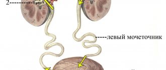
There is no single factor influencing the formation of a cyst in the left kidney, but taking into account statistical data, this type of benign tumors is more often left-sided. As a rule, the prerequisites for the formation of cysts are formed in the human body at the stage of intrauterine development, when the process of formation of renal structures is disrupted.
The nature of the occurrence of a benign tumor is largely determined by the anatomical location of the left kidney, which is located above the right. Another important factor in the appearance of a renal cyst on the left is trauma, as well as previously suffered infectious and inflammatory diseases of the urinary system.
In 70% of cases, a cyst in the left kidney is diagnosed at the stage of serious changes in the paired organ, which is due to the absence of clinical manifestations of the disease.
In urological practice, a separate list of factors is identified, the presence of which leads to an increased risk of developing a cyst on the left kidney. These factors include:
- Vegetovascular dystonia.
- Hypertonic disease.
- Age over 45 years.
- Various injuries to the lumbar region on the left.
- Previous myocardial infarction.
- Pulmonary tuberculosis.
- Urolithiasis disease.
- Previously undergone surgical interventions on the organs of the urinary system.
- Infectious and inflammatory diseases of the kidneys and other organs of the urinary system, including sexually transmitted pathologies.
A left-sided renal cyst forms gradually, and the entire formation cycle consists of several stages. At the initial stage, a cavity is formed, limited by a connective tissue capsule. Subsequently, there is a systematic accumulation of liquid exudate, which can be represented by blood, purulent or serous contents.
The rate of increase in the volume of a benign tumor directly depends on the cause of its occurrence, as well as on the presence of concomitant diseases and the general state of the person’s immunity.
Complications
Complications for this type of cyst are rare , but the following are possible:
- Menstrual dysfunction (delay or prolonged and heavy periods).
- Necrosis, torsion of the cyst, hemorrhage into the cyst cavity into the ovary or abdominal cavity.
- Cyst rupture, bleeding.
Classification
The most durable structures are those neoplasms that are located superficially on the left kidney. Such cysts are called sinus cysts. The sinus cyst capsule consists of connective tissue fibers and has a thickness of 1 to 2 mm. The internal structure of a left-sided sinus cyst can be represented by either a single cavity or multiple cavity chambers.
Based on the location and nature of the clinical manifestations, the following types of cysts on the left kidney are distinguished:
- Pseudocystic neoplasms.
- Complicated cysts.
- Multi-chamber structures.
- Unilocular cysts.
The listed tumors not only have different etiologies and structures, but also require an individual therapeutic approach.
Unilocular cysts
Among the total number of diagnosed renal cysts, single-chamber structures are the most common. The size of such a tumor does not exceed 0.2 cm. Externally, this structure resembles a soap bubble filled with liquid contents. The probable location of a single-chamber renal cyst on the left is the parenchyma of the organ. Less often, the structure is localized in the cortical part of the kidney.
Multilocular cysts
In appearance, such structures have much in common with single-chamber cysts, but the internal structure of such a tumor is represented by several cells filled with liquid contents. Another distinctive feature of a multi-chamber cyst is that each internal chamber is independent and develops at an individual rate.
Pseudocystic formations
When performing examination techniques such as x-rays and ultrasound, pseudocystic neoplasms are visualized as simple tumors. Only a biopsy procedure helps to distinguish pseudocysts from simple neoplasms. The main differences between pseudocysts include the multilayered structure of the capsule, a high potential for malignant degeneration, and a rapid increase in volume.
Complicated cysts
Such structures can have either a single-chamber or multi-chamber structure. In addition, this type of tumor is filled not with serous contents, but with blood or pus. Purulent filling of a complicated cyst occurs if a secondary infection is observed.
Symptoms
Any renal cyst remains asymptomatic for a long time. The patient may not be aware of the development of the disease for a long time. The appearance of clinical symptoms is observed at the moment of a significant increase in the size of the neoplasm, when mechanical compression of adjacent tissues is observed.
Clinical symptoms of a left-sided renal cyst are as follows:
- The appearance of blood fragments in the urine (hematuria).
- Renal arterial hypertension.
- Nagging pain in the lumbar region on the left.
- An increase in the size of the kidney, as a result of which a foreign structure can be easily felt during palpation.
- Aching or nagging pain in the suprapubic region.
Along with general symptoms, patients complain of nausea, thirst, general weakness and malaise, increased body temperature and blood pressure. In the absence of timely treatment, the pathology threatens the occurrence of functional kidney failure.
Stages
The nature of the clinical manifestations, as well as the size of the left kidney cyst, depend on the stage of development of the disease. The entire period of development of a left-sided renal cyst is divided into the following stages:
- Preclinical. At this stage, the patient does not have any changes in his general condition, and the cyst can only be diagnosed by performing an ultrasound examination.
- Compensatory stage. Despite the appearance of such clinical symptoms as fatigue, headache and general weakness, the human body is able to compensate for the changes that occur under the influence of the neoplasm.
- Stage of decompensation. At this stage, there is an accumulation of metabolic and toxic components, the elimination of which was disrupted by the appearance of the cyst. Symptoms of general renal intoxication of the body include instability of blood pressure, nausea, sleep disorders, and intense pain in the lumbar region.
- Terminal stage. Manifestations of the terminal stage make themselves felt if the patient does not receive proper treatment in a timely manner. At this stage, renal failure develops with complete loss of organ functions.
Right ovarian cyst and menstruation
It is necessary to take into account that ovarian cystic formations caused by hormonal reasons lead to MC disorders. The discharge may become scanty or too heavy, and may also have an unusual color.
What types of ovarian cysts are there in women and how do they manifest? Most often, MC disorders are caused by follicular cysts and corpus luteum cysts. If the attending physician suspects the development of a cyst because the patient does not have menstruation, then he will certainly rule out the development of a luteal formation that appears in the first trimester of pregnancy. Since after conception, a woman’s hormonal system begins to function differently, the production of estrogen is reduced in volume, and more progesterone is produced, because it is necessary to maintain pregnancy. The active ovary, which has the main follicle, begins to function more intensively, which is why a cyst appears on it.
A luteal cyst can be called a functional formation, since it resolves on its own, and this happens in the fourteenth week of pregnancy. Progesterone is no longer produced by paired glands, but is localized in the placenta. If pregnancy occurs, but there is a stable absence of the corpus luteum, then there is a risk of its termination, that is, a miscarriage can happen arbitrarily. In addition, a nonfunctional cyst in a pregnant woman is dangerous not only for the development of the embryo, but also for the mother.
The corpus luteum cyst is the main instigator of menstrual irregularities. In addition to the prolonged absence of menstruation and its failure, the patient may experience pain in the lower abdomen. In order to accurately diagnose the presence of a cyst, the gynecologist needs to exclude the possibility of an ectopic pregnancy or other pathologies of the pelvic organs. Therefore, the patient is sent for an ultrasound, and she must also donate blood to be examined for human chorionic gonadotropin.
We recommend you find out: Cyst on the ovary, treatment and what caused it
Diagnostics
If one or more of the previously listed symptoms occurs, each person is advised not to postpone a visit to a nephrologist or urologist. The following examination techniques help confirm or refute a clinical diagnosis:
- Intravenous and survey urography.
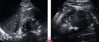
- Ultrasound examination of a paired organ.
- General clinical analysis of blood and urine.
Magnetic resonance imaging helps to identify not only the tumor itself, but also determine its size and location.
Treatment
Drug treatment
If the size of the cyst is small, medical specialists prefer to conduct observational tactics, assessing the dynamics of tumor growth using ultrasound. Regular dynamic monitoring of the cyst is indicated if the tumor diameter does not exceed 5 cm.
Drug therapy for this disease is symptomatic. To relieve the main symptoms of the disease, the following medications are used:
- Baralgin, which allows not only to reduce the intensity of the inflammatory process, but also to relieve pain.
- Drotaverine. This drug belongs to the group of antispasmodics that relax the smooth muscles of the urinary tract.
- Tempalgin. This drug has not only an analgesic, but also an antipyretic effect.
In the event of a secondary infection of a bacterial nature, patients are prescribed a course of antibacterial therapy.
The most effective are antibiotics from the fluoroquinolone group (Ciprofloxacin, Norfloxacin), as well as cephalosporin drugs (Cefaclor, Tercef, Ceftriaxone). In addition, to normalize the process of natural outflow of urine, diuretic (diuretic) drugs are prescribed.
Operation
Measures to eliminate a benign neoplasm are taken if the pathological structure has reached a large size and disrupts the functions of the paired organ.
Volumetric cysts on the left kidney, the size of which exceeds 5 cm in diameter, are subject to surgical removal. Surgical treatment of this pathology is carried out using techniques such as open resection, puncture, laparoscopy or nephrectomy.
Laparoscopic removal of the cyst is indicated if the diameter of the neoplasm does not exceed 10 cm. If the cyst is advanced, open resection of the kidney area along with the neoplasm or radical removal of the paired organ if it is significantly damaged is performed.
After surgery, the patient faces a long rehabilitation period, including restrictive measures and adherence to a special diet.
Diet
Patients who have been diagnosed with a left-sided renal cyst must completely exclude pickled, spicy, fried, smoked and salty foods from their diet.
The daily volume of fluid consumed should not exceed 1.5 liters. In addition, protein foods and table salt are limited to a minimum in the diet.
During the postoperative rehabilitation period, it is necessary to completely stop drinking alcohol and smoking, as well as products containing cocoa and coffee.
Types of cysts
Depending on how the ovarian cyst was formed, the following types are distinguished:
Functional
1Follicular.
Normally, the egg matures in the follicle in a liquid medium. Once the egg has fully matured and is ready for ovulation, the follicle must rupture and allow the release of the egg. If this does not happen for some reason, the unruptured follicle with the egg inside forms a tumor cavity called a cyst. 2 Corpus luteum cyst. After the follicle ruptures and the egg is released, the so-called corpus luteum is formed in its place, which usually goes through several cycles. This type of tumor formation is also called luteal, since the formation of the corpus luteum occurs in the second phase of the cycle, called the luteal. The disease is called luteal cyst of the left ovary when the ovary located on the left is affected. Functional cysts make up the vast majority of all cystic formations and are associated with impaired functional activity, which is where they get their name. As a rule, in most cases, functional cysts do not cause any particular inconvenience and can form and resolve even without the knowledge of their owner.
Endometrioid
Occurs when endometriosis occurs when endometrial cells travel from the uterus to the ovary. Typically, endometrioid cysts occur in mature women. Can be treated with medication or surgery. Otherwise, it can lead to infertility and serious health risks in case of rupture.
Dermoid cyst of the left ovary
It begins to develop in the tissues of the ovary from the very embryo; a predisposition to this arises at the genetic level. When such a cyst is opened, fragments of hair, teeth, fat and nail tissue can be found in it. It does not pose an immediate threat to health, but if detected, it must be removed surgically to eliminate the risk of degeneration into a malignant formation.
Serous cyst
It is a benign tumor formed by epithelial cells. A serous cyst occurs in women between 15 and 70 years of age and is usually located on one side. A serous cyst does not pose an immediate threat to life, and practically does not develop into a malignant one, but it can, accumulating serous fluid, increase in size. If detected, it must be removed surgically.
Paraovarian cyst of the left ovary
Formed in the area between the ovary and the fallopian tube. A future cyst can form even in a girl in the womb under the influence of a host of factors that have not been fully studied, mainly from viral infections. The presence of a paraovarian cyst poses a threat only if the pedicle is twisted and there is a risk of rupture. It is removed surgically.