Uroflowmetry - what is it? This is an ordinary manipulation in which the parameters of the urinary stream are determined. It is carried out in order to find out the condition and check the functioning of the lower urinary tract.
In completely healthy people, the outflow of urine during urination begins slowly, but then the flow rate increases, and then slows down again until the bladder is completely empty. What does it mean if you have been prescribed uroflowmetry? The average price for this type of research is 1,100 rubles.
History of uroflowmetry
The first attempts to measure the speed of a urine stream were made by E. Rehfisch. The scientist designed a device similar to modern uroflometer devices called aeroplethysmograph back in 1897.
In 1948, WM Drake presented a research paper on uroflowmetry. In it, the author described in detail the development and operating principle of a device for mass population research - the uroflowmeter, and proposed the designation uroflowgram for diagnostic graphs.
From that time on, diagnostics gradually began to enter the clinical practice of urologists.
The first specialized 4-channel device is considered to be the Danish system “DISA Electronics”.
Its use has shown that accurate diagnosis has a positive effect on the treatment of patients with urinary problems. Since 1975, the International Committee responsible for the standardization of urodynamic examinations began to function.
The technical capabilities of non-invasive uroflowmetry are constantly being improved; research results allow choosing the most effective and convenient way for the patient and doctor to identify problems with urination.
Code designation in the nomenclature of medical services
In the systematized nomenclature of medical services N 804n, approved on October 13, 2017 by orders of the Ministry of Health of the Russian Federation, uroflowmetry has code A12.28.006, where each value has its own interpretation:
- The letter A denotes a service in medicine with an independent complete meaning, representing a specific intervention for the purpose of diagnosis, therapy, prevention or rehabilitation.
- The number 12 indicates the type of examination of organs using certain manipulations. This also includes physical and drug tests, immune reactions, and studies of a number of blood parameters.
- The number 28 represents the class. In this case, it indicates the urinary system and kidneys.
- 006 is a type of medical service.
The uroflowmetry code simplifies the interaction between structures providing care to the patient and facilitates the collection of statistical data.
What is studied and evaluation indicators
The operating principle of almost all uroflowmeters is similar. The devices record the force of the stream flow at the moment of emptying the bladder, its continuity and speed. The results are displayed as a graphic line. The doctor evaluates the measurements in accordance with accepted standards and based on personal experience.
Uroflowmetry determines activity:
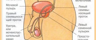
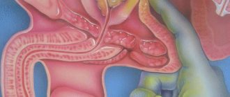
- Detrusor. This is the muscular lining of the bladder, responsible for its contraction and squeezing out accumulated fluid during bowel movements. The muscle, at the moment of filling the organ, transmits signals to the brain, as a result of which the person feels the need to urinate.
- Sphincter. This is a kind of lock that blocks the exit from the bladder to the urethra. The detrusor begins to contract only after the sphincter relaxes. That is, these two muscles work together.
- Urethra. In men, it passes through the entire penis and when obstacles form along this path, certain changes occur during urination.
Before starting the study, the patient must drink a certain amount of water, recording the time.
After this, when the urge occurs, uroflowmetry is performed to determine:
- The total time of urine excretion is the period of time starting from the appearance of the first portion of urine until the completion of urination.
- Maximum speed - the volume of urine released to the maximum per unit of time. Deviation from normative indicators does not always indicate pathology. When assessing the schedule, the doctor must take into account not only the patient’s gender, but also his age and the possibility of detrusor function deterioration. In old age, the maximum speed decreases noticeably.
- Average rate of urine output. It is calculated based on the ratio of urine excreted in ml and the time of urination in seconds.
- The time required to reach the peak velocity of the urine stream. Normally, the curve of the diagram increases sharply and the time to achieve this indicator should not exceed one third compared to the entire length of the uroflowgram.
- Total volume of urine. Diagnostic data are considered the most reliable if the patient managed to extract between 200 and 500 ml of fluid.
- The time it takes for urine to appear from the onset of the urge to emptying the bladder. Normally it should not be more than 10 seconds. This figure increases to minutes in patients with bladder outlet obstruction.
Uroflowmetry is more often used for men. When examining women with dysfunctions of the urinary organs, other methods are more often used, which is primarily due to the anatomical features of the urethra.
Indications - why is such an analysis needed?
Based on the results of uroflowmetry, one can preliminary judge the presence of:
- urethral obstruction – narrowing due to chronic inflammation or injury;
- sclerosis of the cervix - its strictures and scarring (mainly of inflammatory origin);
- sphincter or detrusor atony - usually a malignant or neurological phenomenon;
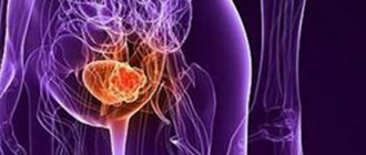
- prostatitis and adenoma - two pathologies manifested by incomplete emptying with a weak, intermittent stream. The prostate gland is located directly below the bladder and surrounds the urethra with its neck just below the sphincter. In nature, this anatomy is designed to prevent urination during sex (taking into account the physiology of sexual intercourse, this is important for a woman’s health). The prostate swells during the production of ejaculate, blocks the flow of urine into the urethra and defecation becomes physically impossible. But its hormonal growth or inflammation as a result of infection also leads to a similar effect.
Among the causes of deviations are polyps and other benign or malignant lesions. Uroflowmetry only allows you to check previously made assumptions. And to clarify the diagnosis, additional studies will definitely be required - cystotonometry, biopsy, ultrasound, etc. Using uroflowmetry, a specialist can also evaluate the results of the intervention or conservative treatment. It allows you to detect and prevent complications, side effects and relapse.
Sources used:
- https://www.tiensmed.ru/news/urofloumetriya1.html
- https://urohelp.guru/diagnostika/metod/urofloumetriya.html
- https://prostatits.ru/prostatit/urofloumetriya.html
- https://uran.help/surveys/urofloumetriya.html
- https://fb.ru/article/350953/urofloumetriya-chto-eto-takoe-pravila-podgotovki-i-provedeniya
- https://prostatu.guru/diagnostika/urofloumetriya-chto-eto-takoe-podgotovka-k-protsedure-i-ee-provedenie.html
- https://edema-club.ru/eyes/skorost-potoka-mochi-u-muzhchin-vozrastnye-normy-urofloumetriya-pri-kakih/
- https://malepotency.ru/urofloumetriya.html
Indications for use
Uroflowmetry is prescribed by doctors if, during a preliminary examination of the patient, based on his complaints and examination, there is a suspicion of the likelihood of developing pathologies such as:
- Adenoma or malignant tumor of the prostate gland. The proliferation of prostate tissue inevitably leads to compression of the urethra, thereby complicating the outflow of urine and causing pathological changes during urination, more details.
- Prostatitis. The disease is characterized by an inflammatory process, which is accompanied by swelling of the gland tissue, which in turn compresses the urethra, interfering with the normal outflow of urine.
- Strictures (narrowing) of the urethra. The causes of formation are inflammatory changes and injury to the organ, leading to the proliferation of connective tissue and, as a consequence, scarring.
- Valves of the urethra. Refers to congenital anomalies. They are characterized by the formation of mucous folds on the inner walls of the bladder, which, at the time of emptying the organ, can block the exit to the urethra, creating an obstacle.
- Stones. They enter the urethra from the bladder or kidneys. Large stones can completely block the flow of urine. In this case, severe pain appears, and the advancement of the stone causes damage to the mucous membrane, which is accompanied by the appearance of blood or urine turning pink.
- Sclerosis of the bladder neck. The cervix is located at the bottom of the organ and passes into the urethra. Cervical sclerosis is a consequence of the proliferation of connective tissue, which in most cases is caused by inflammation.
- Bladder neck spasm. The reason is inflammatory changes, infectious process, neoplasms. Compared to stenosis, spasm is easily eliminated with timely treatment.
- Decreased detrusor tone. It may be caused by changes in the muscle layer or disruption of the innervation of the organ due to neurological pathologies. A characteristic symptom is weak and slow urine output.
- Neurogenic bladder. It occurs as a result of injuries or organic lesions of that part of the brain or peripheral nerves on which the entire process of urination depends. With a neurogenic bladder, there is often no feeling of its filling. As a result, urine accumulates in huge quantities inside the organ and gradually stretches its walls along with the neck. In advanced situations, a gap forms in the cervix through which urine is released uncontrollably.
- Bladder hyperactivity. It manifests itself as frequent urges that occur even with the slightest filling of the organ, and the rapid release of urine. The reason is deterioration in the regulation of the tone of the muscular wall of the bladder.
- Abnormalities of the urethra (curvature or narrowing of the canal). Mostly diagnosed at an early age.
POPULAR WITH READERS: Prostate fibrosis in men, symptoms, causes, treatment methods
Based on the results of uroflowmetry, one can only judge the presence of certain pathologies. To confirm them, the doctor selects advanced diagnostics - ultrasound of the pelvic organs and prostate, cystotonometry, biopsy analysis.
Uroflowmetry is also used to evaluate drug therapy, surgery and a number of other manipulations.
What does it show
Based on the test results, the urologist determines:
- determines the quality of the urinary tract;
- difficulty or slow urination;
- damage to the prostate gland, an increase in the size of an important organ;
- the functionality of the urethral obturator sphincters, preventing uncontrolled urine leakage;
- bladder contractility;
- the severity of obstruction or blockage of the ducts;
- all obstacles that interfere with the normal outflow of urine.
On a note! If serious pathologies of the urinary tract are suspected, the urologist prescribes radionuclide uroflowmetry. In addition to the main indicators, the study shows the volume of residual urine, distal stenosis, vesicoureteral and vesicoureteral reflux.
Signs indicating the need for examination by a urologist
If necessary, uroflometry is prescribed and performed by a urologist.
An examination may be necessary if the patient complains of:
- Pain in the lumbar region and in the projection of the kidneys.
- Painful sensations that occur during emptying of the bladder.
- Any urination disorders - weakness or intermittency of the stream, spontaneous release of urine, frequent or, on the contrary, rare urge.
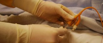
The procedure is completely painless and safe for patients. During this procedure, there is no need to touch the body with instruments or insert manipulators into the urethra.
Despite the fact that uroflowmetry is more often used when examining men, if necessary, it can be performed even on children and pregnant women.
Before prescribing the procedure, the doctor must know exactly what medications, including all kinds of diuretics or other herbs, the patient is taking. If necessary, a number of medications are abandoned several days before uroflowmetry, since their use may distort the chart data.
Contraindications
There are no absolute contraindications to uroflowmetry, but the doctor will refuse the procedure to patients:
- With complete blockage of the urethra. The procedure is allowed after the cause of the obstruction has been eliminated.
- With severe dehydration. It is especially dangerous to carry out analysis using diuretics, since they quickly intensify the patient’s existing water-salt imbalance.
- Early age. Children under 1-2 years of age cannot fully control the process of urine excretion, so uroflowmetry will not provide the necessary information.
Equipment for carrying out
Since the invention of the first uroflowmeter, hundreds of improved modifications of the device have been released onto the market. All modern devices belong to multi-channel and multifunctional technology.
Their design includes a microcomputer and a computing unit with a display, a device for recording and storing the received data, and a printer for printing out the uroflowgram.
The newest UDS, in addition to determining the basic parameters of urination, shows the pressure in the renal pelvis and ureters, the electromyographic activity of the muscular layer of the pelvic floor and sphincters.
Every year, improved models are developed that significantly simplify the diagnostic procedure and at the same time expand its capabilities.
The most accurate devices for uroflowmetry are described below
Solar Blue
The urodynamic system is produced in the Netherlands. This is a stand-mounted device equipped with a wireless flow sensor and puller, as well as an infusion pump.
Designed not only for diagnosing urodynamic disorders, but also for identifying pathologies of the pelvic organs, rectoanal examination of the rectum and anal sphincter and examinations in gynecology.
The Solar Blue system is small in size and portable. The wireless sensor can be placed up to 50 meters from the stand, which significantly increases the comfort of uroflowmetry for the patient. The device is designed in such a way that it can be connected to the general local network of the hospital and integrated with an ultrasound scanner.
SOLAR URO
The SOLAR URO device is also a development of Medical Measurement Systems. In the basic configuration, it allows you to conduct the entire range of urodynamic studies.
Among the additional capabilities of the equipment, it is worth noting the possibility of treating urinary and fecal incontinence, erectile dysfunction in men and women, interstitial cystitis, and nocturnal enuresis in children using biofeedback therapy.
POPULAR WITH READERS: Kegel exercises for men - technique for doing them at home
Nomograms are created automatically in accordance with the requirements of international standards. Any study can be exported for further processing and transferred to the general polyclinic network.
TRITON
Complex for urodynamic diagnostics from the Canadian company Laborie Medical Technologies. The system is based on wireless technology, which allows individual units of equipment to be placed in the most convenient places in the office for the doctor and patient.
As a control module, you can choose not only a desktop computer and laptop, but also portable devices.
In addition to uroflowmetry, the device is used for cystometry, electromyography, cavernosometry, and assessment of erectile dysfunction. There is an option for video-urodynamics.
If necessary, it can be supplemented with a physiotherapy unit for electrical stimulation.
Of the modern uroflowmetric systems, the following also deserve attention:
- Delphis from the Canadian company Laborie.
- Solinette by Laborie.
- Bonito (Laborie).
- Helix.
- Ellipse.
Preparation for the event
It is possible to obtain the most reliable results of uroflowmetry only if the patient is properly prepared for the manipulation.
Necessary:
- Maintain drinking regime. On the eve of the procedure, an adult weighing an average of 70 kg needs to drink from 1.5 to 2 liters of clean water. 30 minutes before the planned test, you need to drink another 200-300 ml of water. The doctor should indicate more accurate figures for the amount of alcohol consumed, based on gender, age, concomitant diagnoses, and weight.
- So that the bubble is filled enough. During the examination, the patient must excrete at least 100 ml of urine, so at least an hour before the diagnosis, you must stop visiting the bathroom. Before uroflowmetry, the subject should have a noticeable urge to empty the bladder.
- Get ready psychologically. A detailed consultation with a urologist on the principles of operation of the device on the eve of diagnosis can help with this. Most modern equipment operates in automatic mode, which means that the patient can be left alone in the office.
- Stop taking certain medications several days before the procedure. Some pharmaceuticals increase or, conversely, slow down urine secretion, so taking them will result in unreliable results.
Uroflowmetry can be performed several times according to indications. Diagnostic data is deciphered only by the attending physician.
Features of the procedure in men
In addition to the main unit, the device is equipped with a funnel with sensors connected to it. During the examination, the patient urinates into a container, and highly sensitive sensors transmit signals to the microprocessor. The received data is processed and displayed on the display, and then, if necessary, in paper form through a printer.
No one supervises the test. But a man must remember that reading all indicators is possible only if he produces at least 150-200 ml of urine. The results of uroflowmetry are known in 5-10 minutes, but interpretation of the analysis may take 2-3 days.
Computer Uroflowmetry
This is a standard study without the use of additional devices.
The patient must answer a number of questions immediately before the procedure, the main ones:
- How much fluid you drank the day before and right now.
- How many urinations were there in the morning and the approximate volume of urine excreted.
- When was the last time you visited the toilet to urinate?
- Is there a urge to defecate at the time of the interview?
- Do you feel thirsty?
- Are any medications taken and their dosage.
If there is no urge to urinate, then the patient is left to wait for their appearance. He is then taken to a separate room for uroflowmetry, where he must, following previously stated instructions, urinate into a funnel.
Evacuation should occur in a person’s normal position and care must be taken to ensure that all excreted urine ends up only in the container. After the emptying is completed, all that remains is to call a health care worker and you can immediately go home.
Pharmacouroflowmetry
It differs from standard uroflowmetry by the use of medications that stimulate urine secretion and accelerate the filling of the organ. The use of pharmaceuticals increases detrusor tone and pressure inside the bladder. If the muscle walls are weak, this does not cause virtually any changes, but with prostatitis or, for example, an adenoma, the speed of urine passing through the urethra increases.
Preparation for pharmacouroflowmetry is no different from preparation for standard manipulation. Initially, the patient undergoes conventional uroflowmetry. Then they give the desired drug (for example, Furosemide).
The medicine begins to act in 30-40 minutes, after the appearance of unbearable urges, the man will need to empty himself into a funnel. After the diagnosis is completed, both results are compared.
With determination of residual urine
The essence of the technique is to determine urine residues and their amount in the bladder after conventional or drug uroflowmetry. Normally, at the end of urination, no more than 10% of the fluid from the volume of urine excreted should remain in the organ. That is, if 200 ml came out, then up to 20 ml may remain, and this will not relate to pathology.
The remaining portion of urine in a larger volume indicates that there is an obstacle in its path. Residual urine is determined using ultrasound diagnostics performed immediately after the main study.
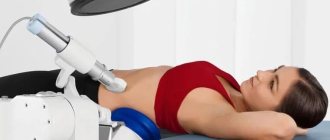
How is the research conducted?
Diagnostics are carried out in a specially designated voiding room. The patient is introduced to the device and shown a button that must be pressed at the beginning of the miction and 5 seconds after its completion. Then the subject is left alone. The device will record urination made into a funnel, which is connected to the device. There is a voiding chair in the office for women; men urinate standing up. The process is recorded automatically. Then the patient is sent to an ultrasound diagnostic room, where the amount of residual urine is determined (normally it should not exceed 30 ml). A catheter is also used for this purpose, but the method is inferior to ultrasound, as it is considered invasive.
Is it possible to perform uroflowmetry at home?
Uroflowmetry, as far as the patient’s actions are concerned, is a simple procedure. But modern systems are quite large and at the same time fragile equipment that requires careful handling. Its use at home is almost impossible and very expensive.
But there are mobile devices that are used to diagnose urodynamic disorders in bedridden patients, in patients with pelvic fractures, etc.
POPULAR WITH READERS: How to diagnose prostatitis in men
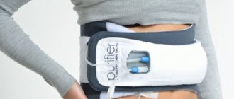
Today, a number of medical equipment manufacturers produce portable devices with wireless sensors. Outwardly, they look like a regular jug with a stand.
During the examination, you need to urinate into a container, and the device will transmit the data to a laptop or smartphone, after which it can be analyzed. Such devices are quite suitable for use at home.
They are especially useful when it is necessary to adjust therapy depending on urinary disorders or monitor changes in the course of the disease in cases of urethral stricture or neurogenic bladder.
In what situations is uroflowmetry necessary?
As a man ages, the condition of his internal organs deteriorates. The urinary system is no exception. The bladder and urethra become depleted and atrophy, which leads to worsening urination.
In addition to age-related changes, the cause of deterioration in urination is bladder outlet obstruction. In other words, a narrowing of the urethra under the bladder. Infravesical obstruction is characteristic of the following changes and diseases:
- prostatitis in various forms;
- stricture of the urinary canal - narrowing;
- sclerosis of the bladder neck;
- benign prostatic hyperplasia;
- neoplasms of various etiologies;
- trauma, inflammation and so on.
The diseases are accompanied by urinary disorders, and uroflowmetry makes it possible to detect pathology even at an early stage.
In the USA and European countries, the diagnostic method is used during the initial visit to a urologist or andrologist. In Russia, the situation is different: uroflowmetry serves as an auxiliary method of instrumental examination. Not all clinics have the opportunity to do this kind of analysis.
Features of uroflowmetry in children
Uroflowmetry in pediatric practice is carried out using the same method as in adults. The only difference is that the parents will have to psychologically prepare the child for the procedure, since many of the children may have difficulty defecating in unfamiliar conditions.
About an hour before the intended manipulation, the child should be given water to drink at the rate of 15 ml per kilogram of weight. Water can be replaced with drinks that stimulate urination - green tea, rosehip infusion, chamomile infusion, berry fruit drinks.
Pharmacouroflowmetry at an early age is not indicated for use, since Furosemide, used to stimulate urine, is contraindicated for use up to 18 years of age.
Features of uroflowmetry in women
When examining women, uroflowmetry is used relatively rarely. The short female urethra is easier to examine with special probes, with which you can quickly determine the location of the narrowing of the canal.
But sometimes the procedure is absolutely necessary. In this case, the woman should be aware of all the features of its conduct and preparation, which are no different from examinations in men.
However, directly during the manipulation, the woman must sit on a special chair with a hole. A funnel is installed in it, and urine should fall into this measuring container.
Where to do
The accuracy of uroflowmetry depends on the quality of the equipment. To carry out the procedure, you can contact diagnostic centers and network laboratories. For example:
- "Invitro": 1500 rub.
- “Men’s and Women’s Health Clinic” (Moscow): 600 rubles.
- Multidisciplinary clinic "EMS" (St. Petersburg): 1100 rubles.
- "Medical on Group" (St. Petersburg): 1580 rubles.
According to reviews from men, it is better to choose private clinics. There, the procedure is carried out delicately, the results are accurate and visual.
Normal parameters, interpretation of graph values
When performing uroflowmetry, the following indicators are assessed:
- V is the total volume in milliliters of urine excreted.
- TWait is the time interval from the sensation of urge to the start of a stream of urine. The indicator determines the tone of the muscles of the urethra, the state of the detrusor muscle, the degree of their readiness to perform the main function. Normally from 10 to 40 seconds.
- TQmax is the length of time required to reach the peak pressure of the urine flow. In the absence of pathologies, the speed increases evenly during the first third of the act of urination. The norm is from 4 to 9 seconds.
- Tmict is the period of time during which the jet speed uniformly decreases. Normally takes up 2/3 of urination.
- Qmax – maximum jet speed. The norm is within 15-20 ml/sec. In men, this uroflowmetry indicator also depends on age.
- Qave – average value of the overall speed. Calculated based on urine volume divided by the combined value of TQmax and Tmict. The normal value is 10-15 ml per second.
If all indicators are within normal limits, then the curve on the graph is a bell shape with a slightly flatter right edge.
If there are pathologies, the curve will be in the form of all kinds of slides, broken lines, teeth, or too low.
An experienced urologist, examining a uroflowgram, can make an accurate diagnosis at the first glance at the graph.
The rate of urine flow slows down with prostatitis and neoplasms. Ups and downs on the graph indicate deterioration of nervous regulation and urethral blockage.
A weak increase in speed may occur when the channel narrows. However, only a doctor can accurately interpret the data, taking into account the patient’s age and all indicators together.
How is uroflowmetry performed?
There is nothing difficult about the procedure. When performing a simplified technique, it is enough to use a glass with a measuring stick and a stopwatch. This method can be used at home, but it does not give one hundred percent results.
The volume of urine is measured using a measuring cup, and the duration of urination is measured with a stopwatch. To find out the speed of urine outflow, you need to divide the volume of urine by seconds. This will be the approximate speed. If you know the normal results of the indicators, then you can judge the presence of pathology.
It is possible to obtain accurate indicators only by using a uroflowmeter.
The device consists of:
- Containers for collecting urine;
- Patrubkov;
- Stands.
The device is connected to the computer, and all data comes here. The result comes out in the form of a graph from the printer. The doctor will be able to decipher the data and get the result and the presence of pathology.
Carrying out the procedure
The procedure is carried out as follows:
- Before starting the session, the specialist will familiarize the patient with the algorithm for its implementation. The uroflow meter consists of a funnel that is connected to a measuring device and a recorder.
- When the patient is ready to urinate, press start, wait 6 seconds and begin urinating. Then start urinating into the funnel. When you finish urinating, you should wait a few seconds and turn off the device.
- During the procedure you need to relax and be still.
Benefits of the survey
The non-invasive examination method is valued due to several obvious advantages:
- Uroflowmetry takes a minimum of time, and the results are visible immediately.
- No special preparation of the patient for the procedure is required.
- The procedure is painless.
- There is no direct contact with the body and internal cavities, which eliminates infection of the body.
Uroflowmetry can be safely performed with a portable device and at home. Innovative devices are compatible with smartphone software, which allows you to see the results on your phone screen and send them to your doctor at any time.
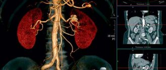
How does the urinary system work?
The body receives organic substances from food and processes it into energy. When the body has received the necessary substances and energy, the breakdown products are eliminated.
The genitourinary system stores the following substances:
- Potassium;
- Sodium;
- Water.
Removes toxins from the body. For example, creatinine and urea. Read a detailed article about creatinine clearance in men and its norm here.