Pathological neoplasms with a round cavity, which are separated by a membrane from the tissues that surround them, are called renal sinus cysts. Often such cysts are formed due to an inflammatory process in the kidneys. The appearance of a neoplasm causes pain in the lumbar region, pressure surges, and hyperthermia of the body. With a prolonged course of the disease, the formation increases in size and impairs the functioning of the kidney. Therefore, when the first symptoms of the disease appear, it is necessary to go to the doctor and apply timely therapeutic measures.
Anatomical features
The kidneys are a paired organ located in the retroperitoneal region of the lumbar region. Perform the following functions:
- excretory;
- endocrine;
- homeostatic;
- participate in the intermediate process of metabolism.
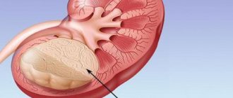
The kidney consists of an upper and lower section and the hilum of the organ. The hilum of the organ carries vessels through itself, and also houses the pelvis with the ureter. The ureter carries urine out. The gate of the organ with its elements is the sinus.
Important. A kidney sinus cyst is a disease in which a cyst forms in the sinus area.
Complications of untreated cysts
Infection of the cyst contents is one of the common complications. It is dangerous due to perforation of the wall of the neoplasm and leakage of pus into the pyelocaliceal system (PLS) of the kidney. Delayed treatment leads to organ abscess and toxic shock.
Possible complications of sinus cysts:
A large sinus cyst of the right kidney damages the renal arteries and veins, which leads to hemorrhages into the pelvis or retroperitoneum. Excessive blood loss is dangerous due to a sharp decrease in blood pressure and brain hypoxia.
How does a renal sinus cyst form?
The sinus cyst of the left kidney, as well as the right one, is formed by separating one of the vessels from the general blood circulation system. Subsequently, serous fluid accumulates in this vessel. Over the entire area of the resulting cavity, a cystic capsule is formed from connective fibers. The diameter of the capsule does not exceed five centimeters.
A sinus cyst of the right kidney is almost immediately accompanied by pronounced symptoms, while a neoplasm in the left kidney can be asymptomatic for a long time. There are cases with simultaneous damage to both kidneys, but it is extremely rare.
Important. This pathology can be acquired at birth or as a result of the influence of negative factors.
Stages of development of entities
In one kidney or in two at once, cysts are located in most cases on the sinus or portal zone of the kidneys. New growths attract fluid, which begins to fill them.
The first stage is characterized by the formation of a “bag”. Its interior has no communication with vessels. Over time, the “bag” becomes separate from the main tissue and creates its own closed system.
The next stage is manifested by filling the cavity with organic liquid. Moreover, fluid can enter the cyst, both at the initial stage of formation and during subsequent development.
Doctors are often unable to accurately diagnose the moment the chamber fills with liquid. But the cyst is almost always filled with contents.
Reasons for education
The following reasons may contribute to the development of pathology:
- predisposition at the genetic level;
- kidney injury: as a result, blood supply is disrupted and an inflammatory reaction occurs;
- the presence of infections in the genitourinary system;
- use of hormonal drugs: such drugs negatively affect the stable functioning of all human organs;
- not following a healthy diet;
- the presence of bad habits: alcohol abuse and smoking, have a detrimental effect on the functioning of the urinary system;
- dangerous and harmful working conditions: according to statistics, this disease is quite common in people whose profession involves working with poisonous and toxic materials.
Causes
It is currently not possible to determine exactly why a cyst appears near the renal sinus. Geneticists offer their vision of the problem, medical practitioners think differently. But, for the most part, experts are inclined to think that the onset of pathology occurs due to disturbances in the development of the fetus during the prenatal period. But genetic indicators can also play a role.
Renal cysts appear even before a person is born. This is due to mutations at the chromosome level. But in order for the cavity to form and fill with contents, a long period of time passes. This period reaches decades and overtakes a person already in adulthood.
In children, this formation is almost impossible to detect. Some causes of renal cysts:
- Prolonged hard work;
- Uncontrolled use of hormone-containing drugs;
- Exposure to harmful chemicals;
- Malignant nature of neoplasms;
- Problems with urinary functions;
- Artery problems;
- Renal tuberculosis;
- Jades;
- Age-related changes.
Symptoms of a kidney sinus cyst
Congenital intrasinus renal cysts occur without symptoms for a long time. Very often, their diagnosis occurs during examination of the body for the presence of completely different pathologies.
An acquired kidney cyst begins to be accompanied by characteristic signs as the tumor grows, which begins to compress the vessels, which leads to blockage of the urinary canals.
The following symptoms are observed:
- Signs of pain. They manifest themselves due to enlargement of the pelvis and impaired fluid removal. The resulting cyst clogs the channel responsible for the outflow of urine, which contributes to the formation of kidney stones.
- High blood pressure. Due to poor circulation in the kidneys and excessive production of renin. This symptom may be accompanied by tachycardia, exhaustion and migraines.
- Presence of blood in urine when urinating. This reaction of the body is caused by the removal of sand and stones, and can also occur due to the destruction of blood vessels.
- Increased body temperature. Occurs due to an inflammatory reaction.
- Kidney failure. It is observed with a significant increase in the kidney sinus cyst, which contributes to the destabilization of the blood supply and can lead to organ atrophy. This symptom is extremely dangerous to human health and can be fatal.
After analyzing the symptoms of the disease, we can conclude where exactly the pathology formed. For pain on the left side, in the spine, with pain radiating to the groin - sinus cyst of the left kidney. For pain syndromes on the right side - sinus cyst of the right kidney.
Diagnostics
The manifestation of the above symptoms is often not taken seriously, being attributed to the presence of other diseases or stress. Renal sinus cysts are a serious condition that requires careful evaluation. Diagnostics will exclude the development of malignant tumors and diseases of the genitourinary system.
Sequential diagnostic examination:
- Laboratory testing of blood and urine.
- Ultrasound;
- Radiography;
- MRI and CT.
A sinus cyst of the left kidney will manifest itself as an increase in protein, creatinine and an increased number of red blood cells. The rest of the examination will help confirm or refute the diagnosis. It is recommended to carry out diagnostics every six months.
Diagnosis of the disease
The disease has many of the same symptoms as other pathologies. To exclude errors in diagnosis, the doctor performs palpation of the kidneys. Analyzes symptom data obtained from the patient.
In the future, laboratory tests of the patient’s tests are studied. Based on the data obtained, a preliminary diagnosis is made.
To confirm it, use the following methods:
- Ultrasound to determine the extent of the lesion and the location of inflammation;
- urography, used when there is doubt about the normal functioning of the outflow of urine or suspicion of renal failure;
- CT scan;
- MRI.
Causes
Modern medicine has not yet fully studied the reasons for which cysts can appear on paired organs, however, there are certain factors that increase the risk of tumor formation. Cysts on the kidneys can be congenital or acquired. Congenital cysts usually appear for the following reasons:
- woman smoking during pregnancy;
- drinking alcoholic beverages during pregnancy;
- poisoning of the expectant mother caused by the action of toxic and chemical substances;
- exposure of a pregnant woman to radiation received during an X-ray examination.
In a child, a cyst can be detected only after he is born. Congenital pathology can affect the left and right kidney, in rare cases, damage to both organs is observed.
Acquired cysts most often appear due to other diseases, such as:
- hydronephrosis;
- tuberculosis of the genitourinary organs;
- hormonal disorders;
- prostate adenoma;
- renal ischemia;
- injuries in the lumbar region;
- stagnation of urine;
- hypothermia of the body;
- various kidney pathologies.
Treatment of kidney sinus cyst
Treatment of renal sinus cysts of the left and right kidney, in the absence of the formation of a low-quality tumor and normal urine output, consists of constant monitoring of the course of the disease.
When viral bacteria are detected in the organ, medical treatment of harmful elements is carried out. In the same way, a person’s blood pressure and urine output are restored. If medications do not stabilize the normal functioning of urine outflow, a drainage method is used. It promotes the pumping of urine from the neoplasm, with the further introduction of a sclerosing agent to restore the walls of the cavity.
This method is accompanied by the risk of infection and sepsis if serous fluid penetrates into the kidney tissue.
The use of surgical treatment methods is necessary in the following situations:
- the sinus cyst of the right kidney stands out in increased size and presses on nearby organs;
- there is hemorrhage in the kidneys;
- the outflow of urine is difficult;
- infection of the cyst with harmful microelements;
- development of cancer.
To surgically remove a cyst, the following methods can be used:
- Removal of serous fluid from the cyst through the skin.
- Laparoscopic surgery. The most gentle method for this disease. Allows you to eliminate the consequences of the disease using computer images. In this case, the surgical incision is from five millimeters to one and a half centimeters, through which the outflow of urine is restored and the harmful formation is removed. A drainage tube is installed at the site where the tumor is removed, followed by suturing.
- In cases of the formation of purulent deposits, mechanical lesions or oncological manifestations, open surgery is used. In this case, it is possible to remove parts of the kidney tissue or the entire organ (in severe cases of the disease).
If blood clotting is poor or if there are other serious diseases that can lead to complications, surgery is contraindicated.
Important. After surgical interventions, a course of antibacterial therapy is used. In some cases, drugs are used to suppress the inflammatory process.
Signs
Congenital cysts are rarely detected in children due to the absence of symptoms. Both in childhood and in adults, when the formation is small, there are no special manifestations. It is diagnosed completely by accident during an examination for a completely different disease.
If the sinus cyst begins to grow and reaches a size of over 5 centimeters, then compression of the vessels and obstruction of the urinary tract occurs. In this case, the patient may present complaints that are not specific, since they may indicate another renal pathology:
Pain syndrome develops. Expansion of the pelvis and disruption of the outflow of fluid from the kidney leads to stretching of its parenchyma, which gives a symptom such as a dull aching pain. If the renal sinus cyst does not allow urine to pass from it to the underlying sections, then congestion leads to the formation of stones. In this case, the patient develops renal colic. These symptoms intensify after physical overload and do not change with changes in body position. There is an increase in pressure due to impaired blood flow in the kidney and an increase in the production of renin. Manifests itself in the form of tachycardia, weakness, headache and cardiac pain. First of all, when measuring pressure, the diastolic indicator increases. Blood appears in the urine due to the release of sand, stones, or a violation of the integrity of blood vessels. The addition of an infectious process leads to an increase in temperature. If the formation reaches a large size, then if left untreated, blood flow in the kidney is disrupted and atrophy develops. In this case, deficiency develops with its typical symptoms - chronic intoxication with metabolic products, edema, polyuria, thirst, sleep disturbance. The pathological process leads to the death of the patient.
By the location of the pain, you can determine which side the formation appeared on. The renal sinus cyst of the right kidney will hurt on the right side in the lumbar region of the spine, and radiate to the inner thigh and groin.
Prevention of pathology
Diet is used as the main preventive measure. To prevent the development of the disease, the following recommendations should be followed:
- reduce fluid intake: it is recommended to limit yourself to one and a half liters per day, taking into account the consumption of various dishes with liquid (soup, borscht, etc.);
- limit salt intake or eliminate it;
- remove fatty foods, various sausages, twists, canned goods, ketchup and mayonnaise from the diet. Use natural juices, compote and purified water for drinking. Significantly reduce the consumption of sweet products;
- reduce protein consumption: this includes dairy products, all types of meat products and legumes. You should use various porridges: buckwheat, millet, rice;
- exclude seafood;
- refrain from consuming alcohol-containing products and smoking tobacco;
- use various vitamin complexes.
Important. It must be remembered that diet does not help cure the disease, but only reduces the harmful effects.
What it is?
A sinus cyst is a banded formation containing serous fluid inside. The localization of a benign tumor occurs in the area of the renal hilum (sinus), can affect the right or left organ, and is less often located on both kidneys. Sinus cysts belong to a number of simple benign formations. They rarely require surgical intervention and cause virtually no discomfort to the patient. The only exception is the pathological growth of the organ, when the tumor compresses the surrounding tissues and disrupts the functioning of the urinary system as a whole.
In nephrology, a sinus cyst is often called a “parapelvic” formation, which is located near the renal pelvis, but, being small in size, does not in any way affect the functionality of the genitourinary system. Cystic growths rarely become large, but when the sinus cyst of the right or left kidney exceeds 5 cm, the clinical picture is most likely to be severe, and the patient will require surgery.
Cysts in the sinus kidneys can develop during fetal development or appear during a person’s life. In the first case, they develop as an anomaly of the urinary system. In children, such formations are difficult to diagnose because they are tiny. Acquired cysts develop against the background of internal disorders, and can be diagnosed completely by accident.
Cysts in the sinus can be present on the right or left; less often, both organs are affected. In practice, a sinus cyst of the right kidney is most often diagnosed, and the clinical picture is more pronounced. A left kidney sinus cyst can go unnoticed for a long time and not cause any signs of disease or internal disorders.
Cystic formations affecting the area of the vascular portal of the kidneys can develop for several reasons, have a congenital or acquired etiology. The reasons for the development of sinus cysts of the left or right kidney are most often hidden in internal disorders of the genitourinary system. If we consider a congenital anomaly, its development can be provoked by the mother’s unhealthy lifestyle and genetic factors. Acquired formations can develop as a result of several causes and predisposing factors, including:
- Lower back bruises.
- Urolithiasis disease.
- Chronic pyelonephritis.
- Prostatitis in men.
- Recurrent cystitis in women.
- Regular stress, depression.
- Age after 50 years.
- Chronic hypertension.
- Internal infections.
- Long-term use of hormonal drugs.
- Alcohol abuse, smoking.
In some cases, it is not possible to determine the etiology of formations, especially when a person has a history of chronic diseases.
Symptoms
Large intrasinus renal cysts provoke the appearance of:
pain syndrome; arterial hypertension; blood in urine; renal failure, which manifests itself as weakness, swelling, thirst, drowsiness during the day and insomnia at night.
Pain from a kidney cyst can appear in the hypochondrium, in the lumbar region and radiate to the groin. Therefore, by the location of the pain, it is possible to determine in which of the two kidneys the cyst is located. If the patient has a sinus cyst in the right kidney, then an aching or dull pain will occur on the right side. If a sinus cyst has formed in the left kidney, pain occurs in the left hypochondrium.
Arterial hypertension occurs due to excess production of the hormone renin, which is synthesized in the kidneys. This hormone interacts with other hormones that regulate blood pressure. As a result, in addition to high blood pressure, the patient experiences headaches, nausea and weakness.
The appearance of blood in the urine indicates serious problems in the functioning of the kidneys. Hematuria can be caused by various kidney diseases, including sinus cysts. Blood in urine can be detected through laboratory tests, and in some cases, even the color of the urine may change due to blood.
Clinical manifestations
A renal sinus cyst of the left kidney often does not manifest itself at all. In the vast majority of cases, pathology is diagnosed accidentally. A person with such an education can live for many years without knowing about his illness. If the formation is localized in the right organ, the symptoms may be erased and manifest as minor disturbances in the functioning of the urinary system. When, for some reason, a tumor-like formation increases in size, reaching 5 or more centimeters, the first pronounced signs may appear:
- Nagging pain on the left or right.
- Frequent surges in blood pressure.
- Periodic headaches.
- Feeling nauseous.
- Increased weakness.
- Temperature increase.
- Swelling of the face and limbs.
- Increased thirst.
- Cloudy urine or presence of blood in urine.
When intrasinus renal cysts are present, the patient’s body temperature rises, general intoxication of the body, problems with urination are observed, and this may indicate an infection.
Treatment
Intrasinus renal cysts are not treated unless they cause severe discomfort. Drug intervention is periodically carried out to relieve the main symptoms: pain, inflammation, normalization of urinary outflow. Antibiotics are prescribed when infection occurs.
Surgery is prescribed in cases where there is a suspicion that a tumor in the kidney has degenerated into a malignant or multicystic lesion of both kidneys. Other reasons:
- tear on the left or right;
- suppuration of the cavity;
- severe hematuria.
A small kidney cyst in the sinus is removed mainly by laparoscopic method, or by performing a skin puncture with a sclerosing agent injected directly into the tumor.
Surgery is necessary when the size of the tumor increases, compresses blood vessels and impairs renal function, as well as in the presence of urolithiasis.
Complications
A sinus cyst of the left kidney, as well as the right one, rarely causes complications, but in rare cases, if it grows pathologically and without proper treatment, it can lead to the development of renal failure. With the rapid growth of the formation, infections of the genitourinary system, there is a risk of developing a purulent process inside the formation, which can serve as a rupture of the cyst. Complications of the pathology include the development of hydronephrosis in the kidney, which ceases to perform its functions. With congenital pathology, there is always a risk of developing multi-chamber intrasinus cysts, which can significantly impair kidney function. This pathology always requires surgical treatment.
Classification of cysts
In nephrology, kidney cysts are classified according to different criteria - location, origin, structure, nature of the contents.
By origin they are:
- congenital - formed during the embryonic development of the fetus;
- acquired – occur against the background of inflammatory diseases, injuries, negative influence of internal or external factors.
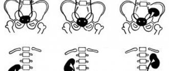
Depending on the location, unilateral and bilateral cysts are distinguished. In the first case, they occur in only one, and in the second, in both kidneys at once.
The classification of formations by structure is important:
- Parapelvical - appear in the renal parenchyma and protrude into the sinus. Cystic vesicles arise on the side of the calyx or pelvis in the region of the renal hilum. In 80% of cases, such cysts are single. They are extremely rarely found in the vestibule of the sinuses of both kidneys. When the capsule grows, the outflow of urine from the organs is disrupted.
- Peripelvic - formed inside the renal hilum. Intrasinus renal cysts have an asymmetrical shape. They deform the sinus, which leads to displacement of the pelvis relative to the renal hilum.
A renal sinus cyst of the left or right kidney is fraught with stagnation of urine, deformation of the pelvis and hydronephrosis.
Treatment methods
Treatment for sinus cysts depends on the size of the cyst. If the formation is small and does not cause any symptoms, treatment is not carried out. It is enough to follow the doctor’s recommendations, regularly visit a nephrologist, and undergo ultrasound diagnostics, which can monitor the dynamics of the disease.
When the first signs of the disease appear, the doctor may prescribe conservative or surgical treatment. Drug therapy will reduce the growth of education, relieve pain and other manifestations of the disease.
- Antispasmodics – reduce pain, relieve spasms: “No-shpa”.
- Anti-inflammatory drugs - relieve pain, relieve inflammation: Ibuprofen, Diclofenac.
- ACE inhibitors - allow you to reduce the risk of vascular pathologies: Monopril.
- Diuretics - improve the flow of urine, prevent the development of stagnant processes: Furosimide, Vershpiron, Lasix.
- Antibiotics - only in cases where a secondary infection has occurred: Emsef, Ceftriaxone.
During the treatment process, it is important to follow a strict diet, which will be prescribed by the doctor individually for each patient.
If the formation is large, when it causes severe symptoms and threatens complications, surgery may be prescribed to remove the cystic growth:
The postoperative period after the operation depends on the chosen technique. With resection, recovery may take more than 3 weeks. After laparoscopy, the patient recovers much sooner – 5 days. The prognosis after surgery is difficult to predict, since everything depends on the patient’s health, age, and the presence or absence of chronic pathologies.
How is the treatment carried out?
If no symptoms are observed and the formation is not larger than 5 centimeters, then treatment is not carried out; such a patient is recommended to undergo a follow-up examination every six months.
If symptoms develop, the principles of assistance are as follows:
- Elimination of pain and inflammation.
- Normalization of blood pressure numbers.
- Restoring the outflow of urine from the kidney.
- Suppression of pathogenic bacterial flora.
The use of traditional methods for treating sinus cysts can only be used as additional therapy and prevention, which does not replace the main treatment and does not have a visible effect on the cause of the pathology.
In acute conditions that can result from a sinus cyst, surgical intervention is performed. Indications for the operation are:
- suppuration;
- gap;
- arterial hypertension;
- disturbance of urine outflow;
- increase in a short period of time;
- hemorrhage into the cavity;
- malignancy, or suspicion of the development of a malignant process.
There is a modern technique for percutaneous puncture of the cyst followed by the introduction of sclerosant. This leads to the collapse of the walls of this formation, and in most cases there is no relapse.
New growths can be removed using laparoscopic surgery. To do this, several incisions are made on the anterior abdominal wall. This technique is minimally invasive, effective, and does not require long-term rehabilitation.
Abdominal surgery is performed in extremely rare cases; the indication for it is the need for complete excision of the kidney, or the presence of a tumor process with the presence of metastases.
Prevention
Some recommendations that every person should adhere to, regardless of gender and age, will help reduce the risk of developing a sinus cyst.
- Balanced and fortified nutrition.
- Adequate fluid intake.
- Quitting alcohol and smoking.
- Reduce consumption of salt, fatty and spicy foods.
- Avoid hypothermia.
An important rule of prevention is timely consultation with a doctor at the first disturbance in the functioning of the urinary system. The sooner a person consults a doctor and undergoes the necessary research methods, the lower the risk of developing not only these pathologies, but also many other diseases.
The cystic cavity can be located anywhere in the kidney. One option is a renal sinus cyst: a fluid cavity in the hilum area where the neurovascular bundle and an important part of the urinary system, the pelvis, are located. Despite the proximity of these anatomical structures, sinus fluid neoplasms are harmless and manifest a minimal number of symptoms. If surgical treatment is necessary, it is optimal to use puncture emptying of the cystic cavity or endoscopic removal.
Therapy prognosis and prevention
The success of treatment depends on the diameter of the formations, their number and the presence of complications. In 79% of patients, small blisters are detected that do not increase in size or grow very slowly. There is no treatment for such forms of pathology. But a person should be examined by a nephrologist at least 2 times a year.
With large cysts in the sinuses, the prognosis worsens. They interfere with the passage of urine, which causes complications - hydronephrosis, renal failure. But with radical treatment, re-formation of tumors rarely occurs.
To prevent kidney cysts, you should:
- promptly treat urological infections;
- be examined by a nephrologist every six months;
- limit consumption of table salt;
- give up bad habits;
- control blood pressure.
Compliance with preventive measures reduces the risk of cyst formation several times. Timely diagnosis and therapy eliminate life-threatening complications.
Sinus cyst of the left kidney is a formation from the category of simple cysts. To understand the reasons for its occurrence, it is necessary to consider the structure of the organ. The kidneys are paired elements located in the posterior wall of the abdominal cavity. They perform excretory, homeostatic and endocrine functions, and take part in intermediate metabolism.
The kidney has an upper and lower pole, a gate through which blood vessels pass, and it also contains a pelvis with a ureter that drains urine. The gate with all its input components is defined as a sine. A cyst is a round, hollow formation separated from surrounding tissue by a membrane. It may consist of one or several chambers and contain a cloudy liquid in its cavity.
A cyst in the renal sinus is a simple formation that develops at the hilum of the kidney, near the renal pelvis; its other name is parapervical.
Symptoms of pathology
The absence of any manifestations during the formation of cysts in the sinuses of the right or left kidney is a completely typical situation: the patient has no complaints, pain or trouble with urination. This usually happens in cases where the size of the tumor does not exceed half a centimeter (5 mm). Most often, the detection of a cystic tumor is an incidental finding during ultrasound examination.
If the size of the right or left kidney increases due to a cystic formation of more than 5 mm, the following symptoms will occur:
- Aching or pressing pain in the side, depending on the location of the fluid cavity (right, left side or entire lower back), intensifying with exercise.
- Tendency to arterial hypertension with an abrupt increase in pressure, which is more typical for the pathology of the left kidney.
- Changes in urine tests due to compression of the pelvis or obstruction of the urinary tract.
The essence of pathology
As a rule, renal sinus cysts do not come into contact with the pelvis itself. The disease most often affects women over 50. Renal sinus cysts can be congenital or acquired.
The first develops with the formation of a kidney in the embryo. An intrauterine disorder in the kidney tissue can develop due to smoking or drinking alcohol during pregnancy. Intrauterine infection can also have a negative impact. In childhood, sinus cysts are diagnosed in rare cases due to their small size. In addition, children do not have a large load on the kidneys, this protects them from inflammatory processes.
A renal sinus cyst, formed as a result of obstruction and caused by the outflow of urine, is acquired. In medical practice, such a formation occurs in both the right and left kidneys. Less commonly, tumors are diagnosed in both organs at the same time. The formation rarely reaches large sizes - on average, its diameter does not exceed 5 cm. Due to the anatomical structure (the right organ is located lower than the left), the cyst of the right kidney manifests clinical symptoms faster, mainly due to the greater load. The formation on the left may go unnoticed: the pathology can have a hidden course and not cause concern for a long time.
Sinus cysts can be single or multiple. Such formations are detected by chance during a diagnostic examination.
In some cases, complications arise and the cyst may increase in size, putting pressure on surrounding tissues. This leads to disruption of the blood supply in the collecting system, which, in turn, forms hemorrhage in the cystic capsule. An enlarged tumor can obstruct the flow of urine and develop kidney failure. The most dangerous complication is malignancy - degeneration into oncology. The transformation of a tumor into cancer is determined by external signs.
Diagnostic methods
The simplest, most accessible and effective method for detecting renal sinus cysts is ultrasound. Echography allows not only to identify small formations, but also to pay attention to possible complications in the form of compression of blood vessels and the pelvis. A significant drawback of ultrasound diagnostics is that the diagnostician may not detect a small tumor.
If you have symptoms of sinus cysts, especially on the left side, you need to undergo a full examination including:
- survey and excretory urography;
- CT scan of the kidneys with contrast;
- MRI.
The advantage of using computed tomography or magnetic resonance imaging with contrast will be:
- obtaining layer-by-layer images of structures with clear and volumetric localization of the cavity with very high information content of the materials;
- the ability to distinguish a cyst in the region of the sinus and renal pelvis;
- assessing the risk of future surgery due to the occurrence of a tumor near the renal vessels and nerves;
- minimal negative impact on the human body.
The doctor will prescribe all general clinical tests, with which you can assess the patient’s condition and prepare for surgery.
Methods for diagnosing cystic formations
Timely diagnosis is difficult due to the long latent (hidden) course. People consult a doctor with unexplained lower back pain, increased blood pressure and urinary problems.
- Analysis of urine. A kidney sinus cyst damages the internal structures of organs, which is why an increased content of protein and red blood cells is found in the urine.
- Kidney MRI. The method is used to differentiate cystic vesicles from non-benign tumors. When examined, cysts look like small balls filled with fluid.
- Ultrasound of the kidneys. Cystic cavities are defined as anechoic objects that do not reflect the ultrasound wave.
In case of complications, the diagnosis is supplemented with functional studies - dynamic scintigraphy, excretory urography. Based on the data obtained, the correct functioning of the kidneys is assessed.
Effective treatment methods
If the cystic sinus cavity is small, constant monitoring by a doctor is required: if the cyst grows, surgical intervention is necessary. The following types of operations are used:
- percutaneous puncture removal of fluid from a sinus cyst, which eliminates the need for traumatic open surgery;
- laparoscopic removal by enucleation of the sinus cyst with mandatory excision of the walls of the fluid cavity;
- endoscopic resection of part of the kidney tissue.
In some rare cases, with a complicated version of the kidney sinus cyst, it is possible to use open surgery, when the doctor has to perform a nephrectomy.