Causes
To understand the reason for the formation of a solitary kidney cyst, you need to know the physiology of the appearance of urine - its primary form is formed in the descending part of the loop of Henle (name by the author). If the tubule becomes clogged, fluid accumulates in it, it begins to swell, and a cavity gradually forms. The following reasons can lead to this condition:
- genetics - with congenital narrowing or partial fusion of the duct;
- age-related changes - can provoke a violation of the integrity of the tubule membrane;
- renal or systemic diseases - the mechanism of pathogenesis is similar to the previous one;
- hormonal changes - can provoke the appearance of cysts during puberty or menopause in women.
This is only a hypothesis; the exact reasons for the appearance of solitary cysts have not yet been studied and are the subject of debate among scientists. But it was clinically established that the patients were in the listed risk groups at the time of detection of the pathology.
Kinds
There are the following types of breast cysts:
- Shape: oval, round, irregular;
- By size – small, medium and large;
- By quantity – single or multiple;
- By the number of cameras - single-chamber or multi-chamber;
- Based on the presence of signs of inflammation - cysts with or without inflammation.
Typical cysts have smooth internal walls, while atypical cysts have growths on the walls. There is an atypical, fibrous, ductal, solitary, multilocular cyst of the mammary glands. If a complex breast cyst contains pinpoint tissue inclusions, doctors conduct in-depth diagnostics.
Atypical breast cyst
An atypical breast cyst is an accumulation of fluid in the cavity, which was formed when the gland duct expanded. It is round or oval in shape, can be of different sizes, and has a fibrous capsule. An atypical cyst is a benign neoplasm with growths that protrude into the cavity.
- An atypical cyst is formed for the following reasons:
- Long period of existence of cystic formation;
- Often recurrent breast cysts:
- Inflammatory processes that occur in the cyst;
- The presence of papillomatous formations in the cystic cavity.
If the cavity of an atypical cyst is small, it does not pose a problem for the patient and does not reveal its presence for a long time. After the cavity enlarges, the first signs of pathology appear - nagging pain in the mammary glands, which intensifies during menstruation. If the neoplasm is large, characteristic bumps are visible on the chest. They can be easily felt with your fingers. A large cyst in the mammary gland deforms a woman's breasts.
Fibrous cyst of the breast
Fibrous cyst of the mammary gland occupies a central place in mammology and is a background disease in the development of breast cancer. There are non-proliferative and proliferative forms of fibrous cyst. Fibrous cysts of the mammary glands develop under the influence of the following provoking factors:
- Hormonal imbalance;
- Weakened immunity;
- Disorders of internal organs (liver, thyroid gland).
The disease manifests itself with characteristic symptoms:
- Increased pain on the eve of menstruation;
- The presence of cluster-shaped nodular seals in the mammary gland;
- Discharge of fluid from the nipples.
A fibrous cyst with dense mammary contents is predominantly expelled by papillomas.
Solitary cyst of the mammary gland
A solitary cyst of the mammary gland is a benign dysplasia that does not degenerate into a malignant tumor. The neoplasm has a round shape and elastic consistency. It is a cavity filled with liquid of various colors and surrounded by a capsule. Brown fluid in a breast cyst may be evidence of atypia or vascular microtrauma. The longer the cyst exists, the denser its capsule. A solitary cyst is located in one breast.
The following reasons for the development of a solitary cyst are distinguished:
- Burdened heredity;
- Neuroendocrine disorders;
- Multiple abortions;
- Age after 35 years;
- Overweight;
- Late birth.
The cause of the disease can be stress, nervous strain, early puberty, complete refusal of breastfeeding, mastitis and trauma to the mammary gland.
A solitary cyst is manifested by pulling, pressing or aching pain in the mammary gland, which intensifies in the second phase of the menstrual cycle. The pain may radiate to the shoulder blade, neck or shoulder. At the end of menstruation, the pain stops. A woman can identify a large solitary cyst on her own by palpation.
Ductal cysts of the mammary glands develop at any age, but are most often detected after 48 years. This disease is diagnosed in 1% of cases among all breast tumors. Ductal cysts are small growths inside the breast. This is a benign tumor that is a precancerous condition. The risk of developing a malignant breast tumor against this background is high.
Ductal cysts develop under the influence of the following provoking factors:
- Hormonal disorders;
- Ovarian dysfunction;
- Abortion;
- Inflammation of the uterine appendages;
- Obesity;
- Diabetes mellitus and other endocrine diseases.
Clinically, this pathology is manifested by discharge from the nipples of a clear, brown, bloody or greenish fluid. A ductal cyst can be palpated if it is present in the area of the milk ducts. It is palpated as a dense, painless formation. The disease is most often discovered by chance, during a routine examination.
Multilocular breast cyst
A multi-chamber cyst develops with an unhealthy lifestyle, alcohol abuse, nicotine and drug use, and poor diet. The cause of the disease may be poor nutrition, metabolic disorders, stress, anxiety, and hormonal imbalances. First, one cystic cavity develops, then new ones are born, after which they merge. This is how a multi-chamber cyst is formed.
A multi-chamber cyst of the mammary glands is a dangerous disease that can develop into a malignant tumor. When felt, cysts resemble soft balloons, but sometimes they are hard. May be round, oval or irregular in shape.
Symptoms and types
Typically, solitary neoplasms occur without pronounced clinical signs; many patients may not even know about their presence until a simple cyst is discovered during a random examination.
Depending on the location, there are three types of solitary cysts; they are listed in the International Classification of Diseases (ICD) 10th revision:
- if the seal is located superficially under the capsule, a subcapsular kidney cyst is exposed;
- if the renal tissue is affected, but the pelvis is not affected, it is done;
- but if the seal penetrates the renal sinus, it means the patient has a parapelvic cyst.
The latter type is the most dangerous, since the neoplasm can interfere with the flow of urine or put pressure on neighboring organs. In this case, a simple kidney cyst provokes the following symptoms:
- soreness in the abdomen, lower back, or pelvic area;
- change in urine color;
- impaired diuresis;
- temperature increase;
- the appearance of swelling.
The listed symptoms of a solar cyst of the kidney cannot be ignored - it is better to carry out restorative therapy in the initial stages than to treat complications for a long time later.
Possible complications
Of the possible complications, the most dangerous is rupture . More often this happens with injury to a large renal cyst.
When ruptured, the contents of the cavity are poured into the perirenal fatty tissue. In case of damage to adjacent tissue, hemorrhage occurs, similar in structure to a kidney bruise. The rupture is accompanied by acute pain. If the kidney tissue is damaged, pain shock may also develop.
Another complication is infection of the contents followed by suppuration . In this case, there is always a risk of the formation of a renal abscess, which can lead to a dangerous septic condition.
In some cases, there is a possibility of cancerous degeneration of kidney tissue cells in the tumor wall. If this diagnosis is confirmed by cytological or histological methods, urgent surgery is indicated.
Diagnostics
A routine examination is rarely prescribed, only if a solitary seal has become a cause for concern. In this case, it is required to detect complications.
The following diagnostic methods will help identify a solitary kidney cyst:
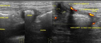
- Ultrasound - allows you to detect a solitary cyst of the right or left kidney, the size of the lesion, and also makes it possible to assess the degree of lost functions;
- CT or MRI are good diagnostic methods, but not everyone can do them due to contraindications;
- duplex scanning - can complement the examination to make an accurate diagnosis;
- Blood and urine tests will allow you to assess the progression of the disease and give a prognosis for the patient.
After the examination, the patient is given a diagnosis. When a solitary neoplasm is identified, its size, location and effect on body functions are indicated.
Diagnostic methods
To confirm or refute the diagnosis, the doctor needs to know about all clinical manifestations. The information will allow for differential diagnosis with other renal tumors.
Symptoms of a right kidney cyst should be distinguished from the symptoms of chronic appendicitis.
Of the instrumental diagnostic methods most often used:
- Ultrasound;
- computed tomography;
- X-ray examination with contrast of blood vessels.
During the examination, a round formation with clear contours and homogeneous content is observed in the renal tissue. This picture is most often characteristic of hollow kidney formations.
When diagnosed by CT, the diagnosis can sometimes be difficult in cases of localization in the peripelvic region. In addition, this method does not detect inclusions in the cyst wall or in its cavity.
The X-ray angiography method is more informative . During the examination, a contrast agent is injected and a series of photographs are taken.
Among non-invasive methods, preference should be given to ultrasound. The method has no contraindications for patients of any age.
Additionally, the patient may be prescribed cystography. During the examination, a special needle is used to fill the cyst cavity with a contrast agent and an X-ray is taken. The procedure is necessary to exclude malignant degeneration.
Treatment
If a solitary kidney cyst causes symptoms, treatment is required to compensate for impaired functions. Surgery is prescribed in extreme cases; usually the doctor is inclined towards conservative therapy. It includes:
- taking diuretics (Veroshpiron, Hypothiazide) – if the patient experiences swelling or increases in blood pressure;
- analgesics (Ketanov, Ibuprofen) – if you are experiencing severe pain or constant discomfort in the lumbar region;
- antispasmodics (Bendazol, Gimecromon) – if you experience spastic pain and there is a contraction of the smooth muscles of neighboring organs;
- diet – limiting salt, spices and fluid intake;
- physiotherapy (SWT, ionizing exposure) – prescribed at the end of treatment to maintain the compensatory functions of the body.
When carrying out conservative therapy, the patient constantly visits the doctor, and the doctor monitors its effectiveness. If the solitary cyst grows or new formations appear, surgical intervention is prescribed.
The given treatment regimen is general. The doctor may prescribe other treatments if appropriate. Self-medication is dangerous to health.
Symptoms of pathology
Symptoms of a solitary cyst in the kidney do not appear immediately. This negatively affects further therapy, because removing a small tumor would bring less stress to the patient’s body. The first signs of the disease appear when the cystic formation reaches 1–2 centimeters in diameter, then the person experiences
- aching, pulling or bursting pain on the side where the cyst is located (if it is in both kidneys, then the discomfort spreads throughout the lower back);
- pulling sensation in the lower back, lower abdomen, less often in the groin area;
- swelling within 2–3 hours after getting up in the morning;
- changes in the physical characteristics of urine (increased turbidity, change in odor and color);
- urinary disturbances, increased frequency of urination;
- pain in the kidneys that occurs when emptying the bladder;
- the appearance of blood in the urine;
- constant increase in blood pressure.
If the solitary cyst has been infected, waste products of pathogenic organisms accumulate in it - the patient’s temperature rises, chills appear, and symptoms of general intoxication are noticeable (nausea, lack of appetite, yellowing of the skin and sclera, bad breath).
Prevention
After discovering a simple cyst in the left or right kidney, the patient begins to adhere to a preventive program. It includes:
- constant diet;
- avoiding hypothermia;
- rejection of bad habits;
- maintaining an active lifestyle;
- constant visits to the doctor.
Prevention is prescribed for life; if followed correctly, exacerbations of the pathology and possible complications are minimized. If you do not deviate from your doctor’s recommendations, you can live your whole life with a solitary cyst without harm to your health.
A solitary cyst is usually asymptomatic and is detected during a random scan. If it does not bother you, the patient is constantly observed by a doctor and does not receive treatment. Therapy or surgery is required when the pathology worsens.
Author: Denis Filin, doctor, especially for Nefrologiya.pro
Prognosis and complications
Complications are rare, occurring in approximately 2–4% of cases. The main ones are:
- hemorrhage (hemorrhagic cyst);
- infection;
- gap
In addition to symptoms, complicated cysts acquire features that overlap with atypical complex cysts. A hemorrhagic cyst, complicated by bleeding, can occur at the site of a pre-existing simple cyst or form due to traumatic hemorrhage into the renal parenchyma.
Approximately 6% of simple cysts are complicated by bleeding, usually as a result of trauma, enlargement, or due to increased bleeding (hemorrhagic diathesis).
As the hemorrhagic cyst resolves, residual calcification develops on its walls. For this reason, the walls of the cyst thicken and internal septa develop. In this case, the cyst becomes multi-chambered, essentially acquiring the features of a complex cyst. Thus, hemorrhagic cysts require careful diagnosis to exclude malignancy and determine the need for surgical intervention. In addition to hemorrhage, simple cysts may become infected or a kidney abscess may resolve into a cyst. The wall of an infected cyst is often very thick and sometimes also calcified.
If the cyst does not enlarge and does not cause complications, then the prognosis for such a patient is good. The neoplasm does not negatively affect human health.
Useful video about kidney cysts
List of sources:
- Abdusalamov A.F. Diagnosis of diseases of the upper urinary tract using virtual endoscopy and three-dimensional reconstruction: dis. Ph.D. honey. Sciences / A.F. Abdusalamov. M., 2004. - P. 177.
- Trapeznikova M.F. Diagnosis and treatment of simple kidney cysts / M.F. Trapeznikova, S.B. Urenkov, U.R. Bah. -M., 1997. 197 p.
- Khitrova A.N. Differential diagnosis of renal sinus cysts and hydronephrosis using complex ultrasound examination: dis. Ph.D. honey. Sciences / A.N. Khitrova. M., 1995. - 150 p.
Difference between fibroadenoma and breast cyst
How is a cyst different from a breast tumor? A cyst is a benign neoplasm surrounded by a capsule and filled with fluid. When palpated, the cyst has a soft-elastic consistency, a smooth surface, and is not fused with the surrounding tissue. The skin over the cystic formation is not changed.
Fibroadenoma also refers to benign neoplasms. It is not fused with the surrounding tissues and skin and grows slowly. It does not metastasize and does not recur after removal.
A malignant breast tumor has a dense consistency and a lumpy surface. The neoplasm is fused with the surrounding tissues and motionless. The skin over a malignant breast tumor resembles a lemon peel. If the malignancy is located under the areola, the nipple will be retracted deeper into the breast. In the presence of metastases, the lymph nodes are the first to be affected.
Characteristics of the disease
In modern medicine, a cyst is any cavity that has walls made of relatively healthy cells. Its sizes may vary. It is typical for benign kidney formations to have different sizes - from a few millimeters to several centimeters. Location: upper or lower part of the renal segment.
The disease most often affects men. Mostly, a solitary cyst of the left kidney occurs, less often the right one. It is easily diagnosed using ultrasound.
What is a solitary cyst?
A simple kidney cyst is a cavity formation surrounded by connective tissue. The cavity contains liquid inside. The size of a solitary cyst can reach 10 cm. In children, a simple cyst has a slightly different clinical picture. It is initially formed during intrauterine development. The body adapts to it. The cyst grows until the kidneys stop functioning normally (one or two). This disease is especially dangerous for children because after treatment of the cyst, the likelihood of relapse is too high.
The disease is more common in adults, especially those over 40 years of age. In addition, it has been found that in men, the kidneys are more often affected by such formations. In the left kidney, a solitary cyst is much more common (according to many years of medical research) than in the right. If there are cysts on both sides, then this condition is called polycystic.
Disease prognosis
With a cystic formation of insignificant size, a person can lead a normal life and throughout his life not even learn about the existing pathology. In case of large sizes, treatment is carried out, which consists of surgery followed by removal of the cystic formation. After surgery, the pathology may disappear once and for all and no longer bother the person, and sometimes a relapse occurs, often in a more complex form.
Radical surgical intervention is performed extremely rarely, only when there is a suspicion of a malignant formation. In this situation, it is difficult to predict the disease, since everything depends on the complexity of the course and the severity of the pathology. In this case, doctors perform partial or complete removal of the organ. If you suspect a solitary cyst, you should urgently consult a doctor in order to influence the pathology in time and avoid complications.
Diet
An important therapeutic method for cystic formations is diet, which helps reduce the load on paired organs and the severity of individual symptoms.
During the treatment period, the patient must reduce or completely stop eating salt. Salt leads to fluid retention in the body, which causes swelling, and atrophy of organ tissue and kidney failure can also occur.
To reduce the severity of swelling and blood pressure, you should stop drinking large amounts of liquid and completely avoid carbonated, sweet and alcoholic drinks.
Reducing the daily volume of protein foods, which require a lot of energy to eliminate, will help reduce the load on the kidneys. In the process of assimilation, protein foods lead to the formation and accumulation of a large number of harmful breakdown products (creatinine, uric acid), which gradually poison the body from the inside.
The causes of this pathology
There is no exact list of possible causes of a simple kidney cyst. Scientists have proven that most often this is a consequence of injuries in the kidney area, the result of tissue damage caused by pyelonephritis or other diseases, including cancer. Congenital solitary cysts are also common. Considering that this pathology does not bother children, they often learn about it in adulthood. In this case, there is a disruption in the connection of the tubules with the urinary tract. Quite often, not one, but several simple cysts are found in the kidney.
Additional risk factors for simple kidney cysts include hypertension, advanced age, and tuberculosis.