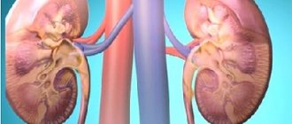
Rapidly progressive extracapillary glomerulonephritis is a special clinical manifestation of the disease, in which changes are detected in the zona glomerulosa of the kidneys. This course of pathology is characterized by the rapid formation and increase of renal failure. The last, terminal stage of the disease quickly occurs, which ends in the death of the patient in the absence of adequate therapy.
Often this clinical manifestation is also called “glomerulonephritis with crescents,” which is the same thing. The disease can be diagnosed as an independent pathology or as a concomitant symptom complicating the underlying systemic disease (vasculitis, lupus erythematosus and other autoimmune diseases). In addition, rapidly progressive glomerulonephritis can be a complication of kidney disease that does not receive timely treatment.
Forms
At the moment, there is no clear opinion about the mechanisms of development of rapidly progressive extracapillary glomerulonephritis, but most doctors agree that damage to the zona glomerulosa of the kidneys in this disease is caused by dysfunction of the immune system. Based on data from many clinical studies conducted, a typological classification was compiled.
Type 1 – anti-BMK
With this development of the disease, the body secretes antibodies that attack the healthy basal cells of the glomeruli of the kidneys. May occur:
- on one's own;
- with Goodpasture's syndrome.
Type 2 – immune complexes
With this course of the disease, the body’s immune system produces special complexes that settle in the tubules of the glomeruli of the kidneys. May occur:
- on one's own;
- with lupus erythematosus;
- after an infection;
- with Schonlein purpura.
Classification of rapidly progressive glomerulonephritisRecommended topic:
Treatment of glomerulonephritis with folk remedies
Recommended topic:
Urinalysis for glomerulonephritis
Type 3 –ANCA
In the blood of patients with this type of disease, special antibodies to the cytoplasm of neutrophil cells are isolated. These protein compounds in the blood plasma actively attack the cells of the basement membranes of the glomeruli of the kidneys. May occur:
- on one's own;
- with Wegener's granulomatosis;
- with microscopic polyangiitis.
In addition to these three forms, recently a fourth type has increasingly been identified, which is a mixture of types 1 and 2. With this form of rapidly progressing extracapillary glomerulonephritis, autoimmune complexes to the basement membrane of the kidney glomeruli and to the cytoplasm of neutrophilic granulocytes are detected in the patient’s blood.
Classification (forms)
In recent years, the classification of chronic glomerulonephritis has been changed. Previously, the disease was divided into types (species) based on symptoms. Today, classifications are based on pathomorphological changes, which are identified by examining a kidney biopsy.
In the CIS countries, the clinical classification , authored by E.M., Tareev. According to this division, there are the following forms of chronic glomerulonephritis:
- Hematuric
- Latent
- Nephrotic
- Hypertensive
- Mixed
According to the classification of Serov V.V. and other authors, based on pathomorphological characteristics the following forms of the disease can be distinguished:
- With half moons
- Diffuse proliferative
- Membranoproliferative
- Mesangioproliferative
- Minimal change nephropathy
- Membranous nephropathy
- Fibroplastic
- Fibrillar-immunotactoid
- Focal segmental glomerulosclerosis
Doctors believe that each of these forms can be acute or chronic.
Instrumental examination of the patient
After interviewing the patient, in which it is necessary to carefully describe all disturbing manifestations, the doctor makes a preliminary diagnosis, which requires confirmation by taking tests and undergoing a series of examinations. It is very important to undergo all tests as quickly as possible and often the patient is offered hospitalization in order to be able to monitor his condition. First of all, laboratory tests of biological fluids are prescribed:
- blood test for antibodies;
- biochemical urine analysis.
Using these two tests, you can determine the correct functioning of the urinary and immune systems, confirm or deny the presence of a subacute inflammatory process in the kidneys or an autoimmune disease.
In parallel with laboratory tests, instrumental diagnostic procedures are prescribed. The most commonly used methods are:
- ultrasound examination of the kidneys and urinary tract;
- X-ray examination of the pelvic organs;
- Magnetic resonance imaging;
- tissue biopsy of the zona glomerulosa of the kidneys;
- diagnostics using radioisotopes;
- urodynamic studies (fixation of the characteristics of the formation and release of fluid from the body);
- uroflowmetry (registration of urine flow parameters).
Such methods of studying the urinary system are used by doctors of urological departments not only for rapidly progressing extracapillary glomerulonephritis, but also for other diseases of the kidneys and excretory tract. Thus, the data obtained after all diagnostic procedures allow the doctor to make the correct diagnosis and prescribe adequate treatment for the patient.
Treatment of the disease
Treatment of glomerulonephritis is aimed at reducing ongoing inflammation at the glomerular level to stop their destruction and usually requires a regimen of immunosuppressive drugs to limit the activity of the immune system:
- For almost all types, corticosteroids remain the main treatment: prednisone and methylprednisolone are nonspecific anti-inflammatory agents that affect many tissues of the body.
- Mycophenolate mofetil is able to achieve partial or complete remission of GN (with the possibility of excluding prednisone and methylprednisolone).
- Highly effective agents in the treatment of glomerulonephritis are cyclosporine and tacrolimus (Prograf).
- Alternatively, drugs prescribed for leukemia are used. Like corticosteroids, they target many cells in the body but therefore also have side effects.
Therapy for primary GN focuses on treating the biological sequelae (edema, proteinuria, hypertension if present, and hyperlipidemia), as well as inducing remission with immunosuppressive drugs:
- Dose escalation of loop diuretics, sometimes with thiazide-type diuretics, may be necessary to maintain euvolemia.
- Angiotensin-converting enzyme inhibitors and angiotensin receptor blockers (prescribed optionally if concomitant hypertension is present) reduce proteinuria to some extent.
- For patients with any renal disorders, antihyperlipidemic agents, including 3-hydroxy-3-methylglutaryl-coenzyme A reductase, are recommended.
With the secondary type of pathology, first of all, the underlying disease that caused the inflammation is treated.
Acute post-infectious GN caused by a bacterial infection such as streptococci should be treated as quickly as possible to prevent it from becoming chronic and causing irreversible kidney damage:
- Antibiotics (eg, penicillins) are used to control local symptoms and prevent infection. It is obvious that antimicrobial therapy does not prevent the development of pathologies, except in cases where treatment is prescribed within the first 36 hours.
- Since patients often have high blood pressure, it must be normalized with antihypertensive drugs, so-called angiotensin-converting enzyme inhibitors. For arterial hypertension or encephalopathy, vasodilators (nitroprusside, nifedipine, hydralazine, diazoxide) are prescribed. The goal of treatment is blood pressure between 125/75 and 130/80 mmHg.
- The accumulation of fluid in tissues (edema) is treated with dehydrating drugs - loop diuretics.
- Glucocorticoids and cytotoxic agents are prescribed for severe cases of poststreptococcal GN.
Since the disease is often caused by an overreaction of the immune system, so-called immunosuppressive therapy, for example with cortisone, is necessary. It slows down the immune system and thereby prevents the progression of the disease.
As a result of inflammation, the filtration function of the kidneys is limited. Therefore, as part of the treatment, nutrition is regulated, which includes a low-protein and low-salt diet and adequate hydration.
Treatment of chronic glomerulonephritis, which leads to kidney failure, may require:
- dialysis;
- kidney transplant
Differential diagnosis
The symptoms of glomerulonephritis with crescents coincide with other systemic kidney diseases, which are characterized by organ failure. Such diseases are:
- relapse of chronic glomerulonephritis;
- an acute form of the same disease, occurring with manifestations of renal failure.
Microscopic examination of biopsy material
It is very important to quickly make the correct final diagnosis and prescribe appropriate treatment, since the outcome of a rapidly developing disease is often unfavorable. However, even after carrying out all the necessary examinations and tests, it is extremely difficult to make a correct diagnosis. A final decision on treatment can only be made after a microscopic examination of a biopsy of renal tissue. Rapidly progressive extracapillary glomerulonephritis is diagnosed only when crescentic formations are found in more than 50% of the glomeruli of the prepared microspecimen. It is very important to collect tissue immediately after suspicion of a fulminant disease.
The pathology can progress rapidly, and great difficulties arise when identifying such a course of the disease. Often hospitals do not have the modern laboratories and diagnostic equipment necessary to make a correct diagnosis.
Treatment of subacute (rapidly progressive) glomerulonephritis
Strict bed rest. Diet, see Acute glomerulonephritis. First, the rice-fruit option, which is a particularly favorable background for antihypertensive and decongestant therapy. It is advisable to systematically use reserpine and hypothiazide, since hypertension can worsen the prognosis of the disease. Due to the severity of the prognosis, treatment with prednisone may be tried in this form of adrenal glycocorticoid disease, with indications of effectiveness (see Acute Glomerulonephritis, Treatment). Corticosteroid therapy should only be used in the early stages of the disease. To achieve a natriuretic effect, treatment should be combined with an infusion of aminophylline 0.24 g in 40 ml of a 40% glucose solution 2-2)/g hours after taking 100 mg of hypothiazide. Hypothiazide at the indicated dose can be combined with aldactone (150 mt per day).
After confirming the diagnosis of subacute (rapidly progressive) glomerulonephritis, it is necessary to immediately begin treatment using therapeutic plasmapheresis, active immunosuppressive therapy, antiplatelet agents, anticoagulants and antihypertensive drugs.
Cyclophosphamide in combination with prednisone is taken orally or, preferably, parenterally. In the form of pulse therapy, cyclophosphamide is used 600 mg intravenously every 4 weeks or more often. When a clinical effect is achieved, the doses of glucocorticoids and cytostatics are reduced to maintenance.
Therapeutic plasmapheresis (10-14 sessions per course), heparin, dipyridamole (400 mg/day) are indicated. For malignant arterial hypertension, a combination of ACE inhibitors and (or) calcium antagonists with diuretics and β-blockers is used.
Treatment of pathology
Quick determination of the cause of the development of the pathological process in the kidneys underlies the positive outcome of the disease. Having made the correct differential diagnosis and found out what caused the body's autoimmune response to the cells of the basement membrane of the glomeruli of the kidneys, the doctor begins treatment.
Who to contact
First of all, the patient turns to the local therapist, who interviews the patient, describes all his complaints, and conducts a physical examination. After identifying the main location of the pathological process, the doctor writes a referral to a specialist.
A urologist treats diseases of the kidneys and urinary tract. He prescribes all the necessary examinations and laboratory tests and suggests hospitalization to the patient to monitor the dynamics of the condition. If glomerulonephritis with crescents is suspected, therapy begins in parallel with diagnostics, since the results may come too late for the patient. 4–6 weeks after the onset of the disease, changes in the zona glomerulosa of the kidneys become irreversible and the prognosis for the patient will be sad.
Methylprednisolone
Treatment of rapidly progressive extracapillary glomerulonephritis requires rapid and active methods. The transient negative dynamics of the patient’s condition and increasing acute renal failure endanger the patient’s life. First of all, the doctor prescribes immunosuppressants and glucocorticoid hormonal drugs. Often such drugs are used in doses significantly higher than recommended. This therapy allows you to quickly stop the development of the pathological autoimmune process occurring in the kidneys and reduce the manifestations of renal failure. Multiple clinical studies show that the most effective drugs for shock therapy for this course of glomerulonephritis are Methylprednisolone and Cyclophosphamide.
After achieving relative stabilization of the patient's condition, intensive renal treatment is begun using plasmapheresis. At the same time, the patient continues to be given immunosuppressive drugs. Sometimes such a procedure can be carried out before shock therapy, provided that the patient was admitted at the very beginning of the disease and is in satisfactory condition, which does not require the use of large doses of drugs.
In addition to plasmapheresis, patients are prescribed hemodialysis. This procedure is very painful and takes place only in the absence of irreversible changes in the kidney structures and tissues. Otherwise, this method will not bring results.
Diet
Diet
After stabilizing the condition of a patient with rapidly progressing extracapillary glomerulonephritis, the urologist prescribes constant medication and gives recommendations on nutrition and maintaining a healthy lifestyle. Naturally, the patient will have to completely give up bad habits (smoking, alcohol abuse), if he has any. Slot machines can provide large payouts if they are not skewed towards the casino, as happens in many dishonest gambling establishments. To get the most out of the slots, you should try Club Vulcan slot machines. They captivate you from the first spin of the reels, give you a lot of positive emotions, and of course fair winnings.
As with all kidney diseases, a patient with a similar form of glomerulonephritis is prescribed diet No. 7a. This diet is aimed at maintaining proper kidney function without unnecessary overload and limits fluid and protein intake. Following such a diet allows you to lower blood pressure, reduce swelling of the limbs and the whole body, and restore the water-mineral balance of the body.
Basic principles of a therapeutic diet:
- Reduce the amount of liquid consumed per day to 0.8 liters.
- Small meals, up to 5 times a day.
- Complete exclusion of salt from the patient’s diet.
- Reducing protein intake to 70 g per day.
It is recommended to limit or completely eliminate:
- black bread;
- broths;
- fatty meat, fish;
- smoked meats, sausages, pickles and canned food;
- beans, mushrooms;
- onions, garlic in any form;
- cocoa, coffee and products containing them;
- strong black tea;
- sodium mineral waters.
Smoked meats
This diet reduces the load on the kidneys and is recommended not only during treatment, but also in later life.
↑ Pathological anatomy
According to the topography of the process, intra- and extracapillary forms are distinguished; according to the nature of inflammation - exudative, proliferative (productive) and mixed.
↑ Intracapillary glomerulonephritis
Intracapillary glomerulonephritis, which is characterized by the development of a pathological process in the vascular glomerulus, can be exudative, proliferative or mixed. Intracapillary exudative glomerulonephritis is spoken of when the mesangium and capillary loops of the glomeruli are infiltrated by neutrophils, and intracapillary proliferative (productive) is when proliferation of endothelial and especially mesangial cells is noted, the glomeruli increase in size and become “fingered.”
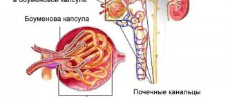
↑ Extracapillary glomerulonephritis
Extracapillary glomerulonephritis, in which inflammation develops not in the vessels, but in the cavity of the glomerular capsule, can also be exudative or proliferative. Extracapillary exudative glomerulonephritis can be serous, fibrinous or hemorrhagic; Extracapillary proliferative glomerulonephritis is characterized by proliferation of glomerular capsule cells (nephrothelium and podocytes) with the formation of characteristic crescents.
Based on the prevalence of the inflammatory process in the glomeruli, diffuse and focal glomerulonephritis are distinguished.
Morphological changes in the kidneys during glomerulonephritis concern not only the glomerular apparatus, although glomerulitis is the leading one in assessing the process, but also other structural elements - tubules, stroma, vessels. In this regard, glomerulonephritis is distinguished with a tubular, tubulo-interstitial or tubulo-interstitial-vascular component. Depending on the nature of the course, as already mentioned, acute, subacute and chronic glomerulonephritis are distinguished. The pathological anatomy of each of them has its own characteristics.
↑ Acute glomerulonephritis
Acute glomerulonephritis, which is usually caused by streptococcus (post-streptococcal, bacterial, glomerulonephritis), and the pathogenesis is associated with circulating immune complexes (immune complex glomerulonephritis), can last 10-12 months. In the first days of the disease, glomerular hyperemia is pronounced, which is quickly joined by infiltration of the mesangium and capillary loops by neutrophils. It reflects the reaction of leukocytes to heterologous complement-containing immune complexes. All renal glomeruli are involved in the process. Soon proliferation of endothelial and especially mesangial cells appears, and the exudative reaction decreases. When leukocytes predominate in the glomeruli, they speak of an exudative phase; when proliferation of glomerular cells is combined with leukocyte infiltration, they speak of an exudative-proliferative phase; with a predominance of proliferation of endothelial cells and mesangiocytes, they speak of a proliferative phase of acute glomerulonephritis.
Sometimes in severe cases, acute glomerulonephritis has morphological changes characteristic of necrotizing glomerulonephritis: fibrinoid necrosis of the capillaries of the glomerulus and afferent arteriole along with capillary thrombosis and infiltration of neutrophilic leukocytes.
In acute glomerulonephritis, the kidneys are slightly enlarged and swollen. The pyramids are dark red, the bark is grayish-brown with small red speckles on the surface and cut or with grayish translucent dots (variegated bud). However, in some cases (death in the first days of the disease), the kidneys at autopsy may appear completely unchanged and only histological examination reveals diffuse glomerulonephritis.
Kidney changes characteristic of acute glomerulonephritis are often completely reversible. However, in some cases they persist for more than a year (acute extended glomerulonephritis) and can transform into chronic.
↑ Subacute glomerulonephritis
Subacute glomerulonephritis develops due to damage to the glomeruli of the kidneys by both circulating immune complexes and antibodies. It progresses quickly (rapidly progressive glomerulonephritis), and renal failure occurs early (malignant glomerulonephritis). Extracapillary productive changes in the glomeruli are characteristic - extracapillary productive glomerulonephritis. As a result of proliferation of the capsule epithelium (nephrothelium), podocytes and macrophages, crescent formations (“crescents”) appear, which compress the glomerulus. The capillary loops undergo necrosis, and fibrin thrombi form in their lumen. Fibrin masses are also found in the capsule cavity, where they enter through microperforations of the capillary membranes. Accumulations of fibrin in the cavity of the glomerular capsule contribute to the transformation of epithelial “crescents” into fibrous adhesions or hyaline fields. Changes in the glomeruli are combined with severe degeneration of nephrocytes, edema and infiltration of the renal stroma. Sclerosis and hyalinosis of the glomeruli, tubular atrophy and fibrosis of the renal stroma occur early.
The kidneys in subacute glomerulonephritis are enlarged, flabby, the cortex is wide, swollen, yellow-gray, dull, speckled with red and well demarcated from the dark red medulla of the kidney (large variegated kidney) or red and merges with the full-blooded pyramids (large red kidney ").
↑ Chronic glomerulonephritis
Chronic glomerulonephritis is an independent disease that occurs latently or with relapses, sometimes stretching for many years and ending in chronic renal failure. The cause of the development of chronic glomerulonephritis is usually unclear, but the mechanism of its development is well studied: in 80-90% of cases it is associated with circulating immune complexes. Chronic glomerulonephritis is represented by two morphological types - mesangial and fibroplastic (sclerosing).
↑ Mesangial glomerulonephritis
Mesangial glomerulonephritis develops due to the proliferation of mesangiocytes in response to deposits under the endothelium and in the mesangium of immune complexes. In this case, there is an expansion of the mesangium of the vascular bundle of the glomeruli and the accumulation of matrix in it. During the proliferation of mesangiocytes, their processes migrate to the periphery of the capillary loops (interposition of mesangium), which leads to “detachment” of the endothelium from the membrane and under a light microscope is defined as thickening, double-circuiting or splitting of the basement membrane of the capillaries. Depending on the severity and nature of changes in both the mesangium and the walls of the glomerular capillaries, mesangioproliferative and mesangiocapillary variants of mesangial glomerulonephritis are distinguished.
In mesangioproliferative glomerulonephritis, the clinical course of which is relatively benign, proliferation of mesangiocytes and expansion of the mesangium are noted without significant changes in the walls of the glomerular capillaries. Mesangiocapillary glomerulonephritis, characterized by the rather rapid development of chronic renal failure, is characterized not only by pronounced proliferation of mesangiocytes, but also by diffuse damage (thickening and splitting) of the glomerular capillary membranes due to mesangial interference. Sometimes, due to the proliferation of mesangiocytes and an increase in the mesangial matrix in the center of the lobules, the capillary loops are shifted to the periphery and compressed, which determines early hyalinosis of the vascular lobules of the glomeruli. In such cases, we speak of lobular glomerulonephritis as a type of mesangiocapillary.
Mesangial glomerulonephritis is characterized by changes not only in the glomeruli, but also in the tubules (dystrophy, atrophy) and stroma (cellular infiltration, sclerosis).
Kidneys with mesangial glomerulonephritis are dense, pale, with yellow spots in the cortex.
↑ Fibroplastic glomerulonephritis
Fibroplastic (sclerosing) glomerulonephritis is a collective form in which sclerosis and hyalinosis of capillary loops and the formation of adhesions in the capsule cavity complete the changes characteristic of other morphological types of nephritis. In cases where the majority of glomeruli undergo fibroplastic transformation, they speak of diffuse, and some of the glomeruli - of focal fibroplastic glomerulonephritis. With fibroplastic glomerulonephritis, especially diffuse, dystrophic and atrophic changes in the tubules, sclerosis of the stroma and blood vessels of the kidneys are pronounced.
Kidneys with fibroplastic (diffuse) glomerulonephritis may be slightly reduced, with small recesses emerging on the surface; they are usually dense, gray-red.
Chronic glomerulonephritis, as a rule, undergoes evolution into secondary (nephritic) shrinkage of the kidneys (secondary shrinkage of the kidneys). The size of the buds decreases, they become dense, their surface is fine-grained (with uniform wrinkling - smooth). The granular nature of the surface of the kidneys is explained by the fact that areas of sclerosis and atrophy (retraction) alternate with areas of hypertrophied nephrons (bulging). On the section, the layer of renal tissue is thin, and the cortical substance is especially thinned; The kidney tissue is dry, anemic, gray in color.
On microscopic examination, atrophy of the glomeruli and tubules and their replacement with connective tissue are noted in the sunken areas. The glomeruli turn into scars (glomerulosclerosis) or hyaline balls (glomerulohalinosis). In the bulging areas, the glomeruli are somewhat hypertrophied, their capsule is thickened, and the capillary loops are sclerotic. The lumen of the tubules is expanded, their epithelium is flattened. Arterioles are sclerotic and hyalinized. In small and medium-sized arteries, fibrosis and intimal hyalinosis are observed, sclerosis and histiolymphocytic infiltration of the stroma are pronounced.
With glomerulonephritis, especially chronic, not only the kidneys are affected; a number of extrarenal changes appear. Because of arterial hypertension, hypertrophy of the heart, mainly the left ventricle, and changes in blood vessels develop - arteries (elastofibrosis, atherosclerosis) and arterioles (arteriolosclerosis) of the brain, heart, kidneys, retina, etc. In this regard, it should be emphasized that changes in the kidneys in chronic glomerulonephritis are associated not only with glomerulitis and its evolution into glomerulosclerosis, but also with nephrogenic arterial hypertension, arteriolosclerosis. With secondary shrinkage of the kidneys, the presence of arterial hypertension, cardiac hypertrophy and vascular sclerosis, the differential diagnosis of chronic glomerulonephritis and the renal form of hypertension is sometimes very difficult.
Consequences and complications
Complications that arise after stabilization of the patient's condition are determined solely after determining the number of glomerular failure affected. Complete remission of the disease is possible, but requires maintenance therapy for the next 2–2.5 years.
As complications after suffering from rapidly progressive extracapillary glomerulonephritis, the following may occur:
- hypertonic disease;
- strokes;
- chronic renal failure;
- possible development of heart failure.
Patients who receive timely treatment can avoid all possible consequences of the disease. If medical care was not provided on time, the patient requires a donor organ transplant.
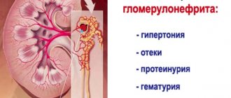
Symptoms and signs of subacute (rapidly progressive) glomerulonephritis
The disease begins suddenly. All clinical syndromes characteristic of acute nephritis are expressed to the maximum. Heart failure is less common. From the first days of the disease, massive swelling, sometimes swelling of the cavities, severe hypertension: systolic pressure approaches 200 mm Hg. Art. or even higher, and diastolic 105-120 mm Hg. Art. and higher. High protevnuria (over 3.5-4 g per day), significant hematuria, sometimes gross hematuria, cylindruria and often leukocyturia, oliguria, more or less pronounced. Already within the first week, a clear decrease in glomerular filtration and azotemia are observed. In the 2nd week, hypertension and swelling do not tend to reverse and even progress. Diuresis does not increase. The specific gravity decreases slightly. The disease is characterized by rapid progression. Death from chronic renal failure is observed after 2 months (3/2-2 years after the onset of the disease).
The onset of the disease is acute, often after a sore throat or hypothermia. The course is rapid with a vivid clinical picture. In some patients, diuresis decreases already in the first days and oliguria may develop. Swelling develops quickly, headaches and insomnia are typical. Thirst and poor appetite are noted.
An objective examination often reveals swelling on the face and lower extremities, and pronounced pallor of the skin. With nephrotic syndrome, there may be anasarca, an accumulation of transudate in the serous cavities (abdominal, pleural and pericardial cavity). Arterial hypertension appears already at the beginning of subacute (rapidly progressive) glomerulonephritis and is usually malignant in nature with a rise in blood pressure of more than 200/110 mm Hg. Art., changes in the fundus in the form of retinopathy and retinal edema. With Goodpasture's syndrome, microscopic polyarteritis, hemoptysis is noted. In older people, symptoms of heart failure are quickly identified.
On X-ray and ultrasound examination, the kidneys are of normal size. Ultrasound reveals parenchymal edema (triangular pyramids). Radioisotope renography reveals symmetrical changes characteristic of chronic renal failure.
Disease prognosis
The general prognosis for the course of the disease is unfavorable. Rapidly progressive morphological changes in the kidneys cannot be fully cured, and serious complications arise that require hemodialysis and other unpleasant procedures. There are cases when doctors decide to remove or transplant a donor kidney. However, even in this case there is no guarantee that a relapse will not occur.
Donor kidney transplant
If the patient does not seek medical help immediately, but the pain becomes unbearable, death is possible. Irreversible changes in the kidney tissue occur as early as 4 weeks of disease development. Thus, the sooner you consult a doctor with disturbing complaints, the greater the chances of successful recovery and minimal consequences and complications.
Diagnostics
Laboratory data • OAM •• Gross hematuria (not always) •• In the urine sediment there are erythrocyte, leukocyte, granular, waxy casts • In blood tests, anemia, leukocytosis, increased concentrations of urea, creatinine • Serological studies •• Increased titer of antibodies to streptococci •• Increase CEC •• Cryoglobulinemia •• Hypocomplementemia •• Abs to BMK •• antineutrophil cytoplasmic Abs.
Instrumental data • Ultrasound and x-ray examination of the kidneys: at the beginning of the disease - an increase in the size of the kidneys, then - a decrease • Kidney biopsy.
Prevention of the fleeting form of the disease
The development of rapidly progressive extracapillary glomerulonephritis is most often associated with causes that are beyond the control of the patient. Autoimmune diseases cannot be prevented, since the structure of the cells of the immune system is encoded at the genetic level. The slightest mutations and changes in the structure of the DNA molecule can lead to the appearance of antibodies that are aggressive to the body’s own tissues.
However, even the frequency of random genetic mutations can be reduced. To do this, you must adhere to several basic recommendations:
- Reduce emotional stress levels.
- Do not spread infectious and viral diseases.
- Reducing the intensity of physical activity, which can deplete the body and reduce the protective properties of the immune system.
- Visiting doctors and regular comprehensive medical examinations.
Such simple rules will prevent the growth of infectious foci in the body, increase the overall tone of the body and help identify the presence of chronic diseases that can lead to dire consequences. It is important to understand that preventing disease is much easier and more pleasant than treating the consequences of neglect of one’s own health.
↑ Pathogenesis
In the pathogenesis of glomerulonephritis, sensitization of the body by a bacterial or non-bacterial antigen with localization of manifestations of hypersensitivity in the vascular glomeruli of the kidneys is of great importance. The role of cooling in the development of diffuse nephritis deserves special attention, since nephritis often develops after repeated or severe single cooling (cold injury). The role of cooling in the development of glomerulonephritis is also evidenced by the seasonal nature of the disease with an increase in acute cases in the winter and spring months. In cases where the development of glomerulonephritis is associated with antigenic stimulation, the formation of antibodies and immune complexes that damage the kidney, we speak of immunologically caused glomerulonephritis. The immunopathological mechanism for the development of kidney changes characteristic of glomerulonephritis is associated in the vast majority of cases with the action of immune complexes (immune complex glomerulonephritis), rarely with the action of antibodies (antibody glomerulonephritis). Immune complexes may contain a heterologous (bacterial) antigen (heterologous immune complexes). Such complexes are associated with the development of immune inflammation in the glomeruli of the kidneys, reflecting an immediate hypersensitivity reaction, which is characteristic of acute and subacute glomerulonephritis. Immune complexes may contain antigen of one’s own organs and tissues (autologous immune complexes). In such cases, morphological manifestations of hypersensitivity of both immediate and delayed types occur in the renal glomeruli. Mesangial forms of glomerulonephritis become an expression of a delayed-type hypersensitivity reaction.
The antibody mechanism leads to the development of autoimmunization, as it is associated with antirenal autoantibodies. Glomerulonephritis in such cases most often has the morphology of extracapillary proliferative, less often mesangial proliferative. A classic example of the antibody mechanism is glomerulonephritis in pneumorenal Goodpasture syndrome, when glomerulonephritis and pneumonia with a pronounced interstitial component and hemorrhages develop simultaneously, which is associated with the commonality of antirenal and antipulmonary antibodies.
Immune reactions that develop on the basement membrane of glomerular capillaries and constitute the pathogenetic basis of glomerulonephritis depend on nervous and humoral influences.