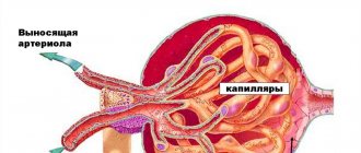
Diffuse changes in the kidney parenchyma are not a disease or a diagnosis, but a statement of disturbances in the structure of the organ. Focal lesions have a specific localization. When they talk about diffuse, this means that changes have occurred in the entire renal structure. They affected the parenchyma (the underlying tissue), the glomeruli, the renal tubules and the sinus (the central internal cavity connected to the pelvis and also the ureter). Blood vessels, lymphatic vessels, and nerve fibers pass through the parenchyma.
Classification of deformities
A diffuse change indicates a disease affecting the functioning and components of the urinary organ. Depending on the location they are divided into:
- changes in the kidney parenchyma;
- deformation of the body and sinuses;
- transformations in the calyces and pelvis.
Clarification of the nature of changes in the structure of the kidney makes a great contribution to further diagnosis. During diagnosis, an enlargement or reduction of the kidney, asymmetry of its contours, thickening or reduction of the parenchyma are detected. Changes in the structure of the collecting system and sinuses, and compactions in the veins of the organ are often detected.
Parenchyma, its functions and norms
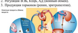
Both layers of parenchyma ensure the normal process of urination, removal of harmful substances from the body and promote internal metabolism. This happens as follows:
Thanks to this, the renal parenchyma performs the following functions:
Diffuse changes in the kidney parenchyma cause problems with these indicators, which, in turn, leads to intoxication and disruption of internal balance and threatens the development of many serious diseases.
The usual dimensions of the organ as a whole and parenchyma are as follows:
Source: https://vsepropechen.ru/pochki/patologii/chto-takoe-parenxima-pochki.html
Detection of abnormalities using ultrasound
Diffuse changes in the kidneys are diagnosed using ultrasound. From the point of view of this method, pathology is divided into clear, unclear, moderate, weak and pronounced changes. An ultrasound machine can observe signs of darkening and blurred contours, anechogenic areas in the parenchyma, zones of hyperechogenicity and anechogenicity in the CL, as well as changes in the contours of the renal pelvis and capsule of the kidney.
Important! In the last months of pregnancy, ultrasound can determine the health status of the fetus's urinary organs.
Features of ultrasound indicators of the kidneys in infants
Diffuse deviations have their own classification. Changes are usually divided into pronounced, weak or moderate, unclear and clear. Diagnosing the pathological condition of infants is not easy, so this type of diagnosis is recommended to be carried out in specialized clinics.
The structure of the kidneys of babies, due to their age, differs from the image of adult organs. A lobulated structure can be interpreted by an inexperienced specialist as a diffuse deviation. Alarming symptoms must be confirmed by other diagnostic methods.
What provokes the pathological condition?
Structural changes in the kidneys are caused by various reasons. During pregnancy, the ureter is compressed by the growing fetus, which leads to hydronephrosis. Congenital pathology of the urinary tract also leads to this outcome. Deformation of the mandibular joint and sinuses of the organ is caused by stones or neoplasms in the system, cysts in the cups and pelvis.
When the sinuses are damaged, a person feels severe pain in the heart muscle and hypertension occurs. Inflammation and sclerosis that develop in a pathological condition cause swelling of the sinus vessels. Incorrect treatment threatens the death of the kidney. In the chronic form of the phenomenon, the organ tends to decrease, and in its acute manifestation, on the contrary, it increases.
Diffuse changes in the kidney parenchyma have a rich symptomatology, because they are provoked by many diseases: tuberculosis, pyelonephritis, nephrosclerosis, glomerulonephritis and cysts.
Causes
Often, diffuse changes in the parenchyma develop during exacerbation of kidney pathologies. Diseases of other organs and systems also provoke negative changes.
Main problems:
Diseases that increase the thickness of kidney tissue
What diffuse changes are has already been discussed above. Consider in more detail the factors causing the pathological condition of the parenchyma.
Polycystic disease
A cyst is an empty cavity or with liquid (sulfur, pus, blood) in the tissue. The cause of formation is inflammation, infection, urolithiasis, tuberculosis, oncology, as well as vascular sclerosis, trauma or genetic predisposition. Depending on the location of the formation, they are divided into sinus and parenchymal. The former are formed from the vessels near the pelvis, the latter - from the kidney canals.
It is difficult to diagnose a renal cyst on your own, since it does not have symptoms. Able to self-remove. But in case of growth and rupture it is very dangerous. Blocks the passage of urine and blood, dilates the pelvis. When ruptured, the contents enter the abdominal cavity, where it causes an inflammatory process. Hemorrhage may begin.
With polycystic parenchyma of both kidneys, the organs are not capable of full functioning.
Acute glomerulonephritis
The cells of the body destroy the capillaries in which the immune complex is deposited during pathology, suspecting them of being harmful. Fibrin is formed in these places, the parenchyma is saturated with leukocytes, and the overlap of the glomeruli creates a problem for their performance. Next, microbleeding, capillary thrombosis, and edema of the urinary organ develop. The disease damages both organs.
Acute type of pyelonephritis
In most cases, the disease affects one organ, and the echogenicity in the renal parenchyma decreases. Accompanying the pathology are changes in the pelvis and sinus. A purulent disease causes foci of inflammation throughout the kidney tissue, alternating them with healthy areas of the parenchyma. A solitary abscess can develop. The kidneys become asymmetrical in size, the tubes and tubules are expanded.
Amyloidosis
Amyloid, accumulating in the kidney tissue, provokes failure of the urinary organs. Upon examination, the cortical zone of the kidney looks expanded, has a red-gray color, and the medullary zone has a greasy appearance. The substance, accumulating in the glomeruli, provokes the death of nephrons, and connective tissue is formed in their revenge.
Diabetic nephropathy
Accumulating in the cells of an organ, glucose poisons the organ. In diabetes, the load on the kidneys increases due to many metabolic processes. Initially, the parenchyma becomes slightly larger. Cystic degeneration is observed. As the disease progresses, at the stage of the patient's need for hemodialysis, the urinary organ decreases.
Causes and symptoms
With diffuse changes in the parenchyma
Often, diffuse changes in the pulmonary function of both kidneys are observed in a child. But this process is not pathological, since up to 3 years of age the child’s kidney is characterized by a lobular structure, and therefore is specifically visualized on ultrasound. The problem is characterized by deterioration of the kidney tissue from the inside. Moderate changes in degree occur regularly when oxygen and nutrients penetrate into the tissues: during eating, breathing exercises. But with pathological destruction, deeper changes occur. At risk are:
Diabetics, overweight people, and smokers are more susceptible to diffuse kidney pathologies.
The following can increase the risk of diffuse changes in the kidneys:
The main reasons why the parenchyma and CLS change diffusely are poor nutrition and alcohol abuse. Common reasons:
The symptoms of the condition are determined by the thickening of the tissue and the resulting asymmetry of the organ in volume. Early signs are swelling, which is caused by problems with capillary permeability and increased blood pressure. As a result, filtering activity towards liquid increases, but its reabsorption weakens. A persistent urination disorder develops, accompanied by pain, burning, and the appearance of blood in the urine. Frequent signs of the onset of diffuse changes are pain in the lower back on both sides, which is caused by an increase in the size and stretching of the capsule of the paired organ.
With diffuse changes in the sinuses of the kidneys
The renal sinus can be indirectly damaged due to stones, atherosclerosis, cystosis, and inflammation in the organ.
Structural changes in the sinuses of the kidneys characterize the development of several pathologies due to the specific location and complexity of the structure of the entrance gate of the paired organ. A compacted renal sinus may indicate the following conditions:
Diffusely heterogeneous changes in the sinuses of the kidneys are accompanied by swelling, pain in the organ and tingling in the heart, problems with blood pressure.
A common cause of diffusion in the sinuses of the kidneys is pedunculitis or fibrolipomatosis - inflammation in the area of the hilum and vascular pedicle of the paired organ. The process is characterized by sclerotic lesions. The reason for the development of fibrolipomatosis is increased renal pressure due to tuberculosis, inflammation of the renal pelvis, the formation of stones in the ureter, and prostate adenomas. When backflowing from the pelvis to the kidneys, urine with toxic decay products enters the intermediate tissue of the body of the kidneys and sinus, leading to inflammation of the lymph nodes and blood vessels with fibrous-sclerotic lesions such as fibrolipomatosis. Against the background of blocking the outflow of lymph in the kidney, congestion worsens, which causes sharp pain in the back, similar to radiculitis.
Diseases that reduce the thickness of kidney tissue
Thinning of the kidney parenchyma occurs due to the progression of chronic pyelonephritis and glomerulonephritis, diabetic nephropathy, nephrosclerosis. Let us consider the features of the development of the pathological condition in individual cases.
Chronic pyelonephritis
It occurs if the disease is not treated correctly, during the transition from the acute to the chronic stage. Pathology destroys the parenchyma and forms scars. When two organs are affected, the kidneys become smaller. In the tissue, it is diagnosed that it is saturated with leukocytes, the death of the tubules and sclerotic zones. An abscess sometimes occurs in the pelvis. Mostly the lesion is one-sided.
Nephrosclerosis
There is a change from parenchyma to connective tissue. It is divided into primary and secondary forms. Primary develops with hypertension. In the secondary type of pathology, the causes of development are varied. As a result of nephrosclerosis, the organ shrinks and the parenchyma undergoes dystrophic changes.
Chronic glomerulonephritis
The diffuse condition is characterized by glomerular hyanilization and tubular atrophy, causing the disappearance of the organ pyramids.
Diffuse changes in the renal tissue of the urinary organ are a symptom of kidney disease. The parenchyma can both thicken and thin. The condition requires medical intervention and treatment of the underlying disease. The causes of anomalies are varied. With the help of ultrasound, you can only ascertain the fact of changes. An accurate diagnosis requires a more thorough examination.
tvoyapochka.ru
Signs and symptoms
With diffuse changes, the walls of the parenchyma thicken, the sinuses of the kidneys enlarge, and the organs differ in size from each other. In the early stages of negative changes, the signs are weak, but as the pathological process develops, symptoms appear that must be paid attention to.
Characteristic manifestations:
Important! Some signs are clearly visible, while other doctors detect them only during an ultrasound or refer the patient for additional examination if urine tests worsen. Annual preventive examinations reduce the risk of advanced renal pathologies.
Based on the results of an ultrasound examination, doctors diagnose the following abnormalities:
Source: https://vseopochkah.com/anatomiya/patologii/diffuznye-izmeneniya.html
Related articles:
- Sorrel kidney stones Causes of formation of kidney stones Nephrolithiasis, as one of the manifestations of urolithiasis, is a widespread pathology. It affects people rich and poor, in cities and villages, living in countries with a high standard of living and in undeveloped economies. Despite its wide representation in the world, [...]
- Ultrasound bruise of the kidney Kidney injury (damage) The kidneys, due to their anatomical position, are to a certain extent protected from external influences. However, they are often damaged by trauma to the abdomen, lumbar region and retroperitoneum. In 70-80%, kidney injury is combined with damage to other organs and systems. IN […]
- Chronic glomerulonephritis what is not possible Chronic glomerulonephritis Chronic glomerulonephritis is a disease of both kidneys, manifested by an extensive inflammatory process that causes changes in the tissues of the organ. Like most chronic diseases, it can go unnoticed for 10-15 years. However, at the same time, […]
- Ultrasound angiolipoma of the kidney ANGIOLIPOMA OF THE KIDNEY Advertising from a consultant: Correspondence consultation is intended only to determine the essence of the problem and ways to solve it, is of a preliminary informative nature and cannot replace a face-to-face consultation with a specialist! Primary diagnosis is not carried out in absentia and specific treatment is not [...]
- Ultrasound signs of dysmetabolic nephropathy Dysmetabolic nephropathy Dysmetabolic nephropathy is a group of diseases that are characterized by kidney damage due to metabolic disorders. Depending on the cause of development, primary and secondary dysmetabolic nephropathies are distinguished. Primary are hereditarily determined [...]
- Herbs for the treatment of chronic pyelonephritis Pyelonephritis and methods of treating it with folk remedies at home There are ready-made pharmaceutical herbal preparations (cyston, canephron, phytolysin). If possible, you can prepare healing infusions yourself. Recipes for getting rid of inflammation of the renal pyelocaliceal system at home […]
- Impact of the kidney symptoms Kidney injury (damage) The kidneys, due to their anatomical position, are to a certain extent protected from external influences. However, they are often damaged by trauma to the abdomen, lumbar region and retroperitoneum. In 70-80%, kidney injury is combined with damage to other organs and systems. IN […]
- Degrees of kidney injury Kidney injury (damage) The kidneys, due to their anatomical position, are to a certain extent protected from external influences. However, they are often damaged by trauma to the abdomen, lumbar region and retroperitoneum. In 70-80%, kidney injury is combined with damage to other organs and systems. IN […]
The structure of the renal parenchyma
- external or outer - cortical
- internal - cerebral
If you examine the parenchyma under a microscope, its outer part consists of small glomeruli, which are entangled with many blood vessels, in which waste liquid is formed - primary urine.
The fluid then enters the medulla, which consists of tubules. They are connected to form a pyramid, through which the processed biological fluid enters the calyces and pelvis. Thanks to the process of reverse absorption (reabsorption), secondary urine is formed when part of the liquid enters back into the blood, and part is excreted from the body along with metabolic products (nitrogenous waste, creatinine, urea, uric acid, etc.).
- cleanses the blood
- removes excess metabolic products
- normalizes water-salt balance
- participates in metabolism
The thickness of the parenchyma changes with age; as a rule, it tends to become thinner.
However, despite all the negative assumptions, we hasten to reassure you, since it can be regenerated and is gradually restored even in those people who have already had some kind of kidney problems. But this largely depends on the immune system and the quality of a person’s metabolism.
This indicator is largely influenced by lipid, carbohydrate, protein, water-salt metabolic processes. If there are any violations, then over time this will have a detrimental effect on the kidneys and their excretory function.
People with diabetes mellitus are at particular risk; even in the earliest stages of endocrine system disease, kidney complications are already possible, which, as a rule, are asymptomatic. These complications of diabetes include
In people aged 15 to 60 years, the thickness of the parenchyma ranges on average from 15 to 22 mm. In older people (over 60 years of age), tissue thinning becomes more pronounced and reaches an average of 11 mm.
Therefore, if the changes occurring in the kidneys are not age-related, then when the patient consults a doctor with complaints about problems associated with the genitourinary system, they may prescribe an ultrasound of the internal organs, through which a more accurate diagnosis is made.
- length from to to 12 cm
- width 5 - 6 cm
- thickness 3 - 4 cm
- weight of 1 kidney is about 200 g
Diffusion treatment
Due to the fact that diffuse changes in the kidneys do not relate to a separate diagnosis, but indicate the development of different types of pathologies, there is no single treatment regimen for the condition. Therefore, based on the results of further research, a final diagnosis is made, determining the stage and form of the pathological process to determine therapeutic tactics. Consequently, the main goal of any treatment regimen is to eliminate the causes that caused diffuse changes in the parenchyma or sinuses of the kidneys.
Diffuse glomerulonephritis, provoked by infection, is treated with specific medications that suppress and eliminate the source of infection. Additional measures include bed rest and a strict diet. Antibacterial medications and hormonal therapy may be required. With pyelonephritis, the problem with the outflow of urine is solved. For this, the patient is prescribed a strict diet, anti-inflammatory medications and diuretics. If nephritis develops due to a blood pressure disorder, taking cardiac and antihypertensive medications and herbal medicine are indicated. Pathologies that cannot be eliminated by conservative methods are treated surgically.
It should be understood that heterogeneous diffusion of the renal parenchyma on one or both sides is not a standard disease that is treated by conventional methods. This requires an integrated approach, both in diagnosis and treatment. Patients with echo signs of diffuse changes are required to be referred to a doctor. It is forbidden to self-medicate or ignore pathology. Such negligent attitude is fraught with the development of acute renal failure.
Source: https://etopochki.ru/bolezni-pochek/inie/diffuziya-pochek.html
Types of parenchyma studies
Despite the ability of the kidneys to regenerate, they are still susceptible to all sorts of pathological formations: cysts, benign, malignant tumors, infections, etc.
The most effective research methods are:
- ultrasonography
- Magnetic resonance imaging
- CT scan
- kidney biopsy
The thickness of the tissue can be used to judge the condition of the organs. If any deviations from the norm are noted, then there is a serious reason to conduct more detailed studies together with testing.
Therapy
Therapy for diffuse changes largely depends on what pathological condition provoked the appearance of such a disorder. If no significant kidney symptoms are observed, the use of renal preparations and other means may be indicated to improve the general condition of this paired organ. Kidney infusions, which include medicinal herbs, allow you to remove harmful substances from tissues and activate the regeneration process. Kidney boron may include the following medicinal herbs:
Among other things, often people who do not suffer from severe symptoms are prescribed a certain diet and drinking regime. These measures, in the absence of chronic or acute diseases, help normalize kidney function and improve their condition.
Considering that often such deviations from the norm are a consequence of diseases such as diabetes mellitus, glomerulonephritis and inflammatory processes of various etiologies, additional diagnostics and targeted treatment of the root cause are required. To determine the cause of the development of pathology, a necessary measure is to conduct laboratory tests of blood and urine, as well as a biopsy of organ tissue. Treatment is further aimed at eliminating the primary disease.
Source: https://apochki.com/drugie-bolezni/diffuznye-izmeneniya-pochek.html
Diffuse kidney changes
Diffuse changes are a very broad spectrum concept. It reflects the final conclusion of the study and is not a diagnosis as such. Signs of chronic organ disorders provoke thinning of the tissue, and infectious effects lead to its thickening and enlargement.
An increase can be provoked by: the presence of fluid in the renal pelvis, a developing abscess, the formation of thrombosis, deformation of the venous structure of the organ or kidney stones.
- blood in urine
- pain when urinating
- pain in the hypochondrium, during an external examination of the patient
The concept of echogenicity is one of the main ultrasound parameters, which reflects the degree of tissue density. Echogenicity decreases as a result of some kind of inflammatory process or swelling.
Increased echogenicity is a consequence of the following diseases and abnormalities:
- diabetic nephropathy
- glomerulonephritis
- metabolic disorder
- inflammation
Normally, the tissue on a kidney ultrasound will be homogeneous.
Additional examinations if suspicious changes are detected
Ultrasound is the main method that determines echo signs of dilated sinuses and areas of increased echogenicity. However, simply stating a fact is not enough, especially for children under 3 years of age. Therefore, to confirm suspicions, they are prescribed a CT scan. Other studies are also being conducted:
- blood tests, urine tests - to determine the level of urea, creatinine,
- daily urine sample according to Zimnitsky to determine the inflammatory process - pyelonephritis,
- CT, MRI - to identify various potential neoplasms,
- excretory urography, which allows assessing the condition of the parenchyma, its structure, and functionality.
After identifying the causes of diffuse changes, the doctor prescribes treatment. It is individual for each patient.
Renal parenchyma cyst
If fluid is retained longer than expected in the nephrons (the part of the kidney that consists of the renal corpuscle, in which filtration occurs and a system of tubules that carry out reverse absorption of fluid - reabsorption), then a cyst may develop in this area.
It has a round shape and reaches sizes from 8 to 10 cm. Due to its size, when it swells, it compresses other renal areas, thereby disrupting the normal functioning of the kidneys.
An ultrasound examination can easily detect any harmful changes, including a cyst, and the following may serve as the basis for referral to an ultrasound: pain in the lumbar region, high blood pressure and detection of blood in a urine test.
But often the disease does not immediately manifest itself and develops asymptomatically, when in general the person feels quite normal.
If you delay and do not start treatment on time, then in the future there will be only one way out - surgical intervention (puncture, open abdominal surgery, etc.)
netdia.ru
Signs of diffusion during ultrasound examination
Diagnostic methods such as CT scanning, MRI and ultrasound help urologists and nephrologists assess kidney health.
Ultrasound is the most common, because it is accessible, does not require complex preparation, and provides a lot of information at a low cost of the procedure.
Signs of diffuse changes in the kidneys and their destructive damage during ultrasound examination:
- It is impossible to determine the veins of the organ;
- The kidney tissue is thickened, expanded, and the size and other indicators can be changed both up and down;
- The echogenicity of the system is weakened;
- The sinus is thinned and an echo signal comes from it;
- The parenchymal tissue has an indistinct contour;
- Blood supply to the system is difficult;
- The tissues of the organ are oversaturated with blood vessels;
- The presence of fluid was detected in the pelvis;
- Identification of echostructures is difficult;
- Reverse blood flow in the arteries of the organ.
Any of these signs may be an indication for a person to be hospitalized. Often diffuse changes in the parenchyma are only a symptom, and the pathology in the system is much more serious. An accurate diagnosis can only be made after a comprehensive study of the health of the human urinary system.
uziwiki.ru
Source: pochki5.ru
Structure
The renal parenchyma consists of two layers:
- cortex , located immediately below the renal capsule. It contains the renal glomeruli, in which urine is formed. The glomeruli are covered with a huge number of vessels. There are more than a million glomeruli themselves in the outer layer of each kidney;
- medulla . Performs an equally important function in transporting urine through a complex system of pyramids and tubules into the calyces and further into the pelvis. There are up to 18 such tubules, grown directly into the outer layer.
One of the main roles of the renal parenchyma is to ensure the water and electrolyte balance of the human body. The contents - vessels, glomeruli, tubules and pyramids - form the nephron, which is the main functional unit of the excretory organ.
The thickness of the renal parenchyma is one of the main indicators of its normal functioning, since it can fluctuate under the negative influence of microbes.
But its size can also change with age, which must be taken into account when conducting an ultrasound examination.
So, in young and middle-aged people, the kidney parenchyma (normal value) is 14-26 mm.
In persons over 55 years of age, the kidney parenchyma (size and normal) is no more than 20 mm. The normal thickness of the kidney parenchyma in old age is up to 11 mm.
Parenchymal tissue has a unique ability to recover, so it is necessary to promptly treat diseases.
general information
When a problem is identified, the size of the bean-shaped organs increases, and a difference between the right and left kidneys is often noticeable. Diffusion is not a disease, it is a tissue condition, a sign indicating the development of negative processes. When deviations are detected, doctors do not treat diffusion, but eliminate the root cause of pathological changes.
Negative symptoms develop in two areas:
Find out about the reasons for the formation of a cyst in the left kidney and about methods of treating the formation.
Read about the characteristic symptoms and treatment methods for nocturia in men on this page.
Study
Diagnostic procedures make it possible to determine the structure of the kidney tissue, examine the internal state of the organ, and timely identify diseases of the urinary system in order to quickly take measures to prevent their spread and aggravation.
Parenchymal tissue can be examined in several ways:
- ultrasonic _ It is carried out for any suspicion of pathological processes. The advantages of the method include the absence of X-ray radiation and contraindications, and the affordable cost of the procedure. Using ultrasound, their number, size, location, shape and condition of the tissue structure are determined. In addition, an ultrasound examination can detect the presence of stones and detect signs of inflammation and neoplasms. Duplex scanning allows you to study renal blood flow;
- CT and MRI. Unlike ultrasound, they are more informative research methods that help identify congenital anomalies, parenchyma cysts of the left and right kidneys, hydronephrosis, and pathologies of blood vessels. They are carried out using contrast enhancement, which has a number of contraindications, so it is prescribed if additional, more in-depth research is necessary;
- biopsy _ It is carried out in stationary conditions. The essence of the method is the examination of microscopic kidney tissue taken from the patient using a special, thin medical needle. A biopsy can reveal: chronic, hidden diseases, nephrotic syndrome, glomerulonephritis, infectious diseases, proteinuria, malignant tumors, cysts. Contraindications: low blood clotting, one working kidney, allergy to novocaine, hydronephrosis, blockage of the renal veins, renal artery aneurysm.
If deviations in the size of parenchymal tissue from the generally accepted norm are detected, it is necessary to contact a specialist for further examination and treatment.
The decision on the choice of diagnostic method should be made by the doctor based on the medical history.
Neoplasms
Sometimes cysts can develop into malignant neoplasms. Unfortunately, this pathology is asymptomatic for a long time and only when the tumor, reaching a large size, compresses the vessels entering the kidney through the sinus, do symptoms appear.
However, all the symptoms will be typical for a disease of the genitourinary system and it is not always possible to identify any of them that are characteristic of a tumor; only the appearance of increasing intoxication, a decrease in the amount of urine excreted and unbearable pain can make the doctor suspect the formation of a tumor.
A characteristic sign of an ultrasound examination of the kidneys, if a malignant formation is suspected, will be diffusely heterogeneous changes in the parenchyma of the organ. This feature is explained by the fact that the tumor-like formation will not have a clear contour, growing gradually, it captures more and more new areas of the parenchyma, sometimes moving to neighboring organs and vessels. Therefore, when you see heterogeneous changes in an organ on the monitor screen, you should think about oncology.
The kidney is a paired organ, therefore, for comparison and to exclude pathology in the other organ, examinations are always performed on both sides. If kidney cancer is suspected, a number of other examinations are prescribed (intravenous urography, MRI, CT, radioisotope examination) and only then a final diagnosis is made.
Diffuse changes in the renal parenchyma
Often, patients are faced with the conclusion of an ultrasound or CT scan: diffuse changes in parenchymal tissue. Don't panic: this is not a diagnosis.
Diffuse means numerous changes in the renal tissue that do not fit within the normal limits. Which ones exactly can only be determined by a doctor after conducting additional examination using tests and monitoring the patient.
Changes may include increased echogenicity of the renal parenchyma, thinning of the renal parenchyma, or vice versa, thickening, fluid accumulation and other pathologies.
Enlargement and swelling of the renal parenchyma may indicate the presence of microliths (stones, calcifications in the renal parenchyma), chronic diseases, and atherosclerosis of the renal vessels. For example, with a parenchyma cyst, tissues are compressed, which negatively affects the processes of formation and excretion of urine from the body.
In most cases, a single cyst does not require treatment, unlike polycystic disease, which is dangerous for the body as a whole.
Multiple parenchymal cysts must be removed surgically.
If the kidney parenchyma is thinned (unless we are talking about elderly patients), it may indicate the presence of advanced chronic diseases. If they were not treated, or the therapy was inadequate, the parenchymal layer becomes thinner and the body is unable to function normally.
To detect diseases at an early stage, do not neglect the diagnostics recommended by your doctor.
Features of sinus pathologies
Any disturbances in the structure of the kidney sinuses, such as compaction, diffuse changes and others, contribute to the disruption of the outflow of urine. There are two types of such diseases:
If upon examination of a patient a lump is detected in the kidney area, this may mean the following:
- presence of stones;
- disruption of the structure of the walls of the pelvis, resulting from a long course of chronic pathology;
- the course of atherosclerosis, which resulted in the formation of characteristic plaques in the vessels that run through the sinus;
- improper development of the organ, in which a change in the fiber near the sinus occurs and an inflammatory process occurs;
- the formation of cysts that compress blood vessels.
When considering the question of what a renal sinus is, it is worth noting the following: if any disturbances occur in its structure, it is impossible to immediately determine the true cause of diffuse changes. This is due to multiple symptoms that occur when this part of the kidneys is damaged.
The diffuse process is accompanied by:
- urinary disorders;
- pain in the kidney area and radiating to the lower back;
- heat.
However, people do not always notice signs indicating pathologies of the renal sinuses.
With diffusely heterogeneous changes in the structure, the following are also sometimes observed:
- pain syndrome;
- swelling;
- tingling sensation in the heart area;
- blood pressure problems.
In some cases, when a pregnant woman is affected by a viral infection, as well as while taking certain medications, the unborn child develops a split in the pelvis. This process can occur in two scenarios:
- the formation of two pelvises, which are connected to the bladder by two ureters;
- the formation of double ureters that enter the bladder as a single unit.
Cleft pelvis is more common in women. Such changes lead to the division of the renal sinus into two sections, as a result of which the organ increases in size. Moreover, each of the formed parts is nourished through its own circulatory system. The symptoms in this case are similar to those described above. Doubling the cups of the pelvis does not lead to serious violations.
- Renal parenchyma: structure, functions, normal indicators and changes in structure
Focal changes
Focal changes are neoplasms that can be either benign or malignant. In particular, a simple cyst is benign, while solid parenchymal tumors and complex cysts are most often carriers of cancer cells.
A neoplasm can be suspected based on several signs:
- blood impurities in the urine;
- pain in the kidney area;
- a tumor noticeable on palpation.
The listed symptoms, if present together, unmistakably indicate the malignant nature of the pathology.
Unfortunately, they usually appear in an advanced stage and indicate global dysfunction.
The diagnosis is made based on research:
- Ultrasound;
- computed tomography;
- nephroscintigraphy;
- biopsies.
Additional methods for studying focal changes that allow us to determine the presence of a blood clot, the location of the tumor, and the type of vascularization necessary for effective surgical treatment:
- aortography;
- arteriography;
- cavography.
X-ray and computed tomography of the bones of the skull, spine, as well as CT of the lungs are auxiliary examination methods if the spread of metastases is suspected.
For malignant tumors in the kidney parenchyma, treatment is usually surgical, which often involves removing the affected organ. For benign tumors, organ-preserving operations are performed, the purpose of which is to excise the tumor with minimal harm. After surgery, cancer patients are given radiation therapy.
Single metastases in the spine and respiratory organs are not a contraindication for nephrectomy, since they can also be excised.
mkb.guru
Hello, dear doctors. I have health problems, complicated by an unbearable quality of life, I wouldn’t wish it on my enemy. I decided to turn to you for advice, relying on your experience in gastroenterology. I am 41 years old. Since I was 20, I have been eating irregularly, very often fried, salty, and spicy. I’ve been smoking since I was 19 (I’m going to quit soon), and until the beginning of 2012 I often didn’t mind drinking beer with smoked fish, which I now really regret. Now I have given up on this and try not to break my diet. A month ago, I went to the gastroenterology department and was discharged with a diagnosis of: Chronic alimentary-toxic pancreatitis, a painful recurrent form with impaired exocrine function, of moderate severity. Hepatomegaly, fatty hepatosis, stage 1 pancreatic lipomatosis, signs of capitate pancreatitis. Superficial gastritis, helicobacter pylori significant amount. Duodenitis. The hospital did not prescribe any treatment, only diet and digestion. enzymes. Moderately diffuse changes in the pancreas. According to ultrasound scan: nonspecific structural changes in both kidneys. Minor pyeloecstasy on the right. Now I am worried about pain in the left kidney. But the biggest problem that poisons life is the constantly elevated body temperature (heat in the body both during exercise and in its complete absence), constant toxic perspiration with a terrible smell of sour fat and dust. I take a bath twice a day and change my clothes. I quit my job, people look at me like I’m an enemy of the people, you can’t get on public transport! It started about three years ago, with mild symptoms, but now it’s unbearable. I wake up 3-4 hours later, my entire forehead is covered with fat with a pungent odor. When I dress and put on the lightest clothes, my body begins to sweat, my legs “burn,” emitting a pungent odor. One friend told me that ““this could be due to diseased kidneys, and your tests are bad.”“ About 6 years ago, at a veterinary clinic, I was treated for gonorrhea, and even earlier for ureaplasmosis, could this be related to this? I don't observe any symptoms. I am enclosing the results of ultrasound and tests and several MSCT images. Please advise, if you know, any solution to the problem, the doctors with whom I spoke cannot say anything. I really hope for your valuable recommendation, for me this is a vital issue.
www.consmed.ru
Glomerulonephritis
As a rule, glomerulonephritis occurs as a result of previous infectious diseases:
- angina;
- otitis;
- scarlet fever;
- pneumonia.
The immune restructuring of the body, provoked by bacterial microflora, causes the kidneys to perceive their own tissues as foreign, exposing them to attack by protective complexes. In a healthy body, immune complexes must be neutralized in the liver, but if this does not happen, the vessels of the renal glomeruli are subject to destructive effects.
With glomerulonephritis, the kidney is usually of normal size, but may be enlarged. The structure of the parenchyma is uneven, due to the enlargement of the renal glomeruli, the vascular system is poorly defined, multiple hemorrhages and microscopic exudative cavities may be present.
Hematuria is one of the diagnostic signs of glomerulonephritis


