Various neoplasms often appear in the kidneys. This happens especially often in people with a genetic predisposition. The most common formations in these organs are cysts. Their types are diverse. One of the most dangerous is a subcapsular cyst of the left or right kidney. It practically does not manifest itself at all and can develop into a malignant tumor.
What is a subcapsular type renal cyst?
Any cyst is a fluid-filled capsule in the thickness of the tissue. Such neoplasms can appear in the thyroid gland, ovaries and other organs. The average size of cysts - depending on the location - ranges from 2 to 4 cm in diameter. Larger specimens require especially close attention.
A subcapsular kidney cyst can reach a diameter of 10 cm or more. In most cases, doctors deal with simple formations, which are divided into 3 types:
- Intraparenchymal, that is, located directly in the thickness of the tissue of the diseased organ;
- Parapelvical - in the area of the kidney gate;
- Subcapsular - under the renal capsule.
As a result, a subcapsular cyst is a simple neoplasm located in a certain area of the kidney. That's what it is. From the inside it is filled with the so-called serous fluid. If the swelling occurs after an injury, the fluid may include particles of pus or blood.
When it comes to the threat of degeneration into a malignant tumor, there are 4 categories:
- The first, also known as primary, accounts for about 70% of all neoplasms. They are benign and do not require immediate medical attention. Easily diagnosed using ultrasound, MRI and other examination techniques.
- The second category is benign tumors that are prone to malignancy. Their main feature is the presence of calcium deposits in the walls. Typically, such structures do not exceed 3-3.5 cm.
- The structure and forecasts of education in the third category are more complex. They already have thick walls and several partitions. The picture is aggravated by the presence of multiple calcium deposits. They are extremely difficult to detect using hardware methods and usually exceed 3 cm in diameter. Such structures require special attention and surgical treatment methods.
- The fourth group is the most dangerous. These are tumors with uneven contours and heterogeneous structure. They have a pronounced tendency to become malignant.
What is the disease?
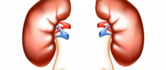
The kidneys are surrounded by fibrous tissue, which serves as a kind of membrane. This type of epithelium is connective and is capable of stretching. Under the influence of certain factors, a growth develops between the fibrous and storage tissue (parenchyma) - a subcapsular cyst. The problem can form in the kidney on the left and right side with equal probability. New growths are usually small - up to 5 cm in diameter. In most cases, the disease does not affect the functioning of the ureter, the vessels inside the organ and the bladder, but if the cyst is the result of an injury, it can become filled with pus, which is no longer so safe.
According to medical statistics, a cyst is a problem that exists in the body of 60% of the population.
Symptoms
Benign tumors appear in the right or left kidney or in two organs at once. Depending on the location, there is a subcapsular cyst of the left kidney and, accordingly, the right kidney. Depending on the location of the tumor, symptoms also appear. If there is a subcapsular cyst of the right kidney, then the pain will be stronger on the right side. And vice versa.
In general, the disease described has the following symptoms:
- Intense dull pain in the right/left side and lower back;
- Problems with urination;
- Increased blood pressure;
- Every now and then emerging infectious diseases of the genitourinary system;
- The presence of some blood in the urine (hematuria);
- Availability of seals.
The kidney, which contains one or more compaction cysts, increases in volume, puts pressure on neighboring tissues and thereby causes constant pain. An increase in blood pressure is also associated with this. Sometimes the diseased organ reaches such a size and density that it can easily be felt from the abdomen.
It is better to talk about localization, symptoms and treatment with your doctor. Without appropriate medical training and a complete examination of the diseased organ, it is a mistake to make any diagnoses.
Subcapsular kidney cyst: treatment of the right and left kidneys
A subcapsular renal cyst is a cavity formation localized under the renal capsule. Inside it is filled with liquid contents. If a neoplasm forms after injury or damage, fragments of pus and blood may be present in the cavity.
In most cases, formations do not exceed 5 cm and do not interfere with the processes of formation and excretion of urine. Cystic formations can form on one or two kidneys.
According to the degree of likelihood of degeneration into cancer, the subcapsular kidney cyst is divided:
- Most formations (up to 70%) are benign subcapsular formations that are not prone to malignancy. Simple neoplasms do not require urgent medical intervention and are accurately detected during diagnostic procedures;
- Neoplasms with the presence of calcium in the walls. They have a low risk of malignancy;
- Formations that have a more complex structure: dense walls with the presence of several cavities, numerous deposits of calcium salts. Cysts are difficult to detect during diagnosis; in most cases, their size does not exceed 3 cm. Surgical treatment is used;
- A complicated type of neoplasm with a multi-cavity structure, unclear contours, and a pronounced tendency to malignancy.
The main factors for the development of subcapsular renal cysts are:
- Excessive growth of epithelial tissue in the tubules of the organ, as a result of which the outflow of urine is disrupted, an increase in the size of the excretory tubule and the cessation of its functioning. A cystic cavity forms in the enlarged renal tubule;
- Necrosis of a section of kidney tissue. In case of death of a tissue fragment, the neoplasm is prone to self-resorption, treatment is not required;
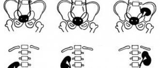
- One of the reasons for the formation of a subcapsular cyst in the kidneys is a congenital factor. Education occurs due to disturbances in fetal development during the embryonic period;
- The formation of a neoplasm can be a consequence of mechanical damage, trauma, complications due to diseases of the urinary system (pyelonephritis, stone formation, glomerulonephritis).
Benign neoplasms develop in one of the organs and can affect 2 kidneys at once. Depending on the location of formation, a subcapsular cyst of the left kidney, a formation of the right organ, or subcapsular neoplasms of both kidneys are distinguished. Localization also determines the main symptoms - pain and heaviness of the affected kidney.
Associated manifestations are:
- Impaired urination. Pain and discomfort occur during urine excretion;
- Increased blood pressure (blood pressure);
- Regularly recurring infections of the genitourinary system;
- There may be blood in the urine.
Particularly large neoplasms can be detected by palpation (from the abdomen). An enlarged cyst puts pressure on nearby tissues, causing pain and blood pressure fluctuations.
The most dangerous complication of a subcapsular cyst is malignancy. Other serious consequences of kidney cysts include:
- As a result of rupture of a large kidney tumor, bleeding occurs and peritonitis (inflammation of the peritoneum) develops. The condition requires immediate medical intervention;
- Regularly occurring infectious lesions of the urinary system;
- Suppuration of a kidney cyst. Associated symptoms: severe pain in the projection of the tumor, fatigue and weakness, fever. Requires surgery and antibiotic therapy;
- An increase in the size of the formation leads to compression of the blood vessels of the organ. The development of renal failure and uremia is possible.
The development of a subcapsular cyst of the right kidney is no different from the formation on the left organ. The location of the neoplasm does not affect its structure or size.
Laboratory studies do not determine the presence of a cystic formation; these diagnostic methods are needed to assess the general condition of organs, identify disturbances in their functioning, and the presence of inflammatory processes.
The most informative methods are:
- Ultrasound examination of the kidneys. The procedure determines the location of the tumor, the degree of damage, and the effect on neighboring tissues;
- Computed tomography with the introduction of a contrast agent to determine the cause of the tumor;
- Magnetic resonance imaging (MRI);
- Radioisotope study to determine the nature of the cyst: benign, malignant.
If the cyst is small in size and does not pose a serious danger, the doctor chooses dynamic monitoring of the pathology. In some cases, subcapsular neoplasms resolve on their own without requiring additional therapeutic measures.
In most cases, drug therapy is prescribed for the treatment of subcapsular renal cysts. Surgical intervention is performed for complicated neoplasms, the development of purulent processes, and a high risk of malignancy.
The main goal of drug treatment is to relieve symptoms, prevent the development of infection, and improve the patient’s condition. Drugs prescribed:
- Lowering blood pressure (Enap, Enalapril). In some cases, a water-salt imbalance develops in the patient’s body, causing an increase in blood pressure. An additional recommendation for taking medications that lower blood pressure is to consume at least 2 liters of fluid per day;
- Painkillers;
- If an infection is detected, antibiotics are prescribed;
- Antimicrobial agents prescribed to relieve inflammation and prevent its further development.
To prevent infection of the urinary system, it is necessary to abandon a number of procedures that may contribute to the penetration and development of infection (gynecological examinations, change of sexual partner, catheterization). It is necessary to avoid hypothermia and observe the rules of intimate hygiene.
The following are used as outpatient treatment methods:
- Puncture sclerotherapy. The procedure is prescribed for small subcapsular formations. During the operation, the contents of the capsule are sucked out through a thin needle. Instead, a sclerosing substance is injected, gluing the walls of the cavity. In most cases, alcohol is used. After the operation, observation in the hospital is not required;
- Retrograde intrarenal surgery. The procedure is performed under general anesthesia. During the operation, an endoscope is inserted through the urethra. The subcapsular cyst is excised, its walls are sutured to nearby tissues.
Removal of a subcapsular renal cyst is necessary in the following cases:
- High probability of malignancy of the subcapsular neoplasm;
- Inability to normalize blood pressure (ineffectiveness of medication;
- Particularly large formations, accompanied by severe pain and serious impairment of renal function;
- Rapid development of neoplasm.
The most common method for removing a subcapsular lesion is laparoscopy. The method is one of the safest and least traumatic; it allows excision of a large subcapsular renal cyst with multiple localizations. During the operation, small incisions (5-7mm) are made on the abdominal wall, through which surgical instruments are inserted.
Abdominal surgery is performed extremely rarely and is prescribed for extensive subcapsular lesions of organs and confirmed malignancy.
The use of traditional methods is acceptable as an additional treatment. Traditional recipes can only be used with the permission and under the supervision of a doctor, as an auxiliary direction of conservative therapy. The most common methods of traditional medicine are the use of burdock and celandine.
To prepare herbal medicine you will need fresh leaves of the plant. The leaves must be washed well and the juice extracted using a juicer. Fresh juice is taken three times a day before meals. The course is 2 months.
A paste made from the leaves of the plant is used as medicine. Young leaves are ground in a blender, the product is taken three times a day, 1 tbsp. before eating.
Celandine
Fresh herbs are crushed in a blender, and the juice is squeezed out of the resulting pulp. Reception begins with 1 drop of juice diluted in 1 teaspoon of water. Each subsequent day, 1 drop is added. The course lasts 10 days.
A serious prognosis is observed with congenital etiology of the disease. The development of multiple cystic formations in most cases does not allow the child to survive until 6 months. Extensive lesions provoke severe complications and pathologies that the immature body of a newborn is unable to cope with.
A favorable prognosis is observed in cases of the development of subcapsular neoplasms in adult patients, with adequate and timely therapy.
Preventive measures for subcapsular renal cysts combine general medical recommendations for strengthening the body’s immune system (feasible physical activity, proper rest, stable psycho-emotional state). Particular attention is paid to diet therapy. The following nutritional rules must be observed:
- The amount of water consumed should be at least 2 liters daily, no earlier than 1.5 hours after meals;
- It is recommended to reduce the amount of salt, in special cases – absolute refusal;
- Smoked foods, fatty and spicy foods, and preservatives are completely excluded;
- Abundant consumption of homemade fruit drinks and compotes. Minimum – coffee and tea;
- Complete cessation of bad habits: smoking, drinking alcohol;
- It is recommended to take vitamin and mineral complexes in courses.
The disease must proceed under the mandatory supervision of a specialist, who must be visited at least once a year. If the patient’s age exceeds 45 years, examination and consultation with a doctor is necessary at least 2 times annually.
Subcapsular kidney cyst: causes and manifestations, treatment methods and prevention Link to main publication
Kidney tumors can appear as cysts; this form of tumor is the most common. As a rule, a cyst forms in the lower or upper part of the kidney. The formation may resemble a ball or an ellipse. Cysts are divided into compound (complex) or simple (single-chamber). Typically, a renal cyst is not life-threatening because it is a benign tumor.
Reasons for education
Most often, a subcapsular renal cyst appears due to excessive growth of epithelial tissue. This is a layer of cells that lines the kidney canals from the inside. If the epithelium grows too quickly, the sloughed cells clog the ducts and interfere with the natural flow of urine. This happens most often due to metabolic disorders.
Since changes in the structure of the organ cause disruptions in its normal functioning, this subsequently leads to the appearance of subcapsular cysts in the left or right kidney.
Other causes of cyst formation are tissue necrosis and congenital changes caused by disorders of embryonic development. In the second case, the appearance of a cyst on the kidney can occur in infancy. Over time, the disease progresses due to the constant flow of fluid through the renal tubules and can progress to subsequent - and more complex - stages.
Causes
In most clinical cases, specialists were able to establish that the main cause of the appearance of a cyst is the active proliferation of epithelial cells. The presented layer lines the inner surface of the kidney. With its active growth, the urinary ducts become clogged, which leads to disruption of the process of urine excretion.
View of healthy kidney epithelial cells. Source: studfiles.net
At the same time, the body experiences abnormal metabolism. Also among the provoking factors, tissue necrosis and congenital changes that were established at the stage of embryo development are stated. In the second case, the formation of a cystic neoplasm occurs in infancy. With gradual progression of the pathology, without proper treatment, the situation becomes more complicated and a benign tumor can become a malignant neoplasm.
Diagnostics
Diagnostic methods for detecting subcapsular cysts are used using hardware. Usually this is an ultrasound, less often an MRI. Interestingly, with the help of ultrasound, the appearance of neoplasms in the fetus can be tracked as early as the 15th week of a woman’s pregnancy. An ultrasound shows that the kidneys have these “capsules” and reads their size, number and location.
If a malignant tumor is suspected, a test such as computed tomography is used. For allergy sufferers who cannot receive a contrast agent, MRI is recommended. This technique allows you to examine a paired organ simultaneously from the left and right sides.
Subcapsular kidney cyst: causes and manifestations, treatment methods and prevention
Congenital developmental defects.
- The tissues of the intratubular epithelium grow rapidly. In this situation, the canal greatly increases in size and no longer participates in the activities of the internal organ. A capsule appears in its place;
- tissue necrosis. Associated with various reasons. Thickening caused by necrosis often does not require therapy because it resolves spontaneously;
- congenital developmental defects. Pathology is formed during the period of intrauterine formation;
- mechanical injuries. A hematoma forms on the kidney, which later transforms into a tumor;
- kidney diseases. The cyst appears after pyelonephritis and other dangerous diseases of the internal organs. It can be caused by urolithiasis.
Under the influence of provoking factors, a cavity is formed filled with liquid, which has virtually no negative effect on the functioning of the urinary system until its size begins to cause concern.
Treatment
Treatment with medications
If the capsule is benign, small in size and simple in structure, drug treatment is used. Often it is symptomatic, that is, it reduces the severity of pain and alleviates the patient’s condition.
The latter is prescribed antimicrobial and painkillers, medications to lower blood pressure and reduce the formation of calcifications in the kidneys. If cystic neoplasms are congenital, it is important for the patient to drink at least 2 liters of water per day. This drinking regime will help maintain the water-salt balance at the proper level.
Medicines affect the entire body, so this technique is effective for both kidneys (both left and right). The doctor determines the type of antibiotics and the course of their administration only after a complete examination of the patient and identification of the nature of the infection present.
And most importantly: the drug therapy described above is applicable only if there is a benign tumor. Malignant neoplasms are treated in a completely different scenario. It is also extremely important for the patient to meticulously follow all the doctor’s instructions. Here's everything you need to know for successful treatment.
Ambulatory treatment
There is no clear answer to the question of how a cyst is treated on an outpatient basis. The doctor chooses the technique depending on the size of the tumor. Usually 2 methods are used:
- Drainage with sclerosis. It is effective in detecting small tumors. In the process, the surgeon inserts an ultra-thin needle into the cavity of the cyst and draws out the serous fluid filling it with a syringe.
- Retrograde intrarenal surgery. Suitable for larger tumors. It is carried out using a laser, which makes a micro-incision on the body of the cyst.
Both operations in modern conditions are considered uncomplicated and quite often practiced.
Surgery
To excise large cysts, as well as to prevent the development of a malignant process, minimally invasive surgery, or laparoscopy, is used. Before removing the tumor, a small incision is made on the abdomen. Through it, a laparoscope is inserted into the internal cavity. Using this instrument and under ultrasound guidance, the cyst is excised.
This operation is minimally traumatic, and the prognosis for patients after it is good. But before cutting anything out, the doctor will examine the existing tumor in detail. Not every tumor/cyst needs surgery. Many can be at the initial stage for years and not bring any suffering to a person. The operation is performed only if there is a real threat of the capsule degenerating into a malignant structure.
Alternative Treatment
Fans of traditional medicine often offer unconventional ways to get rid of kidney tumors. But patients diagnosed with a “subcapsular cyst” should not follow their lead. No infusions or herbs will help get rid of this disease. Addiction to traditional methods of treatment can play a cruel joke and delay the process, which will lead to the formation of a malignant tumor.
Ways to get rid of the disease
Parenchymal disease in the form of a subcapsular neoplasm is treated with various methods. If the formation does not interfere with the functioning of the organ, the patient continues to be observed by the attending physician, periodic examinations are prescribed, and based on the results, a full clinical picture is created. Afterwards treatment is prescribed. This tactic of patient management makes it possible to timely identify the progression of the disease and perform surgery (a last resort).
Treatment with medications
The goal of such therapy is to get rid of symptoms and normalize the patient’s health. The important point is to prevent the development of infection. In case of water-electrolyte imbalance, the doctor establishes a special drinking regimen and prescribes medications that normalize blood pressure.
To eliminate pain, painkillers are prescribed, and if infection develops, antibiotics are prescribed. To prevent an infectious disease, you need to maintain personal hygiene, avoid hypothermia, and it is also recommended to take a course of antimicrobial drugs.
Ambulatory treatment
Outpatient techniques include drainage and retrograde intrarenal surgery. The latter is a manipulation, as a result of which the cyst is excised with a laser. Small formations are treated by puncture (drainage). By inserting a thin needle, the contents are sucked out of the cyst, and instead of it, a substance is injected that glues its walls. This method is the safest way to get rid of a tumor.
Complications
The most important danger is the real risk of the cyst degenerating into a malignant formation, which occurs in 30% of diagnosed cases. This is especially dangerous when different types of tumors simultaneously affect one organ. Thus, along with a subcapsular cyst, a parenchymal cyst, etc. may be present in the kidney.
Other dangerous complications in the presence of a cyst are:
- Internal bleeding;
- Peritonitis (blood poisoning);
- Impaired kidney function;
- Acute intoxication.
All the situations described pose a real threat to the patient’s life and require urgent surgical intervention. In rare cases, treatment with antibiotics is used.
Treatment of the disease
If the cyst is detected by chance and does not bother the patient, then treatment in this case is not indicated, but only dynamic observation once every 6 months.
If the cause of formation is an infectious process, then the following treatment is prescribed:
- Diet, table No. 7 according to Pevzner with limited salt intake.
- Antibacterial therapy taking into account the sensitivity of bacteria to the drug: aminopenicillins - Amoxiclav, Flemoxin, Augmentin, cephalosporins - Ciphalexin, Tsiprolet, Cephalexin, Ceftriaxone, fluoroquinolones - Nolitsin, Moxifloxacin, Norfloxacin.
- Non-steroidal anti-inflammatory drugs - Nurofen, Paracetamol, Ibuklin.
- Antispasmodics – No-shpa, platiphylline, papaverine.
- To normalize the pressure of renal origin - Enalapril, Enap, Lisinopril.
- Diuretics - Veroshpiron, Lasix, Triampur.
- Glucocorticoids – prednisolone, dexamethasone, methylprednesalone.
Surgical treatment is indicated for large tumors or in the presence of a traumatic factor. When deciding on the surgical method of removing a tumor, size is also taken into account. For small sizes, the cyst is drained and the cavity is sclerosed with a special sclerosing agent.
It is possible to perform retrograde intrarenal laser surgery; this procedure is gentle and does not leave a scar on the kidney capsule.
When the size of the cyst is significant, laparoscopic or laparotomy surgery . In this case, the cyst is excised and the capsule is sutured, but the recovery period after such a procedure is longer.
Treatment with folk remedies will be ineffective . Basically, traditional medicine acts as a rehabilitation therapy after the main treatment. The following herbs are used: St. John's wort, chamomile, calendula, rose hips. The pharmacy sells ready-made mixtures, such as kidney tea and diuretic tea. Due to their properties, it is possible to maintain a mild diuretic, hypotensive and antiseptic effect.
Kalanchoe juice has proven itself well . The leaf should be chewed slowly and thoroughly and taken before meals three to four times a day for a month. After a month's break, the course is allowed to be repeated. If after three courses there is no positive effect, then treatment should be stopped.
It is possible to use viburnum juice with honey; for this, the viburnum needs to be crushed and mixed with honey in equal proportions. This mixture can be added to tea or taken separately, 1 teaspoon 3 times a day.
Fresh burdock juice should be drunk for two months three times a day, 1-2 tablespoons. The medicine is taken 30 minutes before meals.
Prognosis and prevention
If the described disease is congenital, then the prognosis is extremely disappointing. Babies with multiple cysts often do not live to see six months. They are at risk of urolithiasis and other complications that a young body often cannot cope with.
If the disease is discovered in adulthood and the necessary measures are taken in a timely manner, then the prognosis is favorable. As a preventative measure, it is recommended to follow a work-rest schedule, avoid hypothermia, and not eat spicy foods that irritate the kidney parenchyma.
Consequences and complications of subcapsular cyst
With delayed diagnosis or lack of treatment, the following complications are possible:
- Rupture of the capsule and the occurrence of peritonitis with infection of the retroperitoneal space and abdominal cavity.
- The transition of a benign formation to a malignant one, according to statistics, 50% develop kidney cancer.
- Internal bleeding.
- Hydronephrosis, due to prolonged pressure on the kidney vessels.
- Disruption of urine outflow with further infection of the kidney.
Symptoms
The formation of a subcapsular cyst far from the site of passage of the urinary ducts in the kidneys leads to an asymptomatic course of the pathological process, which can be detected during routine ultrasound or during diagnostic procedures associated with other diseases. Renal symptoms are practically absent, and their appearance indicates the localization of the cyst near the ureter or significant growth of the cystic cavity. An increase in the size of the subcapsular cyst is accompanied by the appearance of symptoms, the presence of which allows us to suspect the presence of a problem:
- Dull pain appears due to compression of nearby tissues, bundles of nerve fibers by the tumor and an increase in intra-abdominal pressure. If the cyst affects the right kidney, the pain syndrome is often associated with liver pathology, and left-sided pathology requires differential diagnosis with a sluggish course of the infarction.
- The body reacts to the appearance of a neoplasm with increased production of rhinin, an enzyme that takes part in the regulation of blood pressure. With its excessive activity, there is a tendency to hypertension.
- Infectious and inflammatory diseases of the genitourinary system acquire a long course and do not respond well to antibacterial therapy, as a result of which a rise in temperature and signs of general intoxication of the body are often observed.
- Localization of the cyst near the exit site of the ureter leads to disruption of the outflow of urine, swelling increases, and signs of fluid retention appear.
- The growth of the cyst and an increase in the diameter of the kidneys leads to visible changes in the projection area in the lower back. Upon examination and palpation, a compaction with clear contours is detected.
- An increase in pressure in the renal filtration system is accompanied by increased work of the tubules, and visible blood impurities appear in the urine.
In clinical practice, there are cases of complete absence of a clinical picture with a subcapsular renal cyst. The only way to detect a tumor is laboratory and instrumental diagnostic methods.
Subcapsular kidney cyst: causes and manifestations, treatment methods and prevention
The most difficult type of pathology is considered to be congenital. If a cyst is detected in infants, the prognosis is rarely good. In some cases, it causes the death of newborns within a few months after birth.
The development of congenital tumors cannot be predicted, but the likelihood of their occurrence in an adult can be reduced. To do this, you need to follow preventive measures:
- treat identified kidney diseases in a timely manner so that they do not become chronic;
- avoid hypothermia, as well as possible injuries to internal organs;
- follow a gentle diet that excludes some foods;
- Visit your doctor regularly and tell him or her about any worrying symptoms.
A subcapsular cyst is a dangerous neoplasm that requires medical supervision. Its main danger is an increased risk of developing cancer. In 30% of cases, a benign tumor degenerates into a malignant one.
Candidate of Medical Sciences, mammologist-oncologist, surgeon
More articles
Is a parapelvic cyst of the left kidney dangerous?
Parapelvic kidney cysts - what are they? A parapelvic cyst is one of a variety of simple neoplasms in the kidneys. Such cysts are very rare in patients, and according to statistics, they are found in only 1.5% of patients. It is localized in the sinus of the kidney or in the region of the pelvis of the organ. It is worth noting that parapelvic cysts of both kidneys are diagnosed even less frequently than neoplasms in one organ. Due to the fact that it appears in the region of the renal sinus, it is also called a renal sinus cyst.
If this disease is not treated, it can lead to serious complications. Experts note that sometimes it is quite difficult to detect such neoplasms. As a rule, a person becomes aware of their presence when examining other organs or when the following symptoms appear:
- High blood pressure. This occurs as a result of excess renin.
- Problems with urination. The larger the tumor, the more pressure it puts on the ureter.
- Pain. It can be both short-term and sharp, and pulling. Pain appears on the side where the tumors are located.
What it is
First of all, subcapsular renal anomaly is a benign form of neoplasm that develops directly under the kidney capsule. Moreover, inside such a pathology there is serous fluid, and its internal cavity does not have any connections with the ducts.
When the cause of its development is an injury, then in addition to the serous fluid, there will also be blood or pus inside. In addition, a subcapsular renal cyst can simultaneously affect both kidneys, although this is rare.
But when carrying out diagnostics, an indication is always given of where such an anomaly is located.
Drug treatment
If the diagnosis showed the benign nature of the cyst, its small size and simple structure, then medications are prescribed for treatment. Most often, their action is aimed at eliminating unpleasant symptoms and making the patient feel better.
For these purposes, the following types of drugs are used;
- painkillers;
- antimicrobial agents;
- antibiotics;
- drugs to lower blood pressure;
- medications that reduce the concentration of calcifications in the kidneys.
More accurate information about the dosage regimen and dosage of medications is provided by the attending physician based on the established diagnosis.
A congenital pathological process requires control over the water balance in the body. Therefore, a person should drink at least 2 liters of water daily and take medications to lower blood pressure levels.
Drug therapy is acceptable only if the cyst is benign, so experimenting on your own and using medications without the knowledge of a doctor is prohibited.
Outpatient therapy
Based on the size of the formation, it can be drained followed by sclerosis, or retrograde intrarenal surgery can be performed. Thus, the first method is usually used to treat small pathologies. The point of such an intervention is that the doctor pumps out the contents of the cyst through a thin needle, and then injects a substance inside to glue the walls of the formation. It is considered the most gentle method of surgical treatment.
Retrograde intrarenal surgery is performed using a laser, whose role is to cut the membrane of the formation. And in order to ensure that there is no scar left in the place where the incision was, a special thin tube is inserted into the kidney itself.
Laparoscopy
To remove pathologies of significant size, surgical methods with a laparoscope are used. The essence of this operation is to insert a laparoscope and instruments through small incisions, which are used to remove the anomaly. After this, you need to stay in a hospital for a certain time under the supervision of doctors. However, not all cysts need to be removed. Some of them, which are small in size, may only require regular monitoring of their dynamics. Read more about the types of surgical removal of kidney tumors in this article.
Prevention
It is difficult to prevent the development of a subcapsular cyst; specific prevention is impossible due to the small amount of information regarding the exact causes of the formation of the pathological process. General recommendations and standards of behavior related to maintaining health and reducing the functional load on the kidneys are the only preventive way to reduce the risk of developing a cystic kidney tumor:
- Regular scheduled examinations and ultrasound diagnostics once a year help to identify pathology in the early stages.
- Following a gentle diet and rational physical activity help normalize biochemical parameters and increase the body's resistance.
- Timely treatment and prevention of infectious and inflammatory diseases of the genitourinary system reduces the risk of developing subcapsular cystic neoplasms.
- Limiting the weight of heavy objects and eliminating traumatic effects, including those associated with extreme sports.
- Giving up bad habits and reducing the negative impact of toxins serves as a prevention of autoimmune and genetic disorders, and also reduces the risk of malignant degeneration of the cyst.
The main principle of prevention is attention to one’s own health and maximum limitation of exposure to provoking factors.
What are multiple renal cysts?
This designation indicates that a person has many tumors in two kidneys at once. When the number of neoplasms exceeds five, it is generally accepted that these are multiple cysts.
It is worth noting that such neoplasms occur quite often, but patients over 50 are most at risk. There are no very pronounced symptoms of the disease, so it can occur for a long time unnoticed by the carrier. If a person begins to experience discomfort, this means that most cysts have reached more than 5 centimeters in size. Several of them can be noted that may indicate the presence of tumors in the kidneys:
- Severe pain in the lower back.
- Problematic urination.
- Blood in urine.
Subcapsular kidney cyst: treatment of the right and left kidneys
As mentioned above, a kidney cyst can be complex. Simple cysts are differentiated into 3 groups, depending on their location. There are parapelvical, they are located in the area of the renal hilum, intraparenchymatous - located in the renal tissue, and subcapsular - formed under the kidney capsule.
Medicine divides all cysts into 4 categories, each of them reflects the degree of presence of a malignant process in the cystic neoplasm.
- The first category is benign cysts, which are clearly visible using computed tomography, magnetic resonance imaging and ultrasound. Almost 70% of all cysts fall into this group and do not require medical treatment.
- The second category characterizes cysts with insignificant changes. Such a cyst is also benign; it rarely becomes malignant. The second category cyst has walls and calcium deposits in the septa. The neoplasm does not grow more than three centimeters.
- The third category is cysts with a large number of thin septa, which may thicken or contain calcium in the walls. Cysts of this type are difficult to diagnose with ultrasound or x-ray because there is no tissue component. As a rule, such a tumor does not exceed three centimeters. Cysts included in the third category must be under constant observation, and later they are removed surgically.
- Cysts of the fourth category are always removed promptly. Such formations have uneven contours and a lot of fluid inside. A contrast agent accumulates in the cyst, which indicates the presence of malignant cells.
The choice of therapeutic course depends on the size of the cystic formation, as well as on associated complications. Small lumps often do not require treatment. The doctor can take a wait-and-see approach while monitoring the patient's condition. Medications are used in the presence of inflammatory processes and suppuration. Surgery is the last resort if a person’s health or life is at risk.
Medication
Medicines are not able to get rid of the tumor, but they alleviate the person’s condition. First of all, they are aimed at eliminating signs and eliminating inflammatory foci. If the cyst is congenital, then the water-salt balance is usually disturbed. The patient is prescribed solutions that normalize it. In addition, medications are indicated to stabilize blood pressure.
Medicines are not able to get rid of the tumor, but they alleviate the person’s condition.
If the capsule is large and puts pressure on the kidney, causing pain, then painkillers are used. Concomitant infectious diseases are treated with antibacterial agents. Inflammatory processes are controlled with the use of anti-inflammatory medications.
Outpatient
This type of treatment involves puncturing the membrane and removing fluid from it. To do this, in most cases they resort to puncture (if the seal is small). The capsule is pierced with a needle through which the contents are pumped out.
Then a solution is injected inside to help tighten the walls. After the puncture, the patient does not need to stay in the hospital for a long time, since this is a minimally invasive type of intervention.
Surgical
In some cases, drainage of the membrane is impossible, so you need to resort to surgery. This happens in the following cases:
- blood pressure does not decrease even when using strong drugs;
- the process of transformation of a benign tumor into a malignant one has begun;
- the patient suffers from severe pain, and painkillers do not cope;
- the tumor is growing rapidly;
- Kidney activity is impaired.
Excision of tissue using a scalpel.
To remove the capsule, standard tissue excision using a scalpel is used or laparoscopy is used. In this case, small incisions are made through which the cystic formation is removed. After laparoscopy, the patient recovers faster and can leave the medical facility within a few days. Classic abdominal surgery involves a long period of rehabilitation, during which the person feels pain.
Traditional medicine reduces inflammation and slows tumor growth, but this does not apply to subcapsular cysts. With this diagnosis, doctors do not recommend using alternative treatments, as they can worsen the condition. In addition, the patient is wasting his time hoping for a cure. At this time, the cavity increases in size and causes unpleasant complications.
Diagnosis of a cyst in the kidneys
During the examination, the doctor identifies the location of the tumor. The characteristics of the course of the disease do not differ whether it is in the left or right kidney. The doctor indicates which of the paired organs is affected. Laboratory tests do not detect a cyst in the kidney; they are used to evaluate the functioning of the body, including the kidneys.
Informative diagnostic methods are:
- ultrasonography. Detects the size of the kidneys and the number of tumors. You can detect not only a cyst in the kidneys, but also in a fetus developing in the womb from the 15th week;
- CT scan with contrast agent reveals the nature of the tumor;
- MRI. Prescribed if there is an allergy to the contrast agent;
- radioisotope research. Diagnostics determines what type of neoplasm we are dealing with: benign or malignant.
Surgery
In some cases, surgical removal of the cyst cannot be avoided. The likelihood of an operation is determined by the doctor based on the possible risks to the patient’s life.
Main indications:
- high blood pressure levels that cannot be stabilized with medications;
- degeneration of the growth into a malignant formation;
- acute pain that cannot be eliminated;
- critical dysfunction of the affected kidney;
- rapid cyst growth.
Laparoscopy is performed to completely remove the cyst. During the operation, 2 small incisions are made: on the anterior abdominal wall and on the side of the affected kidney. One hole is necessary for inserting a chamber with a lighting device, and the second is for the removal tool. The number of incisions may be greater, at the discretion of the surgeon.
At the end of the procedure, the patient remains in the hospital for another 3-5 days to monitor the dynamics of his well-being.