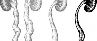
The renal collecting system is dilated, what is it? This is a pathological phenomenon, which almost always indicates the presence of a certain renal pathology. This system, sometimes called the PLS for short, is directly responsible for collecting urine and removing it from the body. In the inner part of the kidney there are small calyces (from six to a dozen), which connect with each other to form larger ones (up to 4 per kidney), and they already exit into the renal pelvis. At the exit site there is a so-called neck - a fairly narrow canal that is easily clogged in various pathologies, causing the expansion of the system components and the growth of the kidneys themselves in size. So, if the renal pelvicalyceal system is dilated - what is it, why does it occur, and how is it treated?
The structure of the pyelocalyceal system
The parenchyma of the CLS consists of an outer part (cortex) and an inner part (medullary substance). The structural units of this system are the calyxes, pelvis and a special structure at their junction - the cervix. Urine is formed due to the filtration of blood plasma in the glomeruli. From there it enters the system of tubules and then into the pyramids, from which it enters first the calyces, and then into the pelvis of the CLS.
Each kidney has 6-12 small calyces, they are connected in groups of 2-3 and merge into large calyces, of which there are 4. The large ones open in the pelvis, where urine accumulates. The pelvis is a funnel-shaped cavity.
Its inner shell consists of tissue that can withstand the aggressive effects of urine. In order for the fluid accumulating in the pelvis to move further into the ureter, smooth muscle tissue is located under the mucosa. They provide peristalsis and urine output.
The neck connecting these two parts of the mandibular joint is quite narrow, so it is vulnerable to urolithiasis, as it can be blocked even by a small stone.
Expansion of the heart rate of both kidneys
The renal collecting system is dilated, what is it? This is a pathological phenomenon, which almost always indicates the presence of a certain renal pathology. This system, sometimes called the PLS for short, is directly responsible for collecting urine and removing it from the body. In the inner part of the kidney there are small calyces (from six to a dozen), which connect with each other to form larger ones (up to 4 per kidney), and they already exit into the renal pelvis. At the exit site there is a so-called neck - a fairly narrow canal that is easily clogged in various pathologies, causing the expansion of the system components and the growth of the kidneys themselves in size. So, if the renal pelvicalyceal system is dilated - what is it, why does it occur, and how is it treated?
Diseases of the organs in question may be acquired or genetically determined.
Congenital diseases are defects of the ureters and the pelvis themselves, these include:
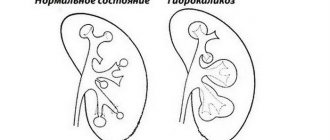
- Hydronephrosis - when the calyces and pelvis are dilated (dilatation of the pelvis), while the kidney parenchyma is atrophied. As a rule, the defect is a consequence of congenital narrowing of the urinary canals; sometimes congenital cases of vesicoureteral reflux (return of urine into the bladder) occur. With it, the structures of the kidneys grow in size, increasing the organs themselves, and the expansion of the central nervous system occurs.
- Narrowing, up to complete fusion, of the ureters, due to intrauterine developmental anomalies. Such phenomena, called strictures, can be either bilateral or develop on one side (for example, the right kidney), and are accompanied by expansions of the entire system. The ureter, like the appendix, ends blindly.
- Doubling the number of pelvis, ureters and calyces. Usually this anomaly does not bother the patient, who may not even be aware of the developmental defect he has all his life;
- Other structural anomalies.
Other reasons:
- kinks of the ureters;
- consequences of kidney ultrasound;
- prolonged retention of the bladder in a full state;
- children may suffer from this pathology if they urinate profusely and infrequently;
- blocking the drainage of urine by kidney stones, inflammatory discharge and other stones;
- excessive drinking;
- urinary tract infections;
- pyelonephritis;
- low tone of the kidney muscles (including due to intoxication);
- urine reflux;
- neurological phenomena;
- other systemic problems, consequences of taking medications, etc.
Disease clinic and diagnosis
An increase in kidney function does not have any specific symptoms, but based on a number of signs, the disease can still be diagnosed quite accurately:
- the patient feels pain in the lower back, groin area;
- frequent, fruitless urges to perform minor necessities;
- slow urination process;
- blood appears in the excreted urine;
- the stomach is bloated;
- the pain is often symmetrical (comes from both kidneys). If it is one-sided, then on the side of the affected organ (for example, the left kidney) it will be much stronger.
In some cases, the patient’s blood pressure increases, fever begins, and tissue swelling appears.
Before starting therapeutic measures, it is necessary to conduct a complete diagnosis:
- Ultrasound of the urinary organs;
- donating blood and urine samples.
This makes it possible to detect pathological changes in organs even in children, including the prenatal period.
Treatment
In cases of expanding inflammatory genesis, the disease is treated symptomatically, the main goal is to stop the inflammatory process.
Medicines prescribed include:
- Indomethacin;
- Diclofenac;
- Voltaren.
They relieve inflammation and relieve pain well.
If a patient experiences muscle spasms in the urinary system, he is additionally prescribed antispasmodics like no-shpa and the like. Patients with a bacterial disease are treated with uroseptics - special antibiotics. This group includes aminoglycosides and fluoroquinolones.
Surgical intervention is indicated if there are difficulties in the outflow of urine. Thus, blockage of the urinary tract is eliminated by crushing the nodules that blocked the ducts using shock wave or contract lithotripsy.
The best treatment for any disease is prevention.
To minimize the risk of developing the disease, you should:
- drink the recommended amount of fluid per day (from one and a half to two liters);
- lead a healthy lifestyle;
- conduct a periodic preventive course of diuretics.
During pregnancy, giving up bad habits and a healthy lifestyle is especially important: this will help prevent the development of congenital defects in the fetus.
What problems may arise
Any pathological process, tissue damage, that is, any deviation from the norm in the condition and functioning of the pyelocaliceal system negatively affects the process of urine excretion and can lead to diseases. In addition, problems may be congenital:
- expansion of the ChLS;
- doubling of kidney structures;
- tissue compaction.
Doubling the heart rate
One of the forms of abnormal kidney development is duplication of the pyelocaliceal system, which is also called incomplete duplication of the kidney. It is not considered a disease, since in most cases people do not make any complaints and often do not even suspect that they have such an anomaly. However, as FLS doubles, the kidney may become more vulnerable to inflammatory diseases.
The anomaly appears during the intrauterine development of the child. There may be a doubling of only one of the structures, or the number of calyces, pelvis and ureters may double. It is possible that from each additional pelvis comes not one, but several ureters, which then merge together, and the common channel flows into the bladder.
The problem arises in cases where the abnormal structure of the organ leads to incomplete excretion of urine from the pelvis, that is, to its stagnation. Disturbances in urodynamics sooner or later lead to the development of diseases; moreover, they create a favorable environment for the proliferation of bacteria, which increases the risk of developing inflammatory processes. With this pathology, the following symptoms are possible:
- lower back pain from an abnormal organ;
- swelling;
- problems with urination;
- blood pressure surges;
- general weakness.
There is no treatment for this pathology as such, and when inflammatory processes develop, symptomatic therapy and the use of antibacterial and anti-inflammatory drugs are prescribed.
Expansion of ChLS
Expansion or scientifically dilatation of the maxillary joint can be congenital, but more often it is acquired for various reasons.
Among the causes of a congenital nature, so-called strictures most often occur, that is, a significant narrowing or fusion of the ureter that occurs in the fetus during pregnancy. In these cases, the ureter is so narrow that urine passes through it with difficulty, or, in general, it ends blindly.
If the renal palsy is enlarged due to other pathological processes, then hydronephrosis is most often diagnosed.
The cause of hydronephrosis is a chronic violation of urine excretion and its stagnation in the kidneys. It is not able to completely pass from the renal pelvis through the ureter into the bladder due to obstacles along the way:
- blockage of one of the structures of the cervical joint with a stone;
- oncological process;
- changes in tissues due to prolonged inflammation;
- traumatic kidney damage.
If the normal movement of urine is disrupted, part of it always remains in the pelvis. As a result, the pressure in the chest joint becomes higher. Because this system is structured with multiple layers of muscle, it can stretch, which initially compensates for overfilling and increased pressure. Gradually, with constant overstretching, the calyx and pelvis are no longer able to return to their normal size. The pathology at the first stage is called calicoectasia and does not yet lead to hydronephrosis.
As the pathological process continues, it spreads to the kidney parenchyma, which leads to deformation of the maxillary joint. Due to constant pressure on the walls of the kidneys, the tissues become thinner, and the process of their blood supply is disrupted. As a result of hydronephrosis, damaged tissues can no longer cope with their functions fully, and renal failure may develop.
The first symptoms of hydronephrosis are:
- aching pain in the lower back, stomach;
- hematuria;
- increased blood pressure;
- swelling.
- developmental anomalies of the pyelocaliceal system;
- kidney injuries;
- urolithiasis disease.
Urolithiasis as a cause of hydronephrosis
Urolithiasis is a urolithiasis, which is one of the reasons for the expansion of the pyelocaliceal system. Particularly dangerous in this regard are stones formed in the pelvis and cervix. The fact is that stones can interfere with the normal outflow of urine, the accumulation of which leads to expansion of the penis and hydronephrosis.
Causes of urolithiasis:
- disruption of metabolic processes in the body;
- insufficient fluid intake;
- unbalanced diet;
- side effects from treatment with certain medications.
Symptoms of kidney stones may not be noticeable as long as urine can pass normally from the kidney. Large stones or the release of stones from the pelvis and blockage of the lumen of the ureter interfere with the flow of urine. This condition is called renal colic. It is accompanied by severe paroxysmal pain, hematuria, and fever.
Inflammatory processes
Inflammatory processes are the most common cause of hardening of the jaw, and one of the most common inflammatory diseases of this organ is pyelonephritis. The process of tissue damage and deformation of the pyelocaliceal system due to inflammation usually occurs gradually with an increase in symptoms and consequences.
Stages of deformation of the maxillary joint during inflammation:
- Alteration is the partial death of epithelial tissue when an infection enters the kidney.
- Exudation is swelling of the walls of the jaw due to the formation of immune complexes.
- Proliferation is an increase in the density of the epithelium in the inflamed area.
Pyelonephritis develops due to the penetration of pathogenic microbes. The likelihood of developing this disease increases with a decrease in immunity, which may be caused by systemic or other inflammatory diseases, hypothermia, and hypovitaminosis. Acute pyelonephritis manifests itself sharply and clearly with severe pain, high temperature, and deterioration in general well-being. With a chronic disease, the symptoms are more subdued.
Main symptoms
In most cases, the expansion of the renal calyx occurs in parallel with pyeloectasia (enlargement of the renal pelvis). But despite the presence of two pathologies, the symptoms are very dull. In young children, even with the dilation of the pelvis over 7 mm, there may be no symptoms of the disease. As mentioned above, hydrocalycosis develops due to stagnation and, as a result, the infectious-inflammatory process begins to actively operate, which leads to the appearance of the following symptoms:
- upon examination, leukocytes and red blood cells are detected in the urine;
- body temperature rises;
- painful sensations in the lumbar region, can be on both the left and right sides;
- pain during urination;
- nausea, sometimes turning into vomiting;
- urine changes color.
The brightness of the clinical picture depends on how dilated the calyx and pelvis are. Most often, symptoms begin to occur when the calyx has exceeded the limit of 4 mm, and the pelvis has exceeded 7 mm. If these indicators are lower, then there are practically no signs of pathology. But if problems with the genitourinary system have previously been observed, then it is worth conducting regular ultrasound examinations of the kidneys.
In young patients, unlike adults, there is such a feature: when the pelvis dilates over 7 mm, clinical manifestations of the disease may be completely absent. Therefore, such patients require careful attention and timely diagnosis.
Diagnosis and treatment
The main diagnostic method is ultrasound examination of the kidneys. During an ultrasound, the doctor will assess the location of the organ, its size, see the compaction of the walls, deformation of the pelvis and cups, as well as the presence of sand and stones. A urine test is also required; if necessary, additional tests are prescribed to help clarify the diagnosis.
Treatment is selected depending on the identified problem. It is quite possible to cope with urolithiasis and pyelonephritis with the help of conservative measures. If there is significant damage to kidney tissue or congenital pathologies, treatment can only be symptomatic. In severe cases, hemodialysis or surgery is indicated.
Basic parameters of the adult kidney
The cortical layer of this most important organ of the adult urinary system is 0.5-0.7 cm thick. The length and width of the kidney in males is much greater than in women.
In the human body, the right and left kidneys differ in size by 5%. Changes in the kidneys occur up to 50 years of age.
In the first year of life, the kidney has a length of 6 cm, at 14-15 years its size increases to 11 cm. After 50 years, the size of the organ decreases, the kidneys droop, and the elasticity of blood vessels changes.
At the initial stages of a child’s life, the kidney is devoid of a fat capsule, and its formation ends at 50 years of age. Then it changes: it becomes thinner or disappears completely.
The thickness of the spherical layer normally ranges from 7 to 12 mm.
The pyramids located in the cortex measure 8-10 mm by 6-8 mm. The cups have a diameter of 5 mm. In adults, the parameters of the pelvis are 25 mm, and in children they are 10 mm. The female organ has dimensions: 7.5-12x10-5 cm, 7.5-10.0 mm - length of the organ: 4.5-5.5 mm - width. The total volume is 300 cm².
The main indicator of kidney function is the thickness of the parenchyma. Normally, it is 20-23 mm, and varies depending on age: at 25 years old - 20±1.5 mm, 56-70 years old ±1.4 mm.
The pyramids located in the cortex measure 8-10 mm by 6-8 mm. The cups have a diameter of 5 mm. In adults, the parameters of the pelvis are 25 mm, and in children they are 10 mm. The female organ has dimensions: 7.5-12x10-5 cm, 7.5-10.0 mm - length of the organ: 4.5-5.5 mm - width. The total volume is 300 cm?.
An ultrasound interpretation gives the most complete picture of the condition of these organs, and can also confirm or refute the doctor’s preliminary diagnosis. In order to correctly decipher ultrasound results, it is important to know the criteria and norms by which the general state of the environment is determined.
The main criteria when interpreting sonography results are the shape and size of the kidneys, as well as the size of the parenchyma (the upper layer of kidney tissue, which performs the task of balancing the internal environment).
Ultrasound examination (ultrasound, sonography) is an effective method for diagnosing various diseases and pathologies of the urinary system. This study guarantees an accurate and reliable diagnosis, and also allows you to clearly visualize the area under study.
Ultrasound of the kidneys is aimed at assessing the organization of the structure, identifying pathologies and abnormalities in volume, shape and contour. Sonography reveals abnormal position of the organ, as well as disorders in the urinary system.
General standard indicators
The norm when deciphering an ultrasound of the kidneys is the presence of 11 main indicators:
- bean-shaped organ;
- the right kidney is slightly smaller than the left one in size;
- the contours of the organs are clearly outlined without dark spots or blurry lines;
- the thickness of the hyperechoic capsule does not exceed 2 mm in size;
- the size of the kidneys of an adult is practically the same (a deviation of no more than 2 cm is permissible);
- during breathing, the kidney can deviate from its location vertically by no more than 4 cm (amplitude from 2 to 3 cm);
- renal echogenicity is lower compared to parenchyma;
- perinephric tissue does not differ from the renal sinus in echogenicity;
- the right kidney is located slightly lower than the left;
- the pelvis must be kept clean; traces of sand or inclusions of stones are not desirable;
- indicators of the rear and front walls should not exceed 1.5 cm;
- renal echogenicity should be slightly lower than liver echogenicity.
When interpreting sonography, a specialist can note such indicators as deviations in the structure (anomalies), echogenicity and structure of space-occupying formations (their presence in general), identification of stones and neoplasms (their size, location and degree of malignancy).
If necessary, factors such as cyst, spongy kidney, hypoplasia or aplasia (if diagnosed) can be noted. Experts are sure that the size directly depends on the person’s body weight: the greater the person’s weight, the higher the organ size indicators (volume, height, height).
The condition of the parenchyma is one of the most important indicators when interpreting sonography. In the normal condition of the patient, it should be of a homogeneous structure, without obvious damage or changes in the structure of the tissue.
The thickness of the parenchyma should not exceed 2.5 cm, but with age, for various reasons (the development of inflammation or atherosclerosis), the thickness of the parenchyma may become smaller, and a thinning process occurs. The normal size of an adult kidney is considered to be the size of the organ, comparable to the size of a fist.
Standards of values when interpreting ultrasound results make it possible to correctly identify many human diseases associated with the urinary system.
Acceptable transcript rates by gender
There are no fundamental differences in decoding by gender, but it is worth noting some nuances of such diagnostics. In the normal state, the size of the organs of men is larger than that of women, which is determined by the larger physique of male representatives. The kidneys of men are large in width, length and thickness. The cortical layer is also larger in men.
Tired of fighting kidney disease?
SWELLING of the face and legs, PAIN in the lower back, CONSTANT weakness and fatigue, painful urination? If you have these symptoms, there is a 95% chance of kidney disease.
If you care about your health , then read the opinion of a urologist with 24 years of experience. In his article he talks about RENON DUO capsules.
This is a fast-acting German remedy for kidney restoration, which has been used all over the world for many years. The uniqueness of the drug lies in:
- Eliminates the cause of pain and brings the kidneys to their original state.
- German capsules eliminate pain already during the first course of use and help to completely cure the disease.
- There are no side effects and no allergic reactions.
source: beregipochki.ru
Symptoms indicating a problem
If the pelvis is slightly dilated, then clinical signs may not be observed at all or may be moderate. Severe pyelocalicoectasis on the right or left is characterized by increased local pressure and the addition of an infectious lesion. The patient complains of stagnation and problematic urine excretion. The clinical picture of pyelocalicoectasia is as follows:
- feeling of heaviness and discomfort in the lower back;
- impaired urinary excretion;
- painful feelings when emptying the bladder;
- change in the smell and color of urine.
It is recommended to consult a physician even if 1-2 symptoms of pyelocalicoectasia are identified, since the disease quickly progresses and leads to complications.
Where is the ChLS located?
The kidney is surrounded on the outside by a fatty layer, where there is a protective fibrous capsule; under the capsule there is renal tissue, which is called parenchyma.
The parenchyma consists of glomeruli and tubules. In the glomeruli, the process of formation of urine occurs, which enters the pyelocaliceal system through the tubules.
The PLS is located inside each kidney, it consists of several cups that pass into the pelvis, the transition point is called the neck, after which they connect to the ureter. The cervix is the narrowest place, so in the presence of stones, even small stones can cause blockage.
If we talk about the pelvis, then it looks like a funnel in which all the urine that is formed in the kidneys accumulates. Its inner mucous membrane is impermeable to urine and the aggressive substances that are contained in it. The movement of urine from the pelvis to the ureter is due to the contraction of smooth muscles.
If a person’s health is normal, then the CLS is a smoothly and reliably operating system for removing urine from the body. If malfunctions occur in one part of it, they are immediately reflected in the other component. As a result of such disorders, pathologies not only appear in the kidneys, but also in the entire urinary system.
Differences in the expansion of the left and right cups
There are no specific symptoms that would indicate an expansion of the calyx of the right or left kidney, but there are several small features. Quite often in medical practice one can encounter cases of severe inflammation on the right side. This is due to the fact that most people are right-handed, so their muscle tone is predominant on the right side. When the first signs of pathology appear, you should immediately contact a specialist.
In the presence of hydrocalycosis on the left side, the amount of renal tissue decreases. This is due to the fact that connective tissue grows in areas of the inflammatory process. And as a result, we can conclude that the more often inflammatory reactions occur in the human body, the greater the likelihood of the formation of areas with connective tissue.
Possible diseases of the collecting system
All kidney diseases, including those of the collecting system, can be congenital or acquired.
Among congenital pathologies, it is worth highlighting defects in the development of the pelvis and ureters, since these two elements are very closely related to each other, and a defect in one of them entails disturbances in the functioning of the other.
Congenital pathologies of renal failure include:
The expansion of the pelvis and calyces, which is accompanied by atrophy of the parenchyma of both kidneys, is called hydronephrosis. This pathology in most cases is secondary and occurs due to the fact that the ureter or urethra is narrowed. In addition, this pathology can be caused by vesicoureteral reflux, in which urine is thrown back or megaureter.
During intrauterine development, narrowing or complete fusion of the ureteric process may occur. This can occur on one or both sides and develops along with hydronephrosis. In such cases, the ureter ends blindly.
During intrauterine development of the fetus, doubling of the pyelocaliceal system may occur, and the number of pelvis, calyces and ureters may increase. There may be an option in which only one ureter emerges from each additional pelvis, but there may be several of them, after which they are brought together into one channel, which flows into the bladder.
Most often, it is the doubling of the renal function that occurs, and in most cases, a person with such a congenital defect can live his entire life without even suspecting that he has this pathology of kidney development.
The development of this pathology occurs due to the fact that a person has chronic disturbances in the outflow of urine, and this can be caused by the following factors:
the exit from the cups or pelvis may be blocked by a stone; if an oncological tumor develops, it gradually blocks the opening of the ureter, and urine flows out worse; with advanced inflammatory processes, structures can develop that will also interfere with the normal outflow of urine; timely and complete outflow of urine from the kidney may be impaired after injury.
Complications
Although enlarged kidneys do not cause pain, they can lead to complications, including those requiring surgical intervention. Dilation of the right and left kidneys with persistently high blood pressure leads to stagnation of urine and a change in its composition. As complications progress, hydronephrosis and kidney failure may occur. There is a high likelihood of stone formation due to changes in the chemical composition and density of urine. At the initial stage and middle stage of development, bilateral pyelocalicoectasia leads to the following complications:
- urination becomes difficult and painful;
- increasing pain in the lumbar region appears;
- the risk of contracting a secondary infection increases;
- in the chronic stage, diffuse damage to the renal parenchyma may be observed.
Return to contents
How the disease develops
At the initial stage of this process, pressure increases in the pelvis and calyces. At first, they can stretch a little due to the presence of smooth muscles, and thus the overflow of the pelvis with urine will be compensated.
At the next stage, the cups expand and the pelvis dilates. This condition is called calicoectasia, and if treatment is started on time, it will not lead to hydronephrosis.
If the necessary treatment measures are not taken, then gradually the tubules and glomeruli of the kidney parenchyma atrophy, due to the fact that they are constantly overflowing with urine and can no longer fully perform their functions. The tissue of this organ begins to decrease, while the dilatation of the jaw increases, and the organ becomes deformed.
The main signs of the development of this pathology will be the appearance of pain in the lumbar region, and it will have a different nature and intensity, and hematuria appears.
In the first stages of the disease, the occurrence of renal colic may be the only symptom, which indicates the development of hydronephrosis of both kidneys. Often patients do not even suspect this, and severe pain that requires taking medications for pain relief is associated with the development of urolithiasis.
At the next stages, when the circulatory system is already significantly expanded, in addition to pain, blood begins to appear in the urine, which is caused by ruptures of the mucous membrane from the action of high pressure and sharp stones.
The appearance of kidney stones can be caused by the following reasons:
improper diet; the person drinks little fluid; excess body weight; diseases causing metabolic disorders; if you take certain medications for a long time.
When a stone blocks the flow of urine, this process is called renal colic and is accompanied by sharp and severe pain.
Most often, the right kidney is enlarged, which is due to the peculiarities of the anatomical structure of the body, but hydronephrosis can also be bilateral, and this first leads to renal failure, and if not treated, then to disability.
Determination that a patient is developing hydronephrosis is made during an ultrasound examination. An experienced doctor sees this by the reduction of the medulla of the kidney and the expansion of the collecting system.
Treatment of this pathology is carried out only in a hospital setting, and the causes that led to the difficulty in the outflow of urine are eliminated.
Diagnostic measures
Among diagnostic procedures, to detect dilation of the calyces of both kidneys, the greatest preference is given to x-ray and ultrasound manipulations. When performing excretory urography, there are two ways to administer a contrast agent: intravenously and through a probe. X-ray methods include intravenous (excretory) urography. This manipulation involves obtaining images of the entire genitourinary system at 7, 15 and 21 minutes after the injection of a special agent, Urografin, into a vein.
Using this manipulation, you can identify the following changes:
Hydrocalycosis of the kidneys during pregnancy
- the process of removing urinary fluid from the body slows down;
- During examination, dilated cups are clearly identified;
- change in the size of the ureter;
- the advancement of the contrast agent in narrow areas of the urethra slows down;
- smooth muscles lose their motor activity.
In the process of carrying out excretory urography, it is worth noting the fact that the contrast agent can be administered not only intravenously, but also using a probe that is inserted through the urethra. For a more thorough and informative diagnosis, Doppler ultrasound of blood vessels is used. Ultrasound diagnostics will help detect only the expansion of the renal calyces. But this method has proven itself very well for monitoring the process in dynamics.
For children, magnetic resonance imaging is used only in extreme cases. For a more thorough study of pathological changes in the genitourinary system, it is necessary to connect Dopplerography of blood vessels. This manipulation will allow you to monitor the state of the circulatory system, namely, determine the absence of blood clots.
When performing an ultrasound, the doctor pays attention to the following symptoms that are observed when the cups expand:
- in the area of changes, from the pelvis and calyces, increased echogenicity is noticeable;
- “Calcium milk” is a phenomenon in urology that combines positive and negative echo signals at the site where dilation and contraction occur.
X-ray and ultrasound diagnostics can be targeted and overview. Survey studies are necessary to find pathology in the human body. Targeted diagnosis is used if the doctor knew in advance which area of the kidney was affected. And therefore, in order to avoid the unnecessary influence of x-ray radiation on the human body, irradiation is directed only to the affected area.
In addition, modern medicine offers a research method such as magnetic resonance imaging (MRI). This procedure allows you to examine the affected organ as accurately as possible. But MRI is used only in extreme cases, namely, when the patient has a pronounced expansion of the cups, when possible complications arise, and also before surgery, in order to determine the scope of work.
MRI is the most informative diagnostic method, but is mainly used in the presence of complications
For young patients, MRI is used only in cases of urgent need, since it is undesirable to irradiate the child’s body again.
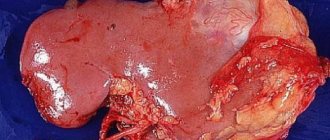
Tumor in the kidneys
One of the common pathologies of the pyelocaliceal system is the development of a tumor. Most often, the right kidney is affected, but this is not necessary; the epithelium of the pelvis, or rather its glandular component, is usually affected.
If the tumor develops quickly, the symptoms will be similar to those of hydronephrosis, blood will appear in the urine and severe pain in the lumbar region. In addition, a person may experience nausea and vomiting, which indicates intoxication of the body, he loses appetite and sharply loses weight.
To diagnose pelvic cancer, an ultrasound examination is also performed, computer diagnostics can be performed, but often a simple objective examination is sufficient.
If there is a suspicion of the development of an oncological tumor, then upon deep palpation, a large compaction is detected near the kidney or the kidney itself increases significantly in size. All studies performed are deciphered and a final diagnosis is made.
If you are often bothered by pain in the kidney area or there is blood in your urine, you should not put off visiting a doctor who will help determine the cause, make the correct diagnosis and prescribe appropriate treatment. The sooner you do this, the less likely it is to develop a complication and the easier, faster and more effective the treatment process is.
Almost always, at the conclusion of an ultrasound scan of the kidneys, the parameters and sizes of the CL are indicated, so the patient immediately has a question, what is it? The renal pyelocaliceal system is the collecting system of the kidney, which serves to collect urine.
Kidney and its functions
Causes
The main cause of the development of hydrocalycosis is considered to be a violation of the circulation of urine in the renal calyces and pelvis.
As a result, a load is created on all nearby organs. Enlargement of the calyx of the right kidney is much less common than the left one, but to a large extent it predominates in patients with excess body weight, since they usually sleep on the right side. Therefore, there is excessive pressure on the kidney, and as a result, deformation of the calyx.
The reason that prevents the complete flow of urine can be hidden on both the left and the right. In this regard, hydrocalycosis of the right and left kidneys is distinguished. The right kidney is most often affected - it is located just below its left “twin” and is more susceptible to negative factors. Sometimes - in about 1/5 of all cases of calicoectasia - bilateral hydrocalycosis is diagnosed.
The causes of hydrocalycosis of the right, left or both kidneys are the same. But there are quite a few of these reasons, so medical scientists divide them into 2 large groups: renal (organic, or direct) and extrarenal (mediated).
Real reasons:
- nephrotuberculosis;
- kidney stones (nephrolithiasis);
- sand and stones in the urinary tract;
- various tumors in the kidneys;
- wandering kidney (nephroptosis);
- kidney prolapse;
- structural abnormalities of the renal vessels;
- urinary system infections;
- injuries and various inflammations of the excretory organs.
Numerous genetic factors can influence the formation of cysts, causing polycystic disease in a child or adult patient. Sometimes the first symptoms of the disease appear in adulthood.
The urinary organ increases in size with the subsequent development of pain and the formation of complications.
Enlargement of both kidneys appears in acute pyelonephritis, nephrotic course of the inflammatory process, glamerulonephritis, amyloidosis.
Diffuse enlargement occurs with a malignant tumor or the presence of multiple metastases. A unilateral change in size occurs in cases where a healthy organ compensates for the work of the diseased area.
The most dangerous is bilateral caliectasis. In this case, the functioning of both kidneys is impaired, and the clinical picture is more pronounced.
Kidney structure
On the outside, the kidney is surrounded by a layer of adipose tissue, and underneath there is a protective fibrous capsule, from which septa extend into the organ.
They divide the internal renal structure into lobes and segments. These partitions also contain blood vessels and nerve fibers.
What does a kidney consist of?
Directly under the capsule is the renal tissue - parenchyma. It consists of the cortex and medulla, which are arranged in layers. The parenchyma contains about a million cells - nephrons.
In their structure, two sections are distinguished: the glomerulus and the tubule system. In the glomerulus, blood plasma is filtered from metabolic products and the breakdown of other compounds.
As a result of this process, urine is formed, which flows into the tubular system. From there it enters the so-called pyramids, which are located in the medulla parenchyma layer. From these, urine enters the CLS.
The first section of the kidneys is the small renal cups, which are shaped like glasses. With a wide edge they cover the papillae of the pyramids, from which urine is secreted. There are from 8 to 12 of them in a healthy kidney.
Such cups are combined into 2 - 3 large ones. They, in turn, merge into the pelvis. This structure is shaped like a funnel, which narrows as it passes into the ureter.
The wall of the kidney joint has the following structure:
inner mucous layer of transitional epithelium; middle layer of smooth muscle; outer layer, which consists of connective tissue.
On the outside, such a formation is covered with a layer of fiber, in which small blood vessels and nerve endings lie.
Complications of bilateral hydrocalycosis
With indicators that are within the normal range of calyx expansion, symptoms of the disease may be completely absent. If no pathological changes occur for a long time, then for several years, sometimes decades, the patient may not be aware of the presence of pathology.
Hydrocalycosis of both kidneys can lead to certain complications, namely:
- pressure on the urethra increases;
- pathogenic microorganisms infect the kidney tissue;
- venous outflow increases;
- An active redistribution of blood flow begins, as a result of which the blood supply to the kidneys increases.
With double dilatation, urinary tract obstruction may occur. This can trigger the renin-angiotensin system to become active, which affects blood pressure.
Pathologies of the collecting system
Almost all pathological processes of the kidneys negatively affect the heart rate. First of all, this is expressed in an increase in their size. In this case, doctors say that the pyelocaliceal system is dilated.
Why does this process occur?
One of the reasons for the expansion of the kidneys is urolithiasis. This disease occurs when stones of various compositions form in the pelvis.
This can be caused by a variety of reasons:
eating disorder; insufficient fluid intake; overweight; various systemic diseases that lead to metabolic disorders; long-term use of certain medications.
When a stone passes from the urinary tract, a mechanical blockage of the ureter most often occurs. But the process of urine formation occurs continuously, and if its outflow is disrupted, the calyces and pelvis of the kidney begin to expand.
This is accompanied by severe pain. This condition is called renal colic.
But sometimes the disruption of the outflow of urine from the kidney can be long-lasting.
This usually occurs due to compression of the walls of the ureter by tumors of nearby organs, and the formation of various scars in the lumen of the urinary tract after surgical interventions.
In this case, a gradual expansion of the heart rate occurs, and this process can be asymptomatic for a long time and reflected only in clinical studies.
This disease is called hydronephrotic transformation of the kidneys.
This disease is extremely dangerous. An enlarged CLS on one side and an inextensible fibrous capsule on the other compress the parenchyma. As a result of circulatory disorders, gradual atrophy and irreversible death of nephrons occurs.
This process can even lead to kidney loss.
Another disease that directly affects CHLS is pyelonephritis. This disease develops due to bacterial inflammation.
Most often, infectious agents enter the pelvis and renal cups ascending from the area of the external genitalia.
Much less often, bacteria are “introduced” into the CLS by the blood flow from foci of inflammation of any location. This usually occurs when the immune system is malfunctioning.
In this case, the expansion of the pyelocaliceal system occurs due to inflammatory exudate. Treatment of such a disease is carried out only with antibacterial drugs.
In addition, there are congenital defects of the cervical spine. The most common is its doubling within one kidney.
Unfortunately, recently, cancerous lesions of the renal structure have also occupied an important place in the structure of urological diseases.
Thus, the size of the renal collecting system plays an extremely important role in the primary diagnosis of many nephrological diseases.
Based on them, doctors can make a preliminary diagnosis and determine a treatment regimen or further examination of the patient.
But sometimes a slight deviation from the norm in the size of the mandibular joint is temporary, and if no further symptoms are observed, the doctor will recommend monitoring over time.
source: gastro-help.ru
The kidneys are located on either side of the spine. The normal size of an adult human organ is 100-120 mm in length and 40-50 mm in width. The left kidney is slightly larger than the right. The size of the organ as a whole depends on the body mass index.
There are different types of kidney diseases. One of them is the expansion of the collecting system. The CLS is located deep in the kidney. It collects and then excretes urine. The system consists of small cups. Using partitions, they are connected to form 2 or 3 large bowls. They form pelvises with a mucous layer inside and a shell of smooth muscle.
ChLS pathologies can be congenital or acquired. Typically, birth defects involve the pelvis and ureters.
From the pelvis, secondary urine flows through narrow channels into the ureter, then into the bladder.
If any abnormalities appear in the kidneys, then in some cases they lead to changes in the functioning of the central nervous system. At the same time, the parenchyma is compressed, the nephrons change. As a result, blood circulation in the organ is disrupted. In advanced cases, the kidney cannot cope with its functions, it stops working.
Congenital pathologies are hydronephrosis, stricture or doubling of the heart rate. Hydronephrosis develops due to narrowing of the ureter or due to reflux during the return of urine. Due to blockage of the ureter with stones or when neoplasms occur, urine also stagnates.
The disease is difficult to diagnose at the initial stage, since there are no clear signs of manifestation. The kidney condition is gradually worsening. The following stages of pyelocalicoectasia are distinguished:
- Elementary.
- Average.
- Chronic.
At the first stage of the development of the disease, there are no external signs. An abnormality of the renal function can only be determined using an ultrasound of the kidneys.
At the middle stage, the CLS continues to expand. The patient experiences difficulty urinating.
At the chronic stage, complications appear. Swelling of the extremities, changes in the color of urine, and back pain are possible.
The expansion of the ventricular system is affected by a disturbance in the flow of urine. The kidneys work under heavy load. Pyelocalicoectasia on the right is more common.
The following main reasons are identified:
- disturbances in the natural outflow of urine;
- reflux;
- accompanying illnesses.
Improper urine flow is possible in pregnant women. Men suffering from prostate enlargement may also have these problems. Pressure is created in the urinary system, urine flows slowly and is delayed. Urine stagnation can range from mild to moderate to severe. Its accumulation causes the kidney to enlarge.
Reverse reflux of urine occurs due to congenital reflux. Urine accumulates in the renal sinus and puts pressure on the pelvis. The kidney is stretched from the inside.
If the calyxes of both paired organs expand, then changes occur in the entire urinary system. Similar disorders occur with prostate adenoma or bladder tumors. Sometimes the expansion of the kidneys in adults is facilitated by injury to the organ, swelling, and infections.
The pelvicalyceal system can be dilated against the background of diseases such as ureteral obstruction, inflammatory processes, enlargement of the urethra and prostate, changes in the blood vessels of the upper urinary tract, and kidney prolapse.
Most often, kidney tumors occur in people over 60–70 years of age, less often in children. The causes of their occurrence may be nephropathy, exposure to harmful chemicals, long-term use of certain medications, and chronic diseases of the urinary organs.
Due to the appearance of a tumor in the kidney tissues, the functioning of the central nervous system may be disrupted. The patient does not immediately understand that changes are occurring in his body. When the tumor is just beginning and forming, the kidneys are still functioning normally. The tumor grows and begins to put pressure on nearby tissues. As a result, the pyelocaliceal system changes. If the tumor is in the ureter, then it closes the passage. The patient will have problems with the outflow of urine. This causes inflammation in the kidneys.
With tumors, in addition to pain and the presence of blood in the urine, nausea, vomiting, weight loss, and general malaise are observed.
Abnormal structure of the CLS
One of the forms of abnormal kidney structure is doubling of the renal function. Usually people with this deviation do not have complaints about their kidney function. They may not be aware of this peculiarity of theirs. When the heart rate doubles, the kidney is more susceptible to inflammation.
Only one structure can be doubled, but the calyces, pelvis, and ureters can be doubled. With abnormal development, several ureters are formed. They merge and exit into the bladder. Due to the abnormal structure of the kidneys, urodynamics are disrupted.
Pregnancy
Enlargement of the renal cups and pelvis is often detected unexpectedly, since the onset of the disease is asymptomatic. A woman undergoes an ultrasound and finds out about abnormalities in the excretory system. The pregnant uterus enlarges and puts pressure on the ureter. Typically, more pressure is applied to one of the kidneys. There is significant expansion on the right or left side.
Another reason is hormonal imbalances. An increase in estrogen affects bladder contractions. The problem can be identified by the results of urine tests.
An alarming factor should be an increase in blood pressure in a pregnant woman. It may also mean that there is a problem with your kidneys.
A woman should monitor the amount of urine excreted and maintain a drinking regime. The doctor prescribes physical exercises that reduce the pressure of the uterus on the kidneys.
Pathology may appear at the embryonic stage. Pregnancy proceeds without any special features. However, the child is born with defects in the structure of the renal pelvis.
Pyelonephritis
An infectious and inflammatory disease affecting the CLS is called pyelonephritis. This disease is caused by pathogenic bacteria. They can enter the body through the bloodstream after illness. The infection may be ascending through the urethra.
Bacteria penetrate into the cups and pelvis of the kidneys. Changes occur in the mucous membrane, it becomes inflamed and thickens. The functioning of the organ is disrupted. There may be an abscess, that is, purulent inflammation of the kidney tissue.
Antibacterial drugs are used to treat inflammation. Therapy is also carried out to restore the flow of urine.
Surgeries for hydronephrosis of the kidneys
A disease such as renal hydronephrosis is rare and ranks sixth among all nephrological pathologies. It is characterized by a violation of the outflow of urine, which leads to complications, often life-threatening. Increasing expansion of the calyces and pelvis followed by death of the kidney parenchyma is called hydronephrosis. Surgery is sometimes the only way to cope with the disease.
When is surgery necessary?
More often, the disease develops in children; one of the kidneys is affected, rarely both. In adults from 20 to 60 years of age, kidney pathology is predominantly registered in women, which is associated with childbearing age, pregnancy and gynecological diseases. Men with prostate adenoma and cancer after 60 years are also at risk.
Hydronephrosis develops due to prolonged congestion.
An increase in urine volume leads to compression of the kidney tissue and its vessels. Pressure increases inside the pelvis and ureter, and glomerular filtration is disrupted. Over time, complications arise, the renal tubules and organ cells atrophy, and necrosis occurs.
Surgery for hydronephrosis is indicated if the benefit from it is greater than the risk of surgery itself. In most patients, surgery can restore function and save the kidney. Removal of an organ is resorted to only as a last resort. Conservative treatment is used as an auxiliary treatment - this is antibacterial therapy and symptomatic care.
Types of surgery
Hydronephrosis can be congenital or acquired. The disease progresses slowly over several periods. The course of the disease can be acute or chronic, aseptic or complicated by the presence of microbial flora. When choosing a treatment method, all features of the patient’s disease must be taken into account.
The culprit of hydronephrosis is most often a violation of the structure of the ureter and its obstruction (blockage) in the area of \u200b\u200bthe exit from the pelvis. Any operation involves restoring the flow of urine. In this case, pyeloplasty is performed for hydronephrosis.
Sometimes during surgery it is discovered that the kidney parenchyma is completely destroyed and cannot be restored. Then the surgeon resorts to removing the organ or its segment. There are several methods for performing organ-conserving surgery and pyeloplasty:
- open surgery under visual control with tissue dissection;
- laparoscopic intervention using instruments and a video camera through punctures in the skin;
- endourological surgery without tissue damage through the urethra; the doctor controls his actions using ultrasound or x-rays.
Nephrectomy - complete removal of an organ - can be performed either during abdominal surgery or laparoscopically. Sometimes plastic surgery is performed for preventive purposes, in order to prevent serious consequences in the future. This is usually the first stage of pathology development. But since there are no signs and diagnosis is difficult, the disease is detected late, when surgery is already vitally necessary.
How the method of operation is selected
The type of surgical intervention is recommended depending on the cause of hydronephrosis. And it is set individually for each patient based on analyzes and the results of all examinations. The laparoscopic method is less traumatic and allows you to quickly return to the patient’s usual lifestyle. The disadvantage of this method is that there is little room for maneuvering in the abdominal cavity, especially for small children.
Plastic surgery is performed if it is possible to preserve the functional abilities of the parenchyma, it is possible to completely eliminate the cause of hydronephrosis in strictures (narrowings). Any type of surgery gives access to the kidney and ureter.
The success of the operation depends on how correctly and completely the outflow of urine is ensured.
No matter how well the operation is performed, tissue swelling and decreased peristalsis of the pelvis and ureter appear after it. The choice of the type of surgical intervention, a successfully selected method of reconstruction of the urinary tract is the key to successful surgical intervention. Therefore, the functioning of the ureter is often maintained by nephrostomy or the placement of a stent after surgery.
Preparatory stage
The main task of the preparatory period is examinations to assess the anatomy of the kidneys and ureter and their functionality. It is necessary to undergo tests, take an X-ray of the OGK and an ECG, and other methods may be needed.
Carrying out an operation if there is an existing infectious process is a last resort. If the patient has all the signs of an inflammatory process, it is necessary to culture the urine for flora and its sensitivity to antibiotics. Depending on the type of bacteria in the urine, a course of treatment with antimicrobial drugs is prescribed.
The gastrointestinal tract will also need to be prepared for surgery. The day before the intervention, you should eat only liquid food. The day before, dinner should take place no later than 18:00. To cleanse the intestines, laxatives are prescribed in the evening. Eating is prohibited on the operating day!
Kidney drainage is an important procedure. It can be performed at the first stage of treatment or during surgery, and is performed in the following cases:
- acute renal failure;
- inflammatory process – pyelonephritis;
- painful shock, life-threatening condition;
- terminal hydronephrosis.
Some conditions require treatment before surgery. With azotemia - an increased content of nitrogenous metabolic products in the blood - the operation is difficult. The patient is prescribed a course of medications and a special diet. Sometimes hemodialysis is required before surgery.
Rehabilitation period
The duration of the recovery period depends on the type of surgery and the form of hydronephrosis. The standard of treatment is open pyeloplasty. It is characterized by a long rehabilitation period and possible complications. The patient spends the first week after surgery under the supervision of medical staff. Regular dressing changes, antibiotics, painkillers and anti-inflammatory drugs are required.
The postoperative period is easier after minimally invasive interventions. Small wounds heal faster and the risk of infection is negligible. It is important to follow all the doctor’s recommendations, reduce physical activity to a minimum, and do not lift heavy objects. Diet will help reduce the load on your kidneys as much as possible.
Dietary nutrition should be followed for three years, and systematic examinations are also necessary during this period. Fatty, fried foods, smoking, marinades, spicy and salty foods are prohibited. You can drink no more than 2 liters of liquid per day. After recovery, sanatorium treatment is recommended.
Development of complications
It happens that after surgery an infection of the urinary system develops and pathogenic microflora appears in the urine. Bleeding may occur during or after surgery. Another problem in the early period is urine leakage, which is associated with a violation of the integrity of the seam. Complications of anesthesia lead to the need for artificial ventilation. The consequences of penetration of gastric contents into the lungs are fraught with the development of pneumonia.
During laparoscopy, circulatory disorders may occur due to changes in pressure in the peritoneum. Damage to neighboring organs occurs due to lack of access. In this regard, an urgent transition to open surgery may be required.
Recurrence of ureteral narrowing may occur in 15% of those operated on. Repeated compression of the ureteral parenchyma by edematous tissue leads to disruption of the outflow of urine. A special flexible tube (stent) must be installed, and it is removed after a few weeks.
Severe complications occur with bilateral renal failure.
If help is not provided in time, this leads to death. Unilateral hydronephrosis can be compensated by the work of a healthy organ. Surgery will eliminate dangerous symptoms and give a chance for a full life.
Diagnosis of the disease
Using ultrasound and x-rays, an increase in the size of the jaw can be diagnosed. Both methods are quite effective.
Currently, devices show not only the disease, but also the cause of the disease.
It is possible to diagnose enlargement of the maxillofacial area during pregnancy. This is at 16-20 weeks. At this time, the fetus has such a size that the renal pelvis is visible. Deviations are more common in boys.
The birth defect may disappear by the age of one, since the newborn has an underdeveloped urinary system.
An ultrasound may show a lump in the joint.
Renal blood flow status
If necessary, in addition to a regular ultrasound, the patient undergoes a duplex scan of the kidneys - Doppler sonography. It makes it possible to diagnose the condition of the renal vessels and see the speed of blood flow. It should not exceed 50-150 centimeters per second.
It is helpful for the layperson to know that a normal Doppler image has more dark tones than bright tones. Bright shades show accelerated blood flow through the arteries of the kidneys (> 200 cm/sec), and this is a symptom of their narrowing - stenosis. It affects your health by high blood pressure (renal hypertension).
ChLS of the kidneys - what this is is not known to all people, this system plays a big role in the process of accumulation and excretion of urine. Violations of its functionality can be caused by various reasons, some of them are easily treatable, while others can lead to irreparable complications. In any case, consultation with a urologist is extremely necessary for problems with CLS.
In this article we will talk about what the penis is and why it is needed, for what reasons does it expand and what it entails, as well as how to detect this disorder using diagnostic methods.
Treatment methods
Therapeutic and surgical methods of treatment are used. To improve blood microcirculation in the kidney, patients are prescribed antispasmodics, antibiotics and other medications.
Special diets and mineral waters with calcium are recommended.
At an early stage of the disease, the structure of the organ is corrected using plastic surgery. The kidneys are returned to their previous size. Slight enlargement of the maxillary joint is easier to treat. An untreated disease can lead to serious complications.
If the kidney has become too large and has lost its function, it is removed. The operation is performed under general anesthesia. If it is possible to use a laparoscopic device, the operation is performed under local anesthesia. The most appropriate method will be chosen by the attending physician.
After kidney correction, the patient has a great chance of recovery. When an organ is removed, long-term rehabilitation and, accordingly, disability are required.
If a baby suffers from expansion of the heart rate, then the doctor chooses a wait-and-see approach in the first year of his life.
Symptoms
This pathological condition has no distinctive individual characteristics. It is detected in the patient in most cases in the later stages of development, when serious medical intervention is required.
The main symptoms are painful urination and nagging pain.
Since many kidney diseases have similar symptoms, it is best to get regular checkups. Urine that stagnates in the pelvis causes infectious processes that can lead to pyelonephritis or the development of an acute abscess.
Prevention
In order for the urinary system to function normally, preventive measures must be used. To do this, tests and ultrasounds are performed annually. It is necessary to empty the bladder in a timely manner to avoid stagnation of urine. For people leading a sedentary lifestyle, regular warm-up is necessary. Healthy eating, avoiding stressful situations, and playing sports are also good prevention.
Thus, enlargement of the kidneys is a serious pathology. It is important to recognize it in time and take the necessary measures.
Types of pathological process
The presence of excessively large kidneys in a person can occur without a clear clinical picture: itching, pain, discomfort at the site of the lesion. This pathology is practically asymptomatic and may not make itself felt for a very long time, while gradually progressing. There are several distinctive symptoms that allow you to determine in which kidney the enlargement occurred.
Kaliectasia is an asymptomatic pathology, so at the first abnormalities in the kidneys, you should consult a doctor. There are several stages of expansion of the calyxes in the kidneys:
- First stage. The pathology manifests itself asymptomatically, no external changes are noticeable, any violations of the pyelocaliceal system can only be detected with the help of ultrasound examination.
- The middle stage involves a significant increase, the patient cannot empty the bladder normally, this process causes him certain difficulties.
- Development of a chronic process. The upper and lower extremities begin to swell, the urine changes color, since the kidneys are not working at full capacity. The process of urination becomes painful. Sometimes the patient feels painful shooting in the lumbar region.
If the inflammatory process progresses in the human body, fever may occur, as well as soreness in the sacral area. If a child has hydrocalycosis, it can be noted that he goes to the toilet more often, and also becomes very capricious. Despite significant swelling, the victim experiences a strong feeling of thirst and drinks water in large quantities.
General information
The normal proportions for an adult kidney are 105 ± 8 mm in length and 45 ± mm in width. When these parameters change, problems with urine drainage occur. Enlargement of the calyces of both kidneys (bilateral pyelocalicoectasia) affects the urinary system and related organs. The disease can develop as a consequence of prostate adenoma or be a consequence of a bladder tumor. In some cases, expansion of both the calyces and the kidneys in adults is observed with cancer, trauma and swelling of the organ, against the background of a kidney infection and during pregnancy. Deformation of the renal pelvis can be congenital, and the predisposition is inherited. In the early stages, the disease does not show clear symptoms, and if disorders in the kidneys are not diagnosed in time, a chronic stage will occur with a whole “bouquet” of complications. If the kidney becomes too large, its size cannot be adjusted and will have to be removed.
Classification and forms of the disease
Abnormally large kidneys do not always have open forms of symptoms, such as pain, itching and pronounced discomfort in the affected area. This is a “silent” disease, gradually worsening and characterized by a smooth change of stages. Right-sided and left-sided pyelocalicoectasia is conventionally divided into several stages and is characterized by a number of clinical differences:
- Elementary. The disease has no external manifestations; the disorder can only be determined using ultrasound.
- Average. The renal collecting system is significantly enlarged, and difficulty urinating is observed.
- Chronic. The patient has noticeable swelling of the extremities, and the color of the urine changes. Walking “small” is painful and difficult. Periodically there is pain in the lumbar region.
In some cases, with a progressive inflammatory process, the patient's body temperature may rise and aching pain may appear in the sacral area. Small children begin to be capricious and ask to go to the toilet more often. Against the background of general swelling of the body, there is a feeling of thirst and increased water consumption. It is necessary to take a urine test and, if abnormalities are detected, consult a doctor.
Dilatation of the renal pelvis
Impaired urine outflow causes an increase in pressure in the renal cavity system. If the process is permanent, the walls of the joint stretch and weaken. The result is an enlargement of the renal pelvis.
What is renal pyeloectasia and how does it differ from hydronephrosis? These terms reflect the same condition.
At the first stage of hydronephrosis, only a change in the size of the renal pelvis is observed. If the CL is deformed, the parenchymal tissue suffers over time, which leads to further progression of hydronephrosis.
- What is doubling of the left kidney?