Pyelonephritis is a disease that is associated with infection in the kidneys and is inflammatory in nature. In another way it is called inflammation of the renal pelvis.
This problem begins when bacteria enter and increase in the urinary system. There is a possibility of developing the disease when receiving an injury or introducing an infection into the body through medical means.
Most often, pyelonephritis, inflammation of the renal pelvis (in Latin Pyelitis), occurs from E. coli. Pathology can appear in different people, regardless of gender and age. But women go to the hospital 3-4 times more often. This is due to the fact that the structure of the female reproductive system is different from the male one, and it is easier for bacteria to enter the body.
Causes of pyelonephritis
Bacteria entering the body through the urethra and multiplying throughout the system up to the kidney is the most common cause of the development of the disease.
Unlike men, the female urinary canal is less protected. The urethra is located close to the anus and reproductive system, which allows bacteria to enter the body almost unhindered.
The male half of humanity gets sick if there are problems with urination: inflammation of the prostate, the formation of kidney stones. When urine stagnates, bacteria multiply in it and move through the urinary canals to the kidneys.
The presence of tumors, stones in the urinary tract, i.e., everything that prevents the release of harmful substances, is one of the main reasons for the development of pyelonephritis in women. But more often, women get sick through the ascending route, when the area near the urethra becomes infected with E. coli.
Prevention of kidney pathology
Preventative measures include adequate fluid intake to ensure regular urination and therefore cleansing of the urinary tract. Women are advised to visit the toilet as soon as possible after sexual intercourse, which can introduce bacteria into the urethra.
Cranberry juice has a positive effect, as it acts against inflammation of the urinary tract.
Attention! The drink should not be consumed by people taking Warfarin and other drugs to reduce blood clotting!
An indispensable preventive factor is personal hygiene.
Other preventative measures include strengthening the immune system, adequate intake of vitamins (fruits, vegetables), regular sleep and rest, and appropriate exercise plays an important role.
Prevention and treatment are 2 things that people suffering from diabetes should pay attention to, in which pyelitis is a fairly common disease.
Manifestation of the disease in women
The manifestation of symptoms of pyelonephritis differs in men and women. This is influenced by differences in the structure of the genitourinary system. Inflammation of the renal pelvis occurs with different consequences, which directly depend on the type of disease and the duration of its course. If the stage of the disease is acute, it can be determined by the following signs:
- sudden increase in body temperature above 37-37.5 ° C;
- manifestation of severe symptoms of poisoning: vomiting, migraines, chills;
- presence of changes in urine parameters: change in color, inclusion of blood clots, unpleasant odor, etc.
If there are any of the deviations listed above, then there is a need to contact a specialist and get tested. First of all, the Pasternatsky test will be performed: light tapping in the kidney area will cause severe pain, and blood will appear in the urine when urinating.
However, if the disease proceeds calmly and without complications, then it can be determined by the following characteristics:
- mild but constant pain in the lumbar area;
- barely noticeable symptoms of poisoning: lethargy, lack of appetite, intermittent headaches;
- the appearance of morning swelling, difficulty urinating.
Inflammation of the kidneys and bladder in women often occur together. But usually the symptoms of cystitis are more pronounced, so it becomes very difficult to determine pyelonephritis in this case.
Common problems
Unfortunately, in medicine there are often cases when damage to the kidney collector system leads to serious health consequences. Common CPS conditions are presented in the sections below.
Pyeelectasia and hydronephrosis
Pyeelectasia is a pathological expansion of the renal pelvis due to congenital and acquired causes. Hydronephrosis is a progressive increase in the size of the jaw, caused by a violation of the outflow of urine, which is accompanied by atrophy of functionally active areas of the organ.
There are a number of reasons for the development of pyelectasia and hydronephrosis, among which the most common are:
- congenital anomalies of kidney development and urinary tract;
- obstruction of the ureter by an additional vessel opening into the lower pole of the kidney;
- incorrect location of the ureter;
- nephrolithiasis and blockage of the ureter with a large stone;
- prostate adenoma, prostatitis in men;
- benign and malignant tumors of the ureter and bladder;
- injuries.
During its course, the disease goes through three successive stages:
Pyeelectasia. Expansion of the pelvis. Pyelocalicoectasia. Expansion of the ChLS in general. Hydronephrosis. The final stage of the disease, accompanied by the development of atrophy of the functional apparatus of the organ and impaired blood filtration in the kidneys.
In addition to the development of persistent progressive chronic renal failure, one of the serious complications of hydronephrosis is rupture (usually unilateral) of the renal pelvis. This condition is seriously life-threatening and requires immediate surgical treatment.
Pyelitis
Pyelitis is a disease of a predominantly infectious nature, characterized by isolated inflammation of the pelvis. Its main etiological factors are nonspecific bacterial flora and reduced reactivity of the body. Often accompanied by the development of hypotension of the renal pelvis.
There are acute and chronic forms of the disease. The latter has a wave-like course, in which periods of exacerbation are followed by relatively favorable remission. Due to frequent inflammatory processes, the filtration and excretory functions of the kidneys are inhibited. This process underlies the formation of chronic renal failure in a patient.
Duplication of the renal pelvis
Medicine is also aware of such an anomaly as doubling the renal function of the kidneys. It could be:
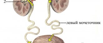
- complete, in which two isolated pelvis located in the kidney continue with two ureters, which subsequently flow into the bladder;
- incomplete, in which the fusion of the two ureters occurs above the point of entry into the bladder.
Thus, the pelvis is a kind of internal cavity of the kidney in which the collection, primary accumulation and further excretion of urine occurs. Many pathologies of its structure and functioning lead to serious disorders of the urinary organs, and therefore require qualified and timely medical care.
pochkizdrav.ru
formed in the nephron structures enters the renal pelvis
.
As they fill and stretch, a threshold of mechanoreceptor irritation is reached, leading to a reflex contraction of the muscles of the pelvis and opening of the ureter. Due to peristaltic contractions of their smooth muscles, urine enters the bladder. The smooth muscles of the pelvis and ureters
have a significant degree of automaticity, and therefore their peristalsis is caused by stretching by the volume of incoming urine.
Bladder -filling
as it accumulates, it begins to stretch its walls, but the tension of the bladder walls does not increase to a certain stretch value, usually corresponding to a urine volume in the bladder of about 400 ml.
The appearance of tension in the bladder wall causes the urge to urinate, since irritation of mechano-receptors leads to the entry of afferent information into the sacral parts of the spinal cord and the formation of a complex reflex act. This act involves not only the spinal structures, but also the central structures located in the brain, which allow voluntary retention of urination or its onset, as well as providing a sensory-emotional reaction. The act of urination
is realized due to the fact that efferent impulses from the spinal center along parasympathetic nerve fibers reach the bladder and urethra, simultaneously ensuring contraction of the smooth muscle of the bladder wall and relaxation of two sphincters - the neck of the bladder and the urethra.
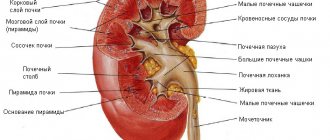
Urine excreted through foramina papillaria
, on its way to the bladder passes through the small calyces, large calyces, renal pelvis and ureter.
Small cups, calyces renales minores,
numbering about 8 - 9, one end covers one - two, less often three renal papillae, the other flows into one of the large cups.
large cups, calyces renales majores
- upper and lower.
Even in the sinus of the kidney, large cups merge into one renal pelvis, pelvis renalis
(Greek pyelos, hence inflammation of the renal pelvis - pyelitis), which exits through the gate behind the renal vessels and, bending down, passes immediately below the kidney gate into the ureter.
Fornical apparatus of the renal calyces.
Each renal calyx encloses a cone-shaped renal papilla, like a double-walled glass.
Due to this, the proximal part of the cup, surrounding the base of the papilla, rises above its apex in the form of a vault, fornix. The wall of the cup vault contains non-striated muscle fibers, m.
sphincter fornicis, which, together with the connective tissue embedded here and adjacent nerves and vessels (blood and lymphatic), constitute the fornical apparatus, which plays a large role in the process of excretion of urine from the kidney parenchyma into the renal cups and prevents the reverse flow of urine from the cups into the urinary tubules.
Due to the close proximity of the vessels to the wall of the fornix, bleeding occurs more easily here than in other places and urine flows into the blood (pyelovenous reflux), which facilitates the penetration of infection. In the wall of the renal cup there are four muscles located above the fornix (m. levator fornicis), around it (m. sphincter fornicis), along the cup (m. longitudinalis calycis) and around the cup (m. spiralis calycis). M. levator fornicis and m.
longitudinalis calycis expand the cavity of the cup, promoting the accumulation of urine (diastole), and m. sphincter fornicis and etc. spiralis calycis narrow the cup, emptying it (systole). The work of the cup is associated with a similar activity of the renal pelvis.
Cups, pelvis and ureter
constitute the macroscopically visible part of the excretory tract of the kidney.
It is possible to distinguish three forms of the excretory tree, which reflect the successive stages of its development (M. G. Prives):
1) embryonic,
when there is a wide saccular pelvis into which the small cups directly flow; there are no large cups;
2) fetal,
when there is a large number of small and large cups that pass directly into the ureter; there is no pelvis;
3) mature,
when there is a small number of small cups merging into two large cups, turning into a moderately pronounced pelvis, which flows further into the ureter. All four components of the excretory tree are present here: small calyces, large calyces, pelvis and ureter. Knowledge of these forms makes it easier to understand the x-ray picture of the excretory tree visible in a living person (during pyelography).
ureter, ureter,
is a tube about 30 cm long.
Its diameter is 4 - 7 mm. From the pelvis, the ureter directly behind the peritoneum goes down and medially into the small pelvis, where it goes to the bottom of the bladder, the wall of which pierces in an oblique direction. In the ureter there are pars abdominalis
- up to the point of its bend through
the linea terminalis
into the
pelvic cavity and pars pelvina
- in this latter.
The lumen of the ureter is not the same everywhere; there are narrowings: 1) near the transition of the pelvis to the ureter, 2) at the border between the partes abdominalis and pelvina
, 3) along
the pars pelvina
and 4) near the wall of the bladder. In a woman, the ureter is 2–3 cm shorter and the relationship of its lower part to the organs is different than in a man. In the female pelvis, the ureter runs along the free edge of the ovary, then at the base of the broad ligament of the uterus it lies lateral to the cervix, penetrates the gap between the vagina and the bladder and pierces the wall of the latter in an oblique direction, like in a man.
Structure.
The wall of the ureter
, as well as the pelvis with cups, consists of three layers: the outer layer is made of connective tissue,
tunica adventitia
, the inner layer is
tunica mucosa
, covered with transitional epithelium, equipped with mucous glands;
between tunica adventitia and tunica mucosa
is located
tunica muscularis
. The latter consists of two layers (inner - longitudinal and outer - circular), which are not connected to the muscles of the bladder and prevent the reverse flow of urine from the bladder into the ureter.
At the point where the ureter enters the bladder there is a third, outermost longitudinal layer of muscles, which is closely connected with the muscles of the bladder and is involved in the ejection of urine into the bladder.
The ureter on an x-ray appears as a long, narrow shadow extending from the kidney to the bladder. Its contours are clear and smooth. The ureter forms curvatures in two planes - sagittal and frontal. Curvatures in the frontal plane are of practical importance: in the lumbar part to the medial side, and in the pelvic part to the lateral side. Sometimes the ureter in the lumbar part is straightened. The curvature of the pelvic part is permanent.
Along the course of the ureter there is
, in addition to the anatomical narrowings described above, a number of physiological narrowings that appear and disappear during peristalsis.
The fornical apparatus is a complex anatomical and functional formation consisting of a calyx with an epithelial cover, a fornical muscular sphincter, a fornical levator, a network of elastic collagen fibers and a rich nervous, vascular and lymphatic system with a large network of interstitial connective tissue. This apparatus is located in the proximal part of the calyx and includes the corresponding papilla, as well as the fornical venous plexus, located in close proximity and around the circumference of the fornix.
The main physiological role of the fornical apparatus is the absorption of water and table salt from the pelvic urine. Under special conditions, absorption of other components of urine, as well as medications or radiopaque substances introduced into the pelvis for pyelographic purposes, is possible. Along with this, the fornix apparatus is involved in the secretion of urine from the tubules into the calyx and pelvis, since in the area of the fornix itself there are muscle and nervous formations that ensure the systolic and diastolic functioning of the calyceal system.
studfiles.net
Kidney (lat. ren) is a paired bean-shaped organ that regulates the chemical homeostasis of the body through the function of urine formation. Part of the urinary system (urinary system) in vertebrates, including humans. In humans, the kidneys are located behind the parietal layer of the peritoneum in the lumbar region on the sides of the last two thoracic and first two lumbar vertebrae. They are adjacent to the posterior abdominal wall in the projection of the 11-12th thoracic - 1-2nd lumbar vertebrae, and the right kidney is normally located slightly lower, since, from above, it borders on the liver (in an adult, the upper pole of the right kidney usually reaches the level of 12- th intercostal space, upper pole of the left - level of the 11th rib). Each kidney is covered with a strong connective tissue fibrous capsule, and consists of parenchyma and a system for storing and excreting urine. The kidney capsule is a dense sheath of connective tissue covering the outside of the kidney. The kidney parenchyma is represented by an outer layer of the cortex and an inner layer of the medulla, which makes up the inner part of the organ.
Average bud dimensions: length 10-12 cm, width about 6 cm, thickness 3-4 cm, average weight 120 g.
· The bud has a bean shape, a smooth surface and a dark red color.
In the kidney there are:
two surfaces – anterior (more convex) and posterior (faciesanterior, posterior);
two poles – rounded upper and pointed lower (extremitassuperior, inferior);
two edges - convex lateral and concave, facing inward, somewhat downward and forward medial (margolateralis, medialis).
· The notch on the medial edge is called the renal hilus (hilus renalis). Through the renal gate, the renal arteries and nerves enter the kidney and exit through the vein, lymphatic vessels and ureter. All these formations (vessels, nerves and ureter) are combined into the concept of the renal pedicle.
· The renal gate “opens” into the renal sinus (sinus renalis). The sinus is filled with blood and lymphatic vessels, nerves, large and small renal calyces, the renal pelvis (see urinary structures of the kidney below) and fatty tissue.
The outside of the kidney is covered (Fig. 3.3):
fibrous capsule (capsulafibrosa) – the innermost, associated with the parenchyma;
fat capsule (capsula adiposa) - located outside the fibrous capsule, represented by a thick layer of loose fatty tissue (especially in the area of the posterior surface and hilum of the kidney).
renal fascia (fasciarenalis) – part of the intra-abdominal fascia, located outside the fat capsule. The renal fascia is connected by fibers to a fibrous capsule and has two layers, anterior and posterior:
§ the anterior layer covers the anterior surface of the kidneys, passing from one kidney to another in front of the renal vessels, aorta and inferior vena cava.
§ The posterior leaf covers the posterior surface of the kidneys and is medially attached to the lateral surfaces of the vertebral bodies (i.e., it is interrupted at the spine).
§ Along the lateral margins of the kidneys, the anterior and posterior layers of the renal fascia grow together.
§ At the upper pole of the kidney, both layers cover the adrenal gland and unite, limiting the movement of the kidneys.
§ The sheets do not merge at the lower poles. The kidneys move down during inhalation, that is, they have physiological mobility.
Kidney fixation
The complex of structures responsible for fixation of the kidney includes:
a) muscular bed of the kidney - formed by the quadratus lumborum muscle;
b) renal vessels - the renal artery and vein - prevent the removal of the kidney from the aorta and inferior vena cava;
c) renal fascia and fat capsule;
d) intra-abdominal pressure - caused by contraction of the abdominal muscles.
The gate continues into the recess in the substance of the kidney - the renal sinus (sinus), which is occupied by:
1. renal calyces (large and small),
2. renal pelvis,
3. vessels and nerves.
All of them are surrounded by fiber.
Small cups - there are 7-10 of them, they are short, wide tubes. One end of them captures the protrusion of the renal substance - the renal papilla (it can capture not 1, but 2-3), and with the other end they continue into a large cup.
Large cups - there are 2-3 of them, merging, they form the renal pelvis, from which the ureter departs.
The wall of the cups and pelvis consists of mucous membrane, smooth muscle and connective tissue layers.
gabiya.ru
Pyelonephritis in pregnant women
About 5% of women experienced inflammation of the renal pelvis in a simple or complex form during pregnancy. Usually the disease is detected at 6-8 months of pregnancy, because the presence of a child increases the pressure of the uterus on the genitourinary system. As a result, urine output decreases, and stagnation develops in the body. Bacteria begin to multiply, which, in turn, further slow down the movement of physiological fluid in the body. Pyelonephritis most often manifests itself in the presence of problems with the bladder.
During pregnancy, the kidneys perform an increased amount of work. They filter, passing through themselves all the liquid that enters the body. If symptoms appear that indicate a malfunction of the urinary system, you should immediately contact a gynecologist who is monitoring the pregnancy and under no circumstances ignore the discomfort. Impaired kidney function leads to intoxication of the body of both the woman and the fetus. This, in turn, can cause miscarriage or serious illness in the unborn baby.
Pyelocalyceal system
The renal papillae, which have the shape of a cone, are surrounded by a calyx, like a vault (in which, in fact, the muscle fibers are located).
The kidneys are a unique filter of the human body, maintaining balance in it, as in a system that removes toxins. The kidneys produce urine, which is then excreted through the urinary tract. These paired organs have a rather complex structure, thanks to which they perform complex functions that preserve human health.
The renal papillae, which have the shape of a cone, are surrounded by a cup, like a vault (in which, in fact, the muscle fibers are located). Here are also located nerve endings, blood vessels and connective tissue, the fornical apparatus, which acts as an important mechanism that allows urine to be removed from the parenchyma into the calyces, preventing its return. Blood vessels are tightly adjacent to the surface of the fornix, which is the main cause of bleeding (pyelovenous reflux). This often causes kidney infection.
Muscle fibers are located in the walls of the cups in different parts in relation to their arch:
- Along the calyx;
- Around the calyx;
- Above the vault;
- Around the vault.
Those muscles that are located along the cup and above the arch, expanding the cavity, allow urine to accumulate. And those that are located around the cup and arch, narrowing the cup, contribute to its emptying. Urine enters the pelvis, then the ureter. All this constitutes the main excretory renal pathway.
The renal excretory tree is divided into three forms:
- Embryonic. It is characterized by a wide pelvis into which the small cups flow directly. Large cups are not formed;
- Fetal. This type has both major and minor calyces, but no pelvis;
- Mature. This type has a correct anatomical structure. Small calyces form large ones, which pass into the pelvis, and it, in turn, passes into the ureter.
Complications
There are a number of complications that appear with inflammation of the renal pelvis:
- anemia (the amount of hemoglobin and red blood cells in the blood decreases);
- renal failure (kidneys cannot function normally);
- sepsis (blood poisoning as a result of the development of an acute pathological process in the kidneys).
Among other things, the risk of premature birth increases, which can lead to the death of the child and mother.
During pregnancy, self-medication is strictly prohibited, as complications may arise that will harm the fetus. Any treatment during pregnancy should be prescribed by a specialist after a thorough examination and tests.
There are three forms of formation of the renal pelvis
embryonic, fetal and mature. In the first form, the large renal calyces are not expressed, so the small renal calyces directly flow into the renal pelvis. In the second form, the existing large renal cups pass into the ureter, and the pelvis is not formed. In the third form, there is the usual number of small renal calyces, which flow into two large renal calyces; the latter pass into the renal pelvis, where the ureter begins. The shape of the renal pelvis is ampullary, tree-like and mixed.
The walls of the pelvis, large and small renal calyces have the same structure. The walls are divided into mucous, muscular and outer adventitia. In the walls of the small renal calyces, in the area of their vault (initial part), smooth muscle cells form a ring-shaped layer - the vault compressor.
Male pyelonephritis
Considering the structure of the male urinary system, kidney inflammation in the stronger sex is recorded by doctors much less frequently. Pyelonephritis progresses due to problems with the outflow of urine caused by the presence of kidney stones and inflammation of the prostate.
Chronic pyelonephritis in men is manifested by symptoms similar to those in women: weakness in the body, loss of appetite, frequent thirst, pain in the lumbar region.
When the kidney stones shift, pyelonephritis will manifest itself with severe pain. A complicated type of inflammation in men more often appears with a descending method of the disease: bacteria enter the body during sore throat or the presence of caries. The acute stage is similar to the female stage.
Treatment
Minor enlargement of the renal calyces does not require specific therapy. This condition usually does not cause any symptoms.
If the clinical picture is pronounced, symptomatic therapy is carried out - painkillers, diuretics. With the development of pyelonephritis, treatment with antibacterial and antimicrobial agents is indicated.
Excessive dilation of the renal calyces can lead to necrosis of the kidney tissue, stone formation, ureteral infection and kidney failure. Timely diagnosis and competent therapy will help avoid surgical intervention and prevent the development of dangerous complications.
Kidney inflammation in children
In childhood, the main symptom of pathology is considered to be a sharp increase in body temperature to 38 °C. If you are in excellent health and there are no other indicators that can characterize a particular disease (ARVI, infections, poisoning), you need to urgently take tests to check for the presence of inflammation.
Characterized by temperature fluctuations, chills, vomiting, and lack of appetite. The child develops drowsiness, abdominal pain, and headache. In this way, the initial stage of inflammation of the renal pelvis in the newborn will appear.
In the chronic form of the disease, symptoms depend on the duration of the pathology. With a latent form of inflammation of the renal pelvis, the kidneys cease to perform their functions in full, which leads to tissue destruction.
What is pyelocalicoectasia (calycopyelectasia) of the kidney?
Calicopyelectasia is considered one of the most serious pathologies of the urinary system. The disease consists of an increase in the size of the renal collecting system. Anatomical changes are associated with a decrease in the lumen of the urinary canal, as a result of which urine is retained in the pelvis and does not have time to be excreted before a new portion arrives. A larger volume of incoming urine causes the pyelocaliceal system to respond by expanding all parts, leading to irreversible pathology.
The disease affects people of all ages, regardless of social status and status. So, often, calicopyelectasia is congenital. During the formation of the genitourinary system, an abnormal deformation of the urinary tract occurs in the fetus, due to which the calyx increases in volume. However, the disease often affects adults as well, as a disease accompanying other pathologies of the urinary system and kidney diseases.
Nephrologists distinguish two types of the disease: unilateral and bilateral. With the latter type, both organs are affected. Unilateral indicates damage to one of the kidneys, usually the right one.
The disease is considered one of the most dangerous due to its difficult diagnosis at an early stage. The diagnosis of pyelocalicoectasia is often made during a general examination. The difficulty of detecting the disease is associated with its asymptomatic course. However, at a later stage the symptoms become more pronounced.
Tests for kidney inflammation
It is necessary to undergo the following tests for inflammation of the renal pelvis:
- biochemical and general analysis of urine and blood;
- check according to Nechiporenko;
- experiments on urine microflora to identify contraindications before prescribing a particular drug.
In case of kidney inflammation, a blood test will show a complete picture of the disease: an increased accumulation of leukocytes, changes in the composition of the blood at the biochemical level.
A urine test will help identify the total number of leukocytes (decrease or increase), the presence of so-called “additives”: blood, pus, protein, increased or decreased salt content.
Diagnostics will also help determine what caused pyelonephritis and select the most appropriate medicine.
An examination of the internal organs is performed using an ultrasound machine and the kidneys, processes of the urinary system, the bladder and its channels are checked. In more advanced cases, a certain amount of contrast agent is injected into the body, and a check is performed using a magnetic resonance imaging machine.
Disease detection method
Diagnosis of diseases of this type is carried out by testing urine and blood. A urine test will show the level of leukocytes, on the basis of which the doctor will make a conclusion about the extent of damage to the organ. Bacterial culture will indicate the type of pathogen and its resistance to antibiotics. To determine a harmful microorganism, the Gram method is sometimes used, which takes less time.
A blood test helps determine the level of leukocytes in it, ESR. An increase in creatinine and urea will indicate the development of complications (renal failure). If it is known that a microbial infection has entered the body through the hematogenous route, the liquid is tested for sterility.
Ultrasound and x-rays are used to exclude the possibility of developing concomitant kidney diseases.
Features of therapy
With inflammation of the renal pelvis, symptoms and treatment are interrelated. Therapy depends on the severity of the disease and is prescribed exclusively by the attending physician.
If inflammation of the renal pelvis is detected in a woman, then she must come to an appointment with the treating gynecologist to determine the source of the spread of bacteria.
If there is inflammation of the bladder or problems with the reproductive system, treatment of pyelonephritis will be inappropriate and will ultimately lead to the development of a chronic form of the disease. Prolonged treatment with antibiotics prescribed to restore kidney function, without eliminating the center of infection, will further weaken immunity to specific microorganisms with a subsequent loss of effect from the medications taken.
A visit to a urologist is necessary for men who have pain in the lumbar region and there is a suspicion of pyelonephritis. It is usually detected in patients over 40 years of age, and more often in a chronic form.
This occurs due to diseases of the urinary system: prostate adenoma, prostatitis, etc. It is necessary to treat the problem of stagnation in order to normalize the outflow of urine, thereby protecting the body from the recurrence of the disease.
In children, the problem most often arises due to birth defects that impair the functioning of internal organs in the urinary system. For this form of the disease, drug treatment is used. But if it does not bring a positive effect, then surgical intervention is used.
Prices
| Disease | Approximate price, $ |
| Prices for examination and treatment for testicular cancer | 3 730 — 39 940 |
| Prices for vaporization of prostate adenoma with a “green laser” | 16 050 |
| Prices for diagnosis and treatment of impotence | 1 320 — 50 000 |
| Prices for diagnostics of the genitourinary system in men | 5 630 |
| Testicular cancer treatment prices | 15 410 |
| Prices for the treatment of urolithiasis | 11 760 — 16 180 |
| Bladder cancer treatment prices | 21 280 — 59 930 |
| Prices for diagnosing prostatitis | 2 720 |
| Prices for diagnosing male infertility | 6 300 |
| Prices for prostate cancer treatment | 23 490 — 66 010 |
| Disease | Approximate price, $ |
| Prices for thyroid cancer screening | 3 850 — 5 740 |
| Prices for examination and treatment for testicular cancer | 3 730 — 39 940 |
| Prices for examinations for stomach cancer | 5 730 |
| Prices for diagnosing esophageal cancer | 14 380 — 18 120 |
| Prices for diagnosis and treatment of ovarian cancer | 5 270 — 5 570 |
| Prices for diagnosing gastrointestinal cancer | 4 700 — 6 200 |
| Prices for breast cancer diagnostics | 650 — 5 820 |
| Prices for diagnosis and treatment of myeloblastic leukemia | 9 600 — 173 000 |
| Prices for treatment of Vater's nipple cancer | 81 600 — 84 620 |
| Prices for treatment of colorectal cancer | 66 990 — 75 790 |
| Prices for treatment of pancreatic cancer | 53 890 — 72 590 |
| Prices for treatment of esophageal cancer | 61 010 — 81 010 |
| Prices for liver cancer treatment | 55 960 — 114 060 |
| Prices for treatment of gallbladder cancer | 7 920 — 26 820 |
| Prices for treatment of stomach cancer | 58 820 |
| Prices for diagnosis and treatment of myelodysplastic syndrome | 9 250 — 29 450 |
| Prices for leukemia treatment | 271 400 — 324 000 |
| Prices for thymoma treatment | 34 530 |
| Prices for lung cancer treatment | 35 600 — 39 700 |
| Prices for melanoma treatment | 32 620 — 57 620 |
| Prices for treatment of basal cell carcinoma | 7 700 — 8 800 |
| Prices for the treatment of malignant skin tumors | 4 420 — 5 420 |
| Prices for treatment of eye melanoma | 8 000 |
| Prices for craniotomy | 43 490 — 44 090 |
| Prices for thyroid cancer treatment | 64 020 — 72 770 |
| Prices for treatment of bone and soft tissue cancer | 61 340 — 72 590 |
| Prices for treatment of laryngeal cancer | 6 170 — 77 000 |
| Testicular cancer treatment prices | 15 410 |
| Bladder cancer treatment prices | 21 280 — 59 930 |
| Prices for cervical cancer treatment | 12 650 — 26 610 |
| Prices for treatment of uterine cancer | 27 550 — 29 110 |
| Prices for treatment of ovarian cancer | 32 140 — 34 340 |
| Prices for treatment of colon cancer | 45 330 |
| Prices for lymphoma treatment | 11 650 — 135 860 |
| Prices for kidney cancer treatment | 28 720 — 32 720 |
| Prices for breast reconstruction after cancer treatment | 41 130 — 59 740 |
| Prices for breast cancer treatment | 26 860 — 28 900 |
| Prices for prostate cancer treatment | 23 490 — 66 010 |