Causes of urinary incontinence
Urinary leakage or incontinence in women is divided into several types depending on the symptoms:
- Stressful. Leakage occurs when intra-abdominal pressure increases, for example, during exercise, sexual intercourse, or during sneezing and coughing. Urine is released spontaneously, there is no urge.
- Urgentnoe. The urge to urinate occurs suddenly and sharply, the woman does not have time to react, or there is no toilet in the immediate vicinity. The urge occurs regardless of the fullness of the bladder; it may be almost empty.
- Mixed. Combines two types. First, tension occurs, for example, when laughing or changing body position, followed by an urge to go to the toilet.
Separately, there is enuresis, when urine is released even at night during sleep, constant urinary incontinence, when the sphincter is disrupted, and dripping, when urine continues to flow after urination.
https://www.youtube.com/watch?v=nCK07IJDrTA
The main cause of incontinence is nervous tension and chronic fatigue. Other reasons:
- Stress incontinence after difficult childbirth. It occurs due to stretching of the pelvic floor muscles and rupture of soft tissues.
- Excess weight. Causes disruption of the anatomical relationships of the pelvic organs and stress incontinence.
- Hormonal changes. The risk group includes women during menopause. The muscles cease to be elastic, the sphincter does not fully perform its functions.
- Surgical treatment of female genital organs.
- Anatomical features of the female body. The urethra in women is wider and shorter, therefore any negative impact on the pelvic floor muscles can lead to incontinence.
- Neurological disorders. Dysfunction of the pelvic organs occurs after a stroke, traumatic brain injury or spinal injuries, circulatory disorders, inflammatory processes in the spinal cord, tumors, diabetes and multiple sclerosis.
- Urogenital infections.
During pregnancy, it is important to differentiate urinary incontinence from amniotic fluid leakage. Urinary incontinence occurs due to the relaxing effects of progesterone on the muscles of the vagina and bladder. Every month its number only increases. As the fetus grows, it puts more and more pressure on the bladder; the sphincter cannot withstand such loads.
Leakage of amniotic fluid is dangerous and requires immediate examination by a doctor. The liquid is transparent and odorless.
Normally, urine accumulates in the bladder and remains there until it is filled, since the walls of the organ are relaxed and the sphincter is tense. When urinating, the situation changes, and urine comes out under pressure. At the end of the process, everything falls into place.
Sometimes this well-established process goes wrong, and some of the urine remains in the urethra. When coughing, physical activity or sudden movement, it comes out drop by drop. If there is little fluid and there are no accompanying symptoms such as pain or bleeding, there is no need to worry about your health. Most likely the phenomenon is caused by natural causes:
- structural features of the urethra and bladder;
- excess weight;
- elderly age;
- pregnancy and postpartum period;
- weakening of the pelvic floor muscles;
- stress.
In old age, due to the aging of the body, it will be difficult to get rid of the problem. In other cases, systematically performing special exercises is most often sufficient. For example, Kegel exercises perfectly strengthen the pelvic muscles through their consistent tension and contraction.
Where does urine come out in women photo
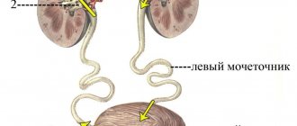
The human urethra, or urinary tract, is a tubular organ that runs from the bladder to the external genitalia. In men and women, it differs in its structure and colonization by microflora.
The organ in representatives of both sexes looks like a soft, elastic tube. Its walls consist of 3 layers:
- external, consisting of connective tissue;
- middle, formed by the muscle layer;
- internal (mucous membrane).
In men, the urinary tract passes through the penis to the outlet and serves to drain urine and release ejaculate during orgasm. In women, it goes from the bladder to the external opening, which is located between the clitoris and vagina, and is needed only for the removal of urine.
The external urethral sphincter is formed as paired muscles. It compresses part of the urethra. In the female body, these muscles are attached to the vaginal area and are capable of compressing it.
The muscles of the urethra in men are connected to the prostate. The internal sphincter has a fairly strong muscle located near the exit from the bladder.
Microflora in the organ
The channel for excreting urine in representatives of different sexes differs in microflora. Immediately after the birth of a child, various microorganisms enter his skin. They gradually penetrate into the body and settle on the mucous membranes and internal organs.
Bacteria cannot penetrate further than the mucous membranes; this process is hampered by the internal secretions of the body, urine, and ciliated epithelium, so they become attached to them. Pathogenic organisms that remain on the mucous membranes become the innate human microflora.
The female urethral mucosa contains several times more bacteria than the male urethral mucosa. It is dominated by lactobacilli and bifidobacteria. They produce acid, creating an acidic environment. If there are few bacteria, then the acidic environment is replaced by an alkaline one, which allows inflammatory processes to develop.
As we grow older, the beneficial microflora in the female urethra becomes coccal. The microflora of the male urethra is represented by streptococci, corynebacteria, and staphylococci; it does not change throughout life.
The composition of the microflora may change depending on the large number of sexual partners. Frequently changing partners introduces dangerous microbes into the body that can cause serious illness.
Men's channel
The male urethra during the embryonic period is similar to the female one, as it consists of the same structures. And once formed, it begins to differ significantly, becomes longer and smaller in diameter, is located inside the penis, and its functions, in addition to urine output, also include ejaculation.
The redistribution of these functions of the male body completely depends on the degree of filling with blood of the cavernous bodies and the spongy body, which surround the male urethra. With erection, the blood supply to the penis occurs, ejaculation occurs, and in the absence of blood supply to the penis, the process of urination occurs.
The male urinary canal has a length of 18-22 cm. In a state of excitement, the length becomes a third longer, in boys before puberty it is a third less.
The urethra in men is divided into posterior (the distance from the internal opening to the beginning of the corpus cavernosum) and anterior (the remotely located part of the canal).
It has two bends in the shape of the letter S:
- The upper (infrapubic) bend bends around the pubic symphysis (half-joint) from below as it passes from top to bottom of the membranous part of the urethra into the cavernous one.
- The lower one (prepubic, prepubic) is located at the point of its transition from the fixed part of the urethra to the mobile one.
When the penis is raised, both curves form one common one, the concavity of which is directed forward and upward. Throughout the male urethra, the diameter of the lumen is not the same, narrow parts alternate with wide ones.
The extensions are located in the prostatic, bulbous part and at the end of the urethral canal (where the scaphoid notch is located). The narrowings are located at the internal opening of the urinary canal, in the area of the urogenital diaphragm, at the external opening of the urethra.
Conventionally, the male urethra is divided into 3 parts:
- Prostatic (prostate). It has a length of 0.5-1.5 cm. It consists of tubules for ejaculate release and 2 ducts (prostatic and sperm excretory).
- Sponginous (spongy). The urethral part is located along the penis in its lower part and has a length of 13-16 cm.
- Cavernous (membranous). The longest section of the male urethra, the length of which is approximately 20 cm. The spongy section contains ducts of numerous small tubules. It is located deep in the perineum, passes through the urogenital diaphragm, which has a muscular sphincter.
The male urethra originates from the urinary sac. Smoothly passing into the prostate area, it crosses this gland and ends at the head of the penis, from where urine and seminal fluid exit. The average size of the urethral lumen in men along its entire length is 4-7 mm, in boys 3-6 mm.
The female urethra is an anteriorly directed, straight tube passing close to the elastic vaginal wall and pubic bone. Its length is 4.8-5 cm, and its diameter is 10 - 15 mm, while it is easily stretched.
Inside, the urinary canal is lined with a mucous membrane, which has the appearance of longitudinal folds, due to which the urethral lumen appears smaller. In the female urethra there is a special obstructing pad consisting of connective tissue, veins, and elastic threads. It closes the duct of the urinary canal.
The female urethra does not perform reproductive functions, although substances are excreted through it, which can be used to determine whether a woman is pregnant or not. The female urethra is surrounded by tissues that are similar in structure to the corpus spongiosum of the penis, and the corpus cavernosum of the clitoris, which is similar to the corpus cavernosum of the penis, is located in front of the urethra.
The urethra itself is hidden in the tissues of the pelvis and therefore does not have mobility. Its anterior surface is adjacent to the tissues that cover the pubic symphysis, and in distant places to the legs of the clitoris. The posterior surface of the external urethral outlet is adjacent to the anterior wall of the vagina.
It is closely connected to the anterior wall of the vagina and is firmly attached to the lower branches of the pubic bones, as well as partially to the ischial bones.
Since it is short and wide in women, located next to the vagina and anus, the danger of bacteria, microbes and other pathogenic microflora entering it is much higher in women than in men. Therefore, they are more susceptible to genitourinary infections.
External hole
In the male half of humanity, the main part of the urethra passes inside the penis, and the outlet is located at the top of its head. If it is not there, the disorder is called hypospadias. If there is partial or complete clefting of the anterior wall of the urethra, the disorder is called epispadias.
The external urethral canal in the fair sex is located between the clitoris (slightly below it by about 3 mm) and the entrance to the vagina.
The location of the external opening may vary. If the lower wall is underdeveloped, it will be located on the anterior wall of the vagina, distant from the entrance.
This process is called hypospadias. The outer hole has a diameter of approximately 0.5 cm, its shape can be round or star-shaped.
Possible diseases
You can understand that dribbling is associated with some kind of pathology by the accompanying symptoms. Depending on the disease they differ:
- Prolapse of the uterus. Most often occurs after childbirth and heavy physical exertion. Women during menopause and overweight women are also at risk. The uterus moves relative to its normal position due to weakening of the muscles and ligaments. The organ begins to put pressure on the bladder, which is why women drip urine after urinating and other urological problems arise. The menstrual cycle is disrupted and white or bloody discharge appears. A nagging pain is concentrated in the lower abdomen.
- Cystitis. Inflammation of the bladder under the influence of pathogens. Patients are bothered by a frequent urge to urinate with little urine output. The physiological fluid becomes darker and more turbid. There may be sediment or blood present. There is a burning sensation and discomfort in the lower abdomen. There is general malaise.
- Urethritis. Infection in the urinary canal area. The clinical picture includes stinging, burning and pain during urination, especially at the very beginning. There is discharge with an unpleasant odor. Often the disease is accompanied by other gynecological and urological disorders.
- Urolithiasis disease. The formation of kidney stones is always accompanied by severe pain. Depending on their size and location, the nature of the pain changes. It can be dull or sharp, concentrated in the lower back or radiate to the labia. The urge to urinate becomes more frequent, and cases of incontinence are common. An increase in temperature, nausea and vomiting may occur.
- Oncology. Tumors in the urethra and bladder interfere with the normal flow of urine. The appearance of neoplasms is characterized by pain, burning and stinging during urination and sexual intercourse. Blood is released from the vagina, small rashes, erosions and ulcers form on the mucous membranes of the vulva. Urethro-genital fistulas are formed.
- Urethral strictures. Pathological narrowing of the lumen of the urinary tract. Usually occurs under the influence of infections or after surgery. The disease manifests itself in the form of a small amount of urine with frequent urges. In this case, there is a feeling of incomplete emptying of the bladder, and after completion of urination, a lot of urine is released. The process of urine outflow itself begins difficultly, after some waiting and straining the abdomen. There is pain and bleeding appears.
- Diseases not related to the genitourinary system. Spinal injuries, neurological diseases and pathologies of the central nervous system lead to disruption of the conduction of nerve impulses. There is increased muscle excitability. Patients lose the ability to control their body. Therefore, incontinence occurs or urine drips after urination in women.
In terms of frequency of occurrence, cystitis ranks first even among young girls aged 15-25 years.
Urinary retention
Difficulty urinating in women leads to hypertrophy of the bladder muscles. Emptying the bladder does not happen in one act of going to the toilet.
The patient has to make more effort and push again with each portion of urine. At first, the patient manages to achieve complete emptying of the bladder, but then this cannot be achieved. Stagnation of urine occurs, and its volume gradually increases - this leads to chronic urinary retention (ischuria).
The concept of chronic urinary retention means that the patient does not completely empty the bladder after urination. This occurs due to an obstruction to the exit of urine. The main causes of urinary retention in the chronic form in women are: tumor formations in the genitourinary organs, which put pressure on the urethral canal.
Further development of the disease can lead to the inability of the sphincter to hold back urine, urine comes out drop by drop from the bladder, such a pathology is called paradoxical ischuria.
Urinary retention in women can become acute. Acute retention occurs unexpectedly to the patient and is characterized by the absolute inability to empty the bladder, despite its complete filling. The acute form can develop against the background of the chronic one.
The reasons for this delay may not be mechanical factors: pathologies of the brain and spinal cord, spinal tumors, diseases of the nervous system. Mechanical factors include:
- urethral compression;
- urethral tumors;
- injuries in the urethral area.
- foreign object.
Diagnostic methods
Already at the first appointment, the urologist can make a preliminary diagnosis. The patient must be told how often incontinence occurs, how much urine is excreted, and how much liquid she drinks.
The gynecologist examines the condition of the muscles and tissues of the pelvic floor, examining whether there is prolapse of the vaginal walls of the uterus. The doctor asks the patient to cough; if urine leaks, this is referred to as stress incontinence.
Tests to determine the cause of incontinence:
- Cystoscopy. Insertion of a thin tube into the ureter and bladder.
- Ultrasound of the pelvic organs.
- Urodynamic examination. Sensors are inserted into the bladder and vagina to record the filling and emptying of the bladder.
A blood and urine test and a smear examination are required.
If your urine is brown and you have back pain, what is it?
Although darkening can be caused by simple dehydration, when combined with pain in the back area, it should alert you to see a doctor. After all, this can be a sign of a number of diseases: jaundice, hepatitis, urolithiasis, urinary tract infection, STDs, cirrhosis, intestinal problems, gastrointestinal bleeding, anemia, liver cancer. If you experience any of the above symptoms, do not self-medicate. Timely diagnosis is a chance for successful treatment.
Treatment Options
Depending on the causes of the problem and the severity of the patient’s condition, treatment methods vary:
- Drug treatment. For bacterial infections, antibiotics are prescribed. Stressful urine production involves taking antidepressants. When the walls and sphincter of the bladder are weakened, the use of drugs that increase muscle tone is indicated.
- Surgical intervention. Necessary for oncology and changes in the structure of the urinary system. Patients have the tumor removed, implants installed, a special gel injected, or laser treatment of the urethra performed.
- Physiotherapy. Aimed at increasing blood flow to the pelvic organs and improving muscle elasticity. For such manipulations, microcurrents, heating or electromagnetic pulses are used.
If after urination a woman produces drops of urine, she should immediately contact a gynecologist, neurologist or urologist. The doctor will help you avoid the need to constantly use pads or diapers and promptly identify the diseases that led to the problem.
Complex treatment includes:
- Non-drug therapy. It consists of performing physical exercises to train the muscles of the pelvic organs. It is recommended to normalize weight and maintain proper nutrition. It is also necessary to accustom the bladder to emptying at certain hours; for this, the doctor draws up a plan, which the patient strictly follows.
- Drug therapy. It consists of taking medications to reduce the contractile activity of the bladder and increase its functional capacity. Antidepressants, antispasmodics, and drugs to regulate irritating impulses from the central nervous system are prescribed.
- Surgical intervention. Used when conservative therapy is ineffective. Minimally invasive and sling surgeries are common.
Traditional methods of combating pathology
Treatment with folk remedies for difficulty urinating is often used. Some patients think that this condition is a consequence of a lack of urine production and try to correct their own condition by taking diuretics and herbal infusions that have a diuretic effect.
Such actions are wrong and can cause irreparable harm. You can resort to the use of medications and herbal compositions only after consultation with a specialist.
To eliminate functional disorders of the bladder, provided there is no organic component of the pathology, the following traditional methods of treatment can be used:
- use of decoctions and infusions of sage leaves;
- drinking freshly squeezed celery juice;
- drinking a decoction of rose hips or fresh juniper berries.
Optimal results in the treatment of pathology can only be achieved with timely consultation with a specialist. The use of improvised means (self-medication) is unacceptable and can cause serious problems.
Diagnosis, treatment and prevention
When seeking medical help, you should tell about all past illnesses, problems or alarming situations. The doctor should conduct a physical examination and order a complete urine and blood test. If necessary, an ultrasound of the abdominal cavity is performed. If no drugs or dyes have been ingested into the body, it is imperative to find out why the urine is brown. This applies to men, women and children. Brown urine is alarming, but without examination by a doctor you should not self-medicate. There is no special treatment, since it is just a symptom of a disease. The person should be further examined, and treatment will be started after the diagnosis is determined. Simple measures can be helpful for many conditions that can cause brown urine. This includes:
The patient must understand that these are just preventative measures. Therefore, if he has doubts about why the urine is brown, it is best to seek qualified help. Simply following the above measures will not eliminate the problem.
Video: What can your urine “tell” about you?
Reasons for deviation from the norm
Impaired urine flow occurs in both women and men. Pathological disturbances in the urination process can be caused by:
- inflammatory phenomena in the urinary organs (cystitis, pyelonephritis);
- neoplasms;
- narrowing of the lumen of the urethra;
- blockage of the ureter or urethra with a stone;
- prostate adenoma in men;
- prostatitis in men;
- cervical cystitis in women;
- pelvic inflammation;
- consequences of injuries.
What factors affect the color of urine?
It is recommended to take a general urine test every six months. This is necessary in order to promptly detect and eliminate various health problems. When studying biomaterial, specialists must take into account the color indicator.
Normally, biological fluid should have a straw color, which is ensured by some pigments present in it:
- urochrome is a substance with a yellow color. Formed from bile pigments during the destruction of hemoglobin;
- urobilin. It is a product of bilirubin metabolism. Gives urine a red or brown tint. The release of richly colored fluid is observed in various pathologies;
- uroerythrin. Gives urine a red tint and is found in small quantities.
If the coloring substances have disappeared for some reason, then the urine becomes colorless.
Periodic changes in its color can be caused by:
- excess fluid intake;
- significant physical activity;
- taking diuretics;
- emotional turmoil;
- severe hypothermia;
- drinking alcohol;
- consumption of certain products.
Colorless urine in women is a sign of excess fluid
After the cessation of exposure to the provoking factor, the color indicator will return to normal. But if the disorder is constantly present and other symptoms appear, this may be a sign of the development of a dysfunctional process.