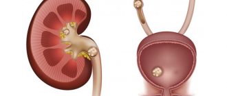
The ureter is part of the urinary canals, these are tubes connecting the bladder and kidneys. As in any organ, a tumor can develop here.
Benign formations are extremely rare - only in children and are expressed as fibroepithelial polyps.
The vast majority of tumor neoplasms are malignant and represent transitional cell carcinoma.
Ureteral oncology accounts for only 2–3% of all diagnosed cancers.
As a rule, detection of a tumor occurs at a very advanced stage, when the neoplasm affects all layers of the organ, adjacent tissues and spreads distant metastases.
What are ureteral tumors?
The content of the article
Neoplasms in the ureter are similar in pathogenesis and etiology to tumors of the renal pelvis. They originate from both epithelium and connective tissue. Connective tissue neoplasms are not very common; neoplasms of epithelial origin - papillomas, as well as squamous cell and papillary cancers - appear much more often.
The primary tumor in the vast majority of cases is located in the lower part of the ureter, in more rare cases - in the middle of the duct. Middle-aged and elderly people are more susceptible to the appearance of tumors of this localization.
Diagnosis of ureteral cancer in the oncology center
A complete health examination is performed using different methods. Ureteral cancer can begin with the formation of squamous cell carcinoma at the in situ stage without the formation of its own blood vessels. Detection at this stage has the best prognosis. At the Sofia Oncology Center, at 2nd Tverskoy-Yamskaya lane 10, the latest diagnostic methods are used to make a diagnosis.
Positron emission and computed tomography
PET/CT can help detect ureteral cancer at different stages. This is one of the main methods of early diagnosis. The clinic uses the latest generation Gemini TF Philips equipment. A high degree of information content of the results is the main advantage in identifying ureteral cancer in patients.
Single photon emission computed tomography
SPECT has high sensitivity. Ureteral cancer in women and men is not so easily determined. SPECT allows you to obtain functional images of organs and tissues of the body at the molecular level. The method works especially well in the early stages of the disease, when there is still time for a complete cure.
Scintigraphy
One of the diagnostic methods. Used to study pathological changes. If ureteral cancer begins to show symptoms, then scintigraphy is recommended in combination with other examination methods. The method is informative based on the results. A procedure is carried out with the introduction of radioactive isotopes into the body.
Laboratory research
Laboratory tests of blood and urine are included in the mandatory examination plan. Ureteral cancer requires comprehensive diagnosis. They also include cytological studies and biopsy. The Sofia Cancer Center has its own laboratory. Analyzes, procedures and research methods are selected individually for each patient.
Causes of ureteral tumors
The urothelium in the ureter is highly sensitive to the chemical composition of urine. Among the factors that increase the risk of developing a ureteral tumor, as well as other neoplasms, smoking is the leading one. Statistics indicate that 70% of men and 40% of women diagnosed with ureteral tumors are long-term smokers.
Urothelial cancer is also more likely to appear in people taking large amounts of analgesics or diuretics. Thus, when treating arterial hypertension, the risk of developing ureteral tumors increases due to the use of diuretic drugs. People whose professional activities take place in oil refining, plastics and plastics production enterprises are also at increased risk.
Doctors say that chronic pyelonephritis, stones in the urinary tract and ureteral injuries slightly increase the risk of neoplasms. Genetic predisposition, Lynch syndrome and malignant tumors of the pelvic organs (uterus, ovaries, intestines, prostate, etc.) also have an impact.
How to treat
To eliminate the symptoms of cancer of this organ, nephroureterectomy is used. Most often, the affected kidney and ureter are surgically eliminated; in particularly severe cases, adjacent tissues and lymph nodes are removed. A person’s continued existence is possible even if he has one kidney, but he will need to regularly see a doctor and use restorative therapy.
At the initial stage of the disease, partial excision of the urinary tract affected by cancer is possible, while restoration of the functioning of the organ is possible only with the help of subsequent prosthetics.
In order to prevent relapse of the disease, courses of immunotherapy and chemotherapy are carried out, and to combat malignant neoplasms, radiation therapy is used with the effect of gamma radiation on the affected tissues. For neighboring organs and tissues, such therapy is not destructive and has the most minimal impact.
In cases with possible damage to the ureter due to genetic predisposition, preventive measures should be taken in advance to prevent the occurrence of cancer of this organ. These include the following actions:
- support for proper nutrition,
- daily drinking of clean liquid in sufficient quantity,
- active lifestyle,
- refusal to work in polluted industries,
- use of herbal preparations,
- Medicines should be taken only in permitted doses and only as prescribed by a doctor,
- When forced into contact with toxic substances, adhere to safety rules.
Types of disease
Neoplasms of the ureter can be classified according to the primary appearance, nature and origin. By nature, benign and malignant neoplasms are distinguished. Based on their origin, they distinguish between primary and secondary, resulting from the spread of a primary tumor from other organs.
Based on their origin, epithelial and connective tissue tumors are distinguished. Epithelial tumors, in turn, are divided into:
- papillomas;
- papillary cancer;
- squamous cell carcinoma.
If connective tissue neoplasms are diagnosed, the presence of the following forms of ureteral tumor can be assumed:
- fibroids;
- sarcomas.
- leiomyomas, etc.
Classification of disease and stage of ureteral cancer
From the moment of onset, ureteral cancer goes through four stages. At the first stage (T1), tumor formation begins. It affects mucous and submucosal tissues. At this stage, it is located locally without germination. As the disease progresses to the second stage (T2), carcinogenic cells are activated. Germination into the muscle layer begins. At the third stage (T3), ureteral cancer affects fatty tissue. At the fourth stage (T4), tumor cells grow into neighboring organs. The peculiarity is that even at the last stage with metastases in the lymph nodes, the prevalence of the disease remains local. All four stages of ureteral cancer are classified according to TNM.
Symptoms of ureteral tumors
If the ureteral tumor is benign, it may not manifest itself for a long time. Among the symptoms of a malignant neoplasm of the ureter, the following characteristic signs of pathology can be identified:
- Hematuria.
It is the presence of blood in the urine that often becomes the reason for a patient to contact a urologist when this pathology develops. More than 90% of all patients with a duct tumor note constant or periodic bleeding in the urine. In 70% of cases, we are not talking about a few drops, which can be easily missed, but about a state of gross hematuria, which usually frightens the patient. - Pain –
approximately half of all patients experiencing this localization of the tumor complain of pain in the lumbar region. The occurrence of pain is associated with blockage of the duct in the ureteropelvic segment. - Dysuria is
a rather specific symptom of ureteral tumors, which can develop in the later stages of the disease. About 10% of patients report severe difficulty urinating, accompanied by pain or discomfort. - General symptoms -
the development of a neoplasm in the body can be accompanied by periodic increases in temperature to low-grade levels, weakness, loss of appetite, weight loss and weakened immunity.
Prolonged ignoring of symptoms can provoke the development of ureterohydronephrosis, which is manifested by expansion of the pyelocaliceal complex with subsequent atrophy of the renal parenchyma. Another complication of a progressive ureteral tumor is the formation of secondary stones.
If the tumor is benign, the main danger will be its degeneration into malignant. For malignant tumors, especially those with a bilateral location, the risk of relapse is very high even with complete treatment of the tumor. In addition, ureteral tumors metastasize early, which significantly complicates the recovery process.
Advantages and disadvantages of various diagnostic methods
Until recently, radiography was the main diagnostic method. To do this, a contrast agent was injected into a vein, and doctors monitored its excretion by the kidneys.
At a certain time during the examination, photographs were taken, which were then studied by specialists. Urologists carried out this diagnostic method independently, monitoring the entire process, every stage.
In modern medicine, computer urography has become the diagnostic standard, which almost 100% detects malignant tumors whose size exceeds 5 mm, and sometimes even 3 mm.
This method evaluates the condition of the ureteral wall. A slightly less sensitive method is magnetic resonance imaging.
Ureteroscopy has also proven itself well. This is a type of endoscopic examination, when a piece of the tumor is taken for histological diagnosis.
In the first stages, a urine sample is taken for this purpose. If cancer cells are detected in it, a decision is made on further examination and treatment.
Diagnosis of the disease
To diagnose a ureteral tumor, a number of laboratory tests are performed, including a general blood test and a urine test with gross hematuria.
An ultrasound examination of the bladder and an ultrasound scan of the pelvis must be performed.
Further may be prescribed: computed tomography, urography, cystoscopy. To make a definitive diagnosis, tissue biopsy is most often used.
Prevention
It is unrealistic to completely protect yourself from the manifestations of ureteral cancer, especially if there is a hereditary predisposition, but any man or woman is quite capable of eliminating the situations that provoke the disease.
General recommendations include the need to maintain a healthy lifestyle, giving up bad habits and normalizing nutrition. You should visit your doctor regularly, and under no circumstances abuse medications that can depress your kidneys by exposing you to toxins.
Measures to avoid illness:
- taking medications only on the recommendation of a doctor and in cases of real need,
- focus on herbal medicines,
- support for a balanced diet,
- physical activity,
- consumption of at least 1.5 liters of clean water per day,
- refusal to work in hazardous industries,
- when in contact with toxic substances, even for a short time, it is imperative to take measures to protect the respiratory tract and skin,
- complete cessation of smoking and alcoholic beverages,
- maintaining a normal rest and wakefulness schedule.
Following all the above tips, it is also recommended to constantly undergo examinations in a medical institution, during which blood, urine and feces are examined in order to timely identify possible pathologies.
Treatment methods
The only way to eliminate a ureteral tumor, regardless of its nature. Is surgical treatment. If the tumor is in an advanced stage and affects surrounding tissues, partial resection of the ureter and the adjacent bladder wall can be performed during the operation.
Tumors of the ureter are resistant to chemotherapy and radiation treatment; after surgery, the patient requires long-term observation in outpatient treatment. Ureteral cancer is extremely rare, according to statistics - from one to four percent of all neoplasms of the upper urinary tract. Therefore, finding a specialist who would be sufficiently qualified to diagnose and treat this disease is not easy.
In our clinic you will find professional doctors, polite staff and the best care. We will provide you with the best conditions for recovery, modern treatment facilities and a comfortable atmosphere.
If you find an error, please select a piece of text and press Ctrl+Enter
Forecast
After surgical treatment, the disease is usually completely cured. Some patients experience relapses. Most often they appear in the bladder. The probability of relapse, depending on the method of surgical treatment used, is 15–50%. Strict monitoring of the patient is required for timely detection of relapse. He undergoes cystoscopy initially every 3 months, then every 6 months, and after 2 years - every 12 months.
Take care of yourself, book a consultation now
Causes and risk factors
The epithelium of the urinary tract is sensitive to the negative effects of chemical factors that have a carcinogenic effect. This group includes smoking and occupational hazards (working with arsenic, gasoline and other industrial poisons).
The next group of predisposing factors includes urolithiasis and inflammatory diseases of the genitourinary system. When concretions (stones) move through the ureters, trauma to the mucous membrane occurs. Frequent disruption of the integrity of the mucosa leads to its hyperplasia, which increases the risk of cellular malignancy.
In addition, the injured membrane, with prolonged contact with urine when it stagnates, is exposed to toxic effects. As a result, an inflammatory process develops, which becomes chronic.
Among other provoking factors, it is worth highlighting increased blood pressure, hereditary history and prolonged use of diuretics.
Recovery prognosis
Ureteral cancer in women can be cured if the pathology is detected early. This is the main criterion for relatively positive forecasts. It all depends on how much the cancer of the pelvis and ureter progresses and whether there is penetration into the urothelial wall. For superficial tumor processes, the prognosis is positive in 90%. With stages 2 and 3, less than 50% of patients live more than 5 years. Age is a key factor, but gender is not. If the tumor has grown and there are metastases, ureteral cancer has an unfavorable prognosis.
Manifestations
As a rule, the disease in the early stages of its development does not have any specific manifestations. Symptoms of the disease appear when the pathological process spreads to the tissue of the ureter or adjacent organs.
When large vessels are damaged by cancer cells, blood in the urine is visible to the naked eye
The most common of them are:
- the appearance of blood in the urine (hematuria is observed in 75–80% of patients, which is due to the destruction of the walls of blood vessels by the growing tumor and their ulceration);
- pain in the lower back on the side of the affected ureter (pain sensations are of a different nature, some patients report only a feeling of discomfort in the back, others complain of unbearable pain);
- dysuric disorders (the lumen of the ureter is often blocked by necrotic masses, blood clots or pieces of tumor, which provokes attacks of pain and cramping when urinating);
- general weakness, unmotivated decrease in working capacity, drowsiness, apathy;
- causeless loss of body weight, loss of appetite, increased sweating during sleep and with minimal physical activity (these signs require special attention, since in most cases they are not given due importance);
- formation palpated in the projection area of the affected ureter.
In advanced cases, palpation of the tumor node through the anterior abdominal wall is possible


