If pain occurs and difficulty urinating, it can be assumed that the patient has stones in the urethra. At the same time, the stream of urine and its color change. If the stones are large, they block the urethra and acute urine retention occurs. With this disease, the bladder, urinary canals, and urethra are affected. The stage of development of the disease is determined by the size of the stones. The choice of treatment method depends on many factors and is determined by the doctor individually for each patient. To prevent surgical intervention, you need to undergo comprehensive diagnostics in a timely manner.
Stones in the urethra can form for several reasons, and their presence will cause discomfort and be fraught with complications.
General information
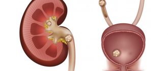
Urethral stones can be primary (that is, initially formed in the urethra) or secondary (formed in the upper urinary tract - the kidneys or bladder and descended into the lumen of the urethra). Specialists in the field of clinical urology are more likely to encounter secondary urethral stones.
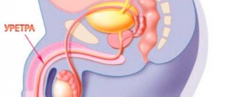
In women, urethral stones are much less common than in men, which is due to the peculiarities of female anatomy: a shorter urethra, its better extensibility and straight direction. In their chemical composition, urethral stones are similar to kidney stones - urate, oxalate, phosphate, carbonate, cystine, protein, cholesterol, etc.
How to remove a stone in the ureter at home?
Treatment at home includes baths with decoctions, hot lotions and compresses, and the use of decoctions and medications.
The most popular types of traditional treatment are::
- Knotweed. 1 tablespoon of herb, poured with boiling water. Leave until completely cool, take on an empty stomach and at night for 2 weeks.
- Rosehip teas are also effective as teas.
- Take steamed flax seeds 4 times a day, 1 tablespoon.
Causes
Primary urethral stones are predominantly found in men. Conditions for their formation may include urethral strictures, fistulas or diverticula of the urethra, prostate adenoma, prostatitis, and chronic urethritis. The stones repeat the configuration of the part of the urethra in which they form and grow. The largest stones form in the diverticula of the urethra.
Secondary stones are a manifestation of urolithiasis (the presence of stones in the kidneys or bladder). Descending from the upper urinary tract, they get stuck in the urethra, causing the corresponding symptoms. The number and shape of urethral stones may vary. Secondary stones often get stuck at the level of the scaphoid fossa, sometimes in the membranous or prostatic part of the organ.
The reasons for the formation of secondary stones may be errors in the food and drink diet, dehydration; disorders of mineral metabolism in the body due to hyperparathyroidism, osteoporosis, bone injuries; gastrointestinal diseases (gastritis, colitis, peptic ulcer); urinary tract infections (pyelonephritis, cystitis), etc.
What influences the formation of stones
The formation of primary stones is rare and is always caused by certain pathologies that disrupt the structure of the urethra. These include: narrowing of the canal lumen, inflammatory processes, fistulas. The appearance of secondary stones, which happens more often, is associated with their migration into the urinary tract, in which they are retained due to its narrowing.
The main causes of stone disease are:
- Severe metabolic disorders due to an unbalanced diet;
- Dehydration of the body;
- Bone pathologies, damage to bone tissue;
- Disturbances in the functioning of the digestive system;
- Inflammatory and infectious diseases of the urinary organs (pyelonephritis, cystitis).
Primary stones take on the shape of the urethral region in which they are located. The largest specimens of stones grow in the diverticular ridges of the urethra. The chemical composition of the formations is identical to kidney stones and is determined by the substances that form them: urates, oxalates, phosphates, carbonates, proteins or cholesterol, and others.
Symptoms
Symptoms of urethral stones and their severity are determined by the location, shape, size, number of stones and the duration of their presence in the organ cavity. Obstruction of the urethral lumen by a stone is accompanied by pain, difficulty urinating, dispersion and weakening of the urine stream, and hematuria. When a stone completely blocks the lumen of the urethra, acute urinary retention develops. Posterior urethral stones cause pain when walking and sitting.
Complications
Due to the prolonged presence of the stone in the urethra, urostasis occurs in the upper parts of the urinary tract, creating conditions for hydronephrosis, urethritis, bedsores in the urethral mucosa, the formation of urethral fistulas and paraurethral abscesses. Urethropuzural stones, partly located in the urethra and partly in the bladder, can cause urinary incontinence. Sexual intercourse is painful; with constant pain, decreased libido and sexual dysfunction may develop.
Relapses
Relapses of nonspecific urethritis occur in the following cases:
- Weakening of the immune system.
- Unprotected sex with an infected woman.
- Repeated infection in the urethra from a source of chronic inflammation in the body (tonsillitis, prostatitis).
- Reproduction of an untreated infection.
- The presence of a constant irritant: diabetes mellitus, urolithiasis, herpes virus.
If symptoms reappear, you should consult a doctor again, since the causative agent may already be different.
Diagnostics
Recognition of urethral calculi is based on characteristic symptoms, palpation data, and the results of instrumental studies. In men, stones are determined by palpation of the hanging part of the urethra or perineum, and if localized in the posterior sections, during a digital rectal examination. In women, a gynecological examination is performed, during which the stone can be felt through the anterior wall of the vagina.
Ultrasound of the bladder visualizes a hyperechoic formation of the urethra, giving an acoustic shadow. A general urine test reveals micro- or macrohematuria, signs of inflammation (leukocyturia), crystalluria, and often an alkaline pH level.
For the purpose of differential diagnosis of stones and foreign bodies of the urethra, they resort to imaging studies - urethroscopy, survey urography, urethrography. The shadow of a calculus can often be seen already on survey urograms. During X-ray contrast examination, the stone is identified as a filling defect or enhanced contrast. Endoscopic and x-ray examination of the urethra allows you to get an idea of the location, size, number of stones and predict methods for their removal.
Symptoms, diagnosis
The clinical picture of stones in the urethra is determined by a number of factors: the number of stones, their shape and size, location and duration of stay. As a rule, an intense pain syndrome quickly develops, especially during urination, which is also accompanied by characteristic difficulties in the form of a sharp weakening and/or its breaking into several streams, “leakage” of urine into the underwear outside the act of urination, and the appearance of blood. In many cases, pain radiates to adjacent areas (head of the penis, perineum, anal area, etc.). With posterior urethral localization, pain may occur when walking or in a sitting position.
In addition, it should be understood that stones in the urethra can be not only the result of a prolonged inflammatory process, but also, conversely, the cause of urethritis with a probable upward spread.
In some cases (for example, when the stone is located in a diverticular protrusion of the urethra), symptoms may be very “smoothed out” or completely absent. In other, much more frequent cases, the stone gets stuck at the very “exit” and can lead to such a serious and emergency condition as acute urinary retention.
In men, urethral stones are usually diagnosed during the collection of complaints, examination and palpation (if not direct, i.e. when palpating the urethra through the penis, then during rectal - standard urological palpation through the rectum). In women, as well as in unclear cases, a bougie is sometimes used - a special probe that dilates the urethra and makes it possible to localize the calculus simply mechanically. For the purpose of differential diagnosis (to differentiate from urethral stricture, prostate adenoma and other suspected types of pathology with similar symptoms), urethroscopy, various modifications of radiographic examination, ultrasound, etc. are used.
Treatment of urethral stones
Most often, stones located in the urethra are pushed into the bladder during urethroscopy. In this case, the next step is lithotripsy. In rare cases, stones wedged into the urethra go away on their own after the administration of antispasmodics, water load, sitz baths, etc.
If attempts at conservative and instrumental removal of stones are unsuccessful, external urethrolithotomy and stone removal are indicated. Surgical removal of stones from the posterior urethra is performed through an open bladder. During the operation, an epicystostomy is performed to divert urine. At the same time or as the next step after removing a urethral stone, surgical removal of the cause of stone formation is required - urethral stricture, diverticulum, prostate adenoma, bladder or kidney stones, etc.
Rehabilitation period
After removal of stones from the kidneys and ureter, the patient undergoes antibiotic therapy to prevent infection. For each patient after surgery, doctors prescribe diuretics (diuretic drugs) and increase the amount of fluid consumed during the day.
Particular attention should be paid to nutrition, since poor nutrition is the main cause of stone formation. For urolithiasis, there is a special diet that excludes the consumption of certain foods. It is worth adhering to it not only during the rehabilitation period, but after the first signs of the disease appear.
When a patient has stones removed using a remote or endoscopic technique, the body recovers quite quickly. When performing a classic operation, the recovery period depends on many different factors.
Regardless of which method was chosen to remove stones from the kidneys and urinary tract, patients must adhere to all medical recommendations. The body will recover faster and the risk of relapse is minimized.
Prognosis and prevention
The prognosis is favorable. Timely removal of urethral stones prevents serious complications (paraurethritis, urethritis, prostatitis, the formation of fistulas and bedsores of the urethra). To exclude the possibility of recurrent stone formation in the urethra, correction of eating behavior and drinking regimen, and elimination of the causes of urolithiasis are required. If there are pathologies of the urinary system, regular monitoring by a urologist is required.
The manifestation of the formation of stones in the genitourinary system may be accompanied by their occurrence in the excretory channel of the bladder, that is, the urethra. The disease is accompanied by pain, difficulty passing urine, in some cases, and even complete retention.
Typically, doctors can diagnose primary stones that can form directly in the urethra, but this occurs only in rare cases. And secondary, they are concentrated in the upper parts of the genitourinary system, that is, in the bladder and kidneys.
This is a more common type of urethral stone formation, and the male population is more susceptible to it. Due to the unique structure of the female genitourinary system, such a pathology rarely occurs in women.
Causes
Typically, the primary development of stones in the urethra occurs in the stronger sex, and the reason for this can be various negative phenomena, these are:
- Fistulas in the urethra.
- Prostatitis.
- BPH.
Stones can form in any part of the urethra, taking its shape and growing, leading to inflammation and pain.
Secondary stones descend into the ureter from the bladder or kidneys, getting stuck in the urethra, leading to the corresponding symptoms. They can be of different sizes, smaller ones can come out along with urine, and large ones, as a rule, get stuck in the scaphoid fossa, and sometimes in the prostatic ureter. The reasons in this case may be:
- Diseases of the gastrointestinal tract.
- Poor nutrition.
- Failure to comply with the drinking regime.
- Violation of mineral metabolism.
- As well as various diseases, such as pyelonephritis, cystitis and so on.
Stones in the urethra in men
Acute pain in the urethra, blood in the urine, discharge of blood from the external opening of the urethra, a sluggish, barely noticeable stream of urine, all these manifestations allow the urologist to suspect a stone in the pendulous urethra. This condition is an emergency and requires immediate qualified assistance. In practice, most often we encounter patients with a stone in the scaphoid fossa of the urethra. If a patient is treated by a non-specialized doctor, there is a risk of misdiagnosis; often they try to treat such a patient for urethritis.
Causes of stones in the urethra (urethra)
Most often it is a consequence of urination problems. They represent stones from 1-2 mm to 1 cm or more. As a rule, in 50% of cases stones with a diameter of less than 5 mm come out on their own. According to this criterion, they are classified into primary and secondary. The first are formed directly in the urethra due to difficulty in the outflow of urine. They are more common in men due to the occurrence of various pathological changes - strictures, diverticulum of the excretory duct, chronic inflammation. The second is detected due to the entry of a calculus from the bladder into the urethra and gets stuck in it.
Symptoms of urethral stone in men
In our practice, formations most often occur in the scaphoid fossa.
Symptoms – difficulty in the outflow of urine; – pain in the penis; – pain at the end of urination; – weakening of the jet; – complete absence of urination (narrowing of the lumen); – spraying urine; – the appearance of blood in urine and semen; – incontinence; – increased frequency of urination at night; - temperature increase; – fatigue, nausea; – pain during sexual intercourse; – lack of erection (or pain during erection); – decreased libido; – pain when sitting or walking in the lumbar region; – pain during defecation, extending into the scrotum and excretory duct.
As a worst-case scenario, when stones remain in the urethra for a long time, bedsores, urostasis, hydronephrosis, urethritis, fistulas, abscesses form, and infertility develops. Many of these symptoms may indicate other diseases, such as prostatitis, urethritis, cystitis, urolithiasis, prostate adenoma and many others.
The reasons for the formation of primary stones can be chronic inflammation in the urethra (urethritis), strictures (tissue scarring and narrowing of the lumen of the urethra), prostatitis, adenoma, diverticula (protrusion of the walls of the urethra, where the largest stones are formed). The basis for the formation of secondary stones is considered to be a violation of water balance, nutritional errors, diseases of the gastrointestinal tract, inflammatory processes and infections of the urinary system (prostatitis, urethritis, pyelonephritis, cystitis and others). It can also be constant stress, overwork, bad habits, violation of personal hygiene rules, hypothermia, injuries to the genital organs, irregular and unprotected sex life. The fastest thing that can be done to diagnose inflammatory processes in the urinary system is to take: - a general blood test (with inflammation, the number of leukocytes and ESR is increased); – general urinalysis (increased values of red blood cells and white blood cells).
Diagnosis by a urologist
Firstly, the palpation method is used as an emergency measure. A digital examination of the canal from the outside (palpation of the penis) or a rectal digital examination is possible.
Secondly, the ultrasound diagnostic method (ultrasound) is used, in this case the image shows a hyperechoic inclusion in the urethra.
Thirdly, excretory urography - the X-ray method is used with the introduction of a contrast agent into the patient’s vein (or urethrocystography - the introduction of contrast into the area of the bladder and excretory duct).
Fourthly, a urethrogram is used to assess the filling defect, as well as enhance contrast;
Fifthly, urethrocystoscopy - examination of the urethra using a cystoscope (insertion of a tube with an optical part and lighting). Treatment
Initially, conservative methods are used:
1. Antibiotic therapy is used to reduce inflammation;
2. Anti-inflammatory drugs are prescribed to reduce pain;
3. Antispasmodics are necessary to expand the ducts and improve patency;
Symptoms
When stones appear in the urethra, the symptoms are almost the same in men and women. The pain syndrome will be expressed more strongly or weakly, depending on their number and size, as well as the duration of stay in the urinary canal.
The longer the stones remain in the urethra, the more dangerous it becomes to the patient's health. This can lead to various inflammatory processes, the development of bedsores in the mucous membrane, fistulas, as well as paraurethral abscesses.
They prevent normal urine flow, and when the lumen is completely blocked, they cause severe pain and interfere with normal movement. In some cases, this can cause urinary incontinence. The accumulation of stones in the urethra can lead to a decrease in sexual activity, since during intimacy with a partner, one experiences pain and discomfort.
Prevention
Measures to prevent bacterial nonspecific urethritis:
- Use of barrier contraception for any type of intimate contact.
- Timely treatment of inflammatory foci in the body.
- Increasing immunity, healthy lifestyle.
- Prevention of prostatitis.
A balanced diet and monitoring the acidity of urine to prevent the formation of salt deposits and stones will help to avoid the development of nonspecific urethritis. If you have diabetes, it is important to maintain normal blood glucose levels. You should also pay attention to regular bowel cleansing and perform daily exercises to improve blood circulation in the pelvic area to avoid stagnation.
We provide examples of such exercises in the article “Symptoms of blood stagnation in the pelvis in men and methods of its treatment.”
Diagnostics
If a stone is stuck in the urethra, you should contact a urologist who will prescribe the appropriate examinations. First of all, palpation of the hanging part of the urethra and perineum is carried out, as well as instrumental examinations.
If stone formation occurs in the posterior part of the urethra, then a rectal examination is used. Women undergo a gynecological examination of the anterior wall of the vagina, as a result, with the help of palpation, the location of stones can be determined.
In addition, patients are prescribed an ultrasound examination and urine tests, which help identify signs of inflammatory processes. Urethral dilation, urethroscopy, urography and urethrography may also be prescribed. All these examinations of the urethra help to create a complete picture of the disease, that is, it will establish the location of the stones, their sizes and shapes. After this, the doctor decides on how to remove the stones.
Diagnosis of stones in the canal
Stones in the urethra are detected in the presence of characteristic symptoms, using palpation and examination with instruments.
In men, palpation of the urethra and rectal examination are performed. And in women, by examining the vaginal walls by a gynecologist.
An ultrasound examination and a general blood test are also performed. Urethroscopy, urethrography, endoscopy, and x-rays are prescribed.
Ultrasound and x-rays help determine the exact location of stones, their size and method of treatment.
| Code | Name | Price |
| 03.00 | Initial appointment with a urologist (candidate of medical sciences) | 2,000 rub. |
| 03.04 | Repeated appointment with a urologist (candidate of medical sciences) | 1,200 rub. |
| 03.60 | Initial appointment with a urologist (MD) | 5,000 rub. |
| 03.61 | Repeated appointment with a urologist (MD) | 3,000 rub. |
| All prices of the men's and women's health clinic | ||
Treatment
Depending on the complexity of the disease, the doctor chooses a specific treatment method, which can be carried out conservatively or surgically.
They can improve blood circulation in the urethra, remove spasms in the muscles of the pelvis and urethra, increase the volume of urine excreted and enhance peristalsis, which helps the passage of stones.
In addition to these drugs, to facilitate the passage of stones through the urethra, special solutions of papaverine, glycerin or novocaine are injected into the urethra.
If the stones are large, then non-invasive laser crushing is used to crush them in the ureter, these are ultrasound and electro-hydraulic methods.
Timely removal of stones from the urethra, and subsequent proper treatment, can give a positive result. But to avoid a recurrence of this situation, patients need systematic monitoring by a doctor.
If pain occurs and difficulty urinating, it can be assumed that the patient has stones in the urethra. At the same time, the stream of urine and its color change. If the stones are large, they block the urethra and acute urine retention occurs. With this disease, the bladder, urinary canals, and urethra are affected. The stage of development of the disease is determined by the size of the stones. The choice of treatment method depends on many factors and is determined by the doctor individually for each patient. To prevent surgical intervention, you need to undergo comprehensive diagnostics in a timely manner.
What should I do to get a stone out of the ureter?
Thanks to advances in medicine, there are a variety of methods that can be used to remove a stone from the ureter.
The following methods are known:
- Conservative treatment - in this case, the doctor adjusts the diet based on the type of stones and selects an individual water load. This option is suitable for small stones that can come out on their own or dissolve. In addition, various drugs based on herbal components and herbal teas are prescribed.
- Lithotripsy is a method of crushing stones in the ureter without damaging the skin using shock waves. The stone breaks into small pieces that can come out on their own. In order to precisely hit the stone, X-rays or ultrasound are used. The price of this method is about 10-80 thousand rubles. The method is contraindicated for inflammatory and purulent diseases of the genitourinary system, pregnancy, menstruation, aortic aneurysm, urate stones, spinal deformity, obesity, cavernous tuberculosis or kidney tumor, as well as for impaired renal function and insufficient blood clotting. Depending on the type of shock waves, lithotripsy may be:
- electromagnetic;
- piezoelectric;
- electrohydraulic.
- Ureteroscopy - removal of stones through the urinary tract. The instructions for this method are based on the introduction of medical instruments into the ureter, through the urethra and bladder, and further removal or crushing of the stone. The procedure is carried out only for stones whose size does not exceed 10 mm. Ureteroscopy is also performed to clarify the condition of the urethra, which makes it possible to identify other pathological processes. Before the procedure, the patient must undergo a series of tests and studies, and stop taking medications that contain acetylsalicylic acid for a week. The intervention is carried out on an empty stomach.
- Abdominal surgery is the most serious way to remove stones and can cause complications. Most often, this type of intervention is resorted to if the size of the stone is extremely large and removal by other means is impossible, and without emergency surgery the death of the kidney may occur. Varieties of open surgery are ureterolithotomy (an incision is made into the ureter) or pyelolithotomy.
What to do if a stone is stuck in the ureter and which method to choose should be decided by a specialist. Forget about self-medication and advice like “run up and down the steps, if the pain does not go away, repeat after 10 minutes” or “sit in a hot bath and drink burdock root tincture at the same time.” Such methods will not bring any benefit, and will most likely only cause harm.
Together with the doctor, the most appropriate method of stone removal is selected. A stone in the ureter can be detected by ultrasound. Open surgical interventions are used only in extreme cases. Lithotripsy can be performed remotely. Medicinal herbs can be used only after agreement with the doctor. Stones up to 2-3 mm can be removed without medical intervention.
How to determine the presence of a stone in the ureter
In most cases, the formation of stones occurs unnoticed, and patients do not even realize the presence of this disease, and the pain is attributed to excessive physical activity and overwork. Only when frequent pain and problems with urination occur does a person consult a doctor.
Modern medicine can diagnose the disease in a timely manner, which plays an important role in preventing serious complications such as injuries to the mucous membranes of the ureter, fistulas or various inflammations. In addition, one of the complications may be internal bleeding that occurs due to blocked outflow of fluid in the kidneys.
In order for a specialist to be able to determine whether a stone is stuck in the ureter, they resort to the following methods:
- OAM;
- internal examination of the bladder with a cystoscope;
- CT scan of the ureters;
- Ultrasound with examination of the urinary ducts;
- panoramic x-ray of the urinary tract;
- cystogram with the introduction of a contrast agent;
- MRI.
In addition to various types of research, do not forget about being examined by a doctor.
As we already know, the majority of patients have severe signs of renal colic. In this case, if the doctor palpates the kidneys, severe pain will be felt, and the reaction after tapping will be sharply positive. After this, the patient will need to urinate so that the doctor can determine whether there is blood in the urine. (Pasternatsky's symptom).
Thus, the selection of both diagnostic methods and methods for removing stones from the ureter remains with the attending physician. It is extremely important to seek qualified help in a timely manner and not attempt to get rid of the stone on your own.
Adequate water consumption is a good prevention of urolithiasis
From the photos and videos in this article, we learned what sensations arise if a stone is found in the ureter, and also became familiar with the types of interventions that allow you to get rid of the stone.
How to recognize urethral stones?
The causes of stones in the urinary system are varied and numerous. Pathological processes occurring in the human body in any of the organ systems can negatively affect the urinary system as a whole and on individual elements, such as the urinary canal. It is important to understand that there may be several reasons and in order to eliminate the problem you need to remove the stone and eliminate the cause of its formation, otherwise there will be relapses. Reasons for the formation of stones:
- bowel disease;
- kidney disease;
- urinary canal fistulas;
- diseases of the prostate and urethra;
- mental condition;
- presence of stress;
- metabolic disease;
- disturbance of water-salt balance due to low fluid intake;
- poor nutrition;
- peptic ulcer, colitis and gastritis;
- diseases of the gastrointestinal tract in general.
Return to contents
Stones in the urethra are divided into primary and secondary. Their description is presented in the table:
| View | Description | Visibility on ultrasound | Difficulty in surgical removal |
| Primary | Formed in the urinary canal | Poorly visible | Difficult to remove |
| Secondary | Descend into the urinary canal from the upper parts of the bladder | Clearly visible | Easy to remove |
| Causes severe discomfort and pain |
Return to contents
Symptoms of the disease appear even when the formations are small.
The first symptoms that you definitely need to pay attention to are:
- intense pain during urination;
- pain in the kidney area;
- regular aching pain in the lower abdomen and head.
If, at the first symptoms, a person does not respond to the body’s signals in time and does not seek help, the disease worsens and the primary symptoms intensify. During this period, doctors note that patients have additional symptoms such as vomiting and dizziness. When the stones are located in the back of the urethra and bladder, the patient feels sharp pain in the pelvis when walking and sitting. If a stone is stuck in the urethra, it completely blocks the urinary canal and causes sudden urinary retention. If you frequently visit the toilet and are unable to control the urge to urinate frequently, doctors suspect that the stones are located directly in the bladder and are large in size. To determine the exact size and location, diagnostics and tests are needed.
general information
When a stone has formed immediately in the urinary canal, it is called primary. If the stone descended into the urethra from the kidney or bladder, then the formation is considered secondary. As a rule, cases of treatment of secondary stones in men are common in urology. This is explained by the specific anatomy of the bladder duct, which is different from that of women.
Causes of the disease in men and women
The causes of stone formation in the human body initially lie in impaired salt metabolism. Urolithiasis, which leads to the appearance of stones in the urethra, is considered a complex disease of various organs: intestines, kidneys, pancreas. Primary stones formed directly in the urethra are mainly diagnosed in men. In shape they resemble the section of the duct in which they grew. Possible reasons for the appearance are:
- chronic urethritis;
- prostatitis or prostate adenoma;
- fistulas, diverticula and urethral strictures.
Secondary stones initially form in the kidney, then entering the bladder, and from there into the urethra. If the stone is small enough that the person does not experience much pain as it passes through the ducts, then the urethra is where the stone is likely to become lodged. This often occurs in men because their urinary tract is longer, thinner and less distensible than in women. Usually the calculus stops in the scaphoid fossa of the urethra. The causes of urolithiasis, which leads to secondary stones, are varied. Basic:
- diseases of the gastrointestinal tract;
- chronic infections of the urinary system;
- salt imbalance due to poor nutrition;
- dehydration;
- disturbance of mineral and hormonal metabolism due to osteoporosis, thyroid problems.
Classification of stones
In the urethra, stones may vary in composition, but those that caused urolithiasis predominate.
There are many types of stones, but the main ones in composition are considered to be three: phosphate or calcium salts, formed due to impaired absorption of calcium or phosphorus by the body; urates - from crystallized urine salts; and oxalates accumulated due to the abuse of plant foods with oxalic acid.
Symptoms
Depending on the shape, quantity and location of stones in the urinary canal, and the length of stay, the severity of symptoms varies. Secondary stones are often felt on the approach to the urethra. This is a pronounced, severe pain in the lower back and abdomen, aggravated by movement and bending. When a stone gets stuck in the urethra, it causes complete or partial blockage of the canal, which causes a weakening of the flow of urine or a complete cessation of outflow. This extremely dangerous condition requires immediate medical attention.
Prolonged presence of a calculus in the urethra, in addition to pain, also causes an inflammatory process, sometimes accompanied by infection. The appearance of blood in the urine is another symptom of the presence of stones in the urinary tract. Secondary stones located near the bladder cause urinary incontinence and pain when walking or sitting. Painful sensations associated with this disease complicate sexual intercourse. Erectile dysfunction may develop.
Doctors know how to help
Correct, comprehensive and timely diagnosis is very important to make a diagnosis and detect the exact location of the stones. In addition to hardware diagnostic methods, examination is carried out by palpation. This procedure is only available for men due to their body structure. To completely remove all existing stones, it is important to carry out the full range of available studies, which are described in the table:
| Procedure name | Description |
| Ultrasound of the urinary system | Designed to detect stones in the bladder and urethra |
| Helps determine the size and location of stones | |
| Overview shots | Determine the location of stones in the urethra, urinary canal and posterior parts of the urinary canal |
| Endoscopy | Determines the size of detected formations |
| Urethrogram | Used to determine the composition of the stone and determine the type of origin of the stone |
| Survey urography | Designed to determine the presence of large formations in the kidneys and urinary tract |
| Helps determine location and approximate size |
Return to contents
The choice of stone extraction method depends on their number, location, mobility and pain in the patient. The stone removal procedure itself takes little time. The operation is performed under local anesthesia and is considered a relatively easy procedure. It is important to understand that any treatment is carried out under the supervision of a doctor, who decides which method of removing stones is most safe and effective.
For small stones, treatment is prescribed with drugs that can dissolve the stones and turn them into small grains. This sand can easily and painlessly come out on its own, without causing severe discomfort to the patient. When choosing this method, the doctor prescribes the use of antispasmodics, since the process of crushing and dissolving causes discomfort. Herbal infusions are also used for bathing. It is important to understand that the patient should not prescribe this gentle method to himself. Such an act can cause complications and inflammation. It is applied only after all factors have been compared by the attending physician and may not be applicable in some cases.
The choice of method and instruments during surgery depends on the size and location of the stone. Anesthesia is used during the procedure. When a stone is stuck near the outlet of the urinary canal, it is removed using an endoscope and medical tweezers. If the stone is large and located deeper, the lithotripsy method is used. This procedure involves crushing the stone using electromagnetic shock waves. The crushed stone is removed using forceps or left to come out naturally.
Medical centers use laser lithotripsy, which is gentle on the stone and causes less discomfort when crushing it. The method of deep surgical intervention is used by doctors when inflammation occurs and purulent masses begin to form, or when it is impossible to achieve a positive result with conservative treatment methods.
Stone formation can be prevented
If you do not start the disease and seek help when the first symptoms occur, removing a stone from any part of the urinary system is simple and quick. It is worth remembering that the formation of stones is a manifestation of abnormalities in the functioning of not only the urinary system, but also other organs, and simply removing the consequences is not enough. To avoid relapses, you need to find the cause of the deviations and approach treatment comprehensively. After removing stones or for prevention, it is important to maintain normal water-salt balance in the body. After determining the type of formations extracted, you need to choose the right diet.
Stone in the ureter
Treatment for urolithiasis depends on the type of stone:
Urats
The reason for the formation is the large consumption of meat, legumes and protein products. Dark in color, often triangular in shape. Treatment is medication. Under the influence of a certain substance, which is concentrated in the urine, dissolves and is excreted along with the urethra.
Oxalates and phosphates
Diagnosed more often. It is formed due to the consumption of large amounts of greens, spinach, mineral water, sorrel. Treatment is crushing or medication.
Struvite
With a high percentage of magnesium content. The result of education is infectious diseases of the genitourinary system. Treatment is surgical removal, antibacterial therapy.
Complex
They are formed from different components. They occur against the background of hormonal changes. Treatment is removal and anti-inflammatory therapy.