Description
Urolithiasis is a disease in which stones of various nature, shape and composition are formed in the organs of the urinary system. Kidney stones can be a consequence of impaired metabolism, which leads to the sedimentation of salts and their combination into conglomerates. Insufficient fluid intake disrupts the urination process; with a lack of urine, it is not possible to completely cleanse the kidneys, bladder and urethra of salts. Poor nutrition, endocrine problems, sedentary lifestyle, genitourinary infections are only a small part of the reasons that form stones.
Classification of kidney stones depending on the number: single or multiple. Depending on the side of the lesion, stones can be diagnosed in the left, right, or both kidneys.
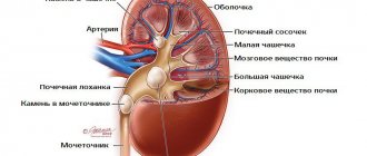
Varieties of stones regarding their shape: coral-shaped, flat, with spikes, round or with edges. Types of kidney stones depending on location in the organs of the urinary system: kidneys, bladder and ureter. The size of the stones can be small, medium and large.
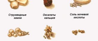
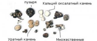
The composition of stones is: struvite, oxalate, urate, phosphate, carbonate. There is also an organic classification, according to which protein, xanthine, cystine and cholesterol stones are found.
Diagnostics
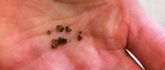
How to determine which kidney stones are present? For this purpose, the doctor directs the patient to undergo a general clinical urine test to determine its acidity and salt composition. If the stone has begun to move from the kidneys, stone fragments or sand may be found in the urine, which greatly simplifies the study of the chemical and organic composition.
How else can you find out what kind of kidney stones you have? To do this, you can perform ultrasound, hysterography and x-ray diagnostics. These methods allow you to determine what the stones look like, their density and quantity. Using ultrasound, urate, xanthine and cystine stones are clearly visible. An x-ray will show the presence of oxalates.
Urats
Urate stones are a type of stone that can only be determined using ultrasound examination. Urine tests and x-rays do not indicate their presence in the urinary organs. Localization varies: in one or both kidneys, ureters or bladder. Such neoplasms can be diagnosed in patients of any age category.
Urate types of kidney stones form when uric acid levels are high. Excess acid is caused by insufficient activity, lack of B vitamins, pathologies of the digestive system, gout, poor nutrition with insufficient fluid intake and an excess of purine. Abuse of sour and salty foods causes an increase in the level of urea in urine.
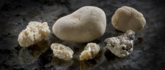
Physical characteristics of urates: smooth surface, round shape with a loose structure, yellow or brown color.
Uric acid stones require medical treatment using drugs aimed at crushing and excretion. An important therapeutic role is played by a diet with the exclusion of sour, salty, smoked, and canned foods. The diet should be rich in vegetables and fruits.
It is believed that this type of stones is dangerous for patients of younger and childhood age - uric acid, with significant accumulation in the kidneys, spreads throughout the body and leads to asthma, allergies, and constipation. In older age, urate is the cause of gout.
Oxalates
The next type is oxalate stones. They are formed as a result of a lack of magnesium and vitamin B. The causes are endocrine disorders, diabetes mellitus, impaired metabolism, kidney inflammation and Crohn's disease.
They are considered the most dangerous type, since such salt deposits cannot be treated with medications or traditional medicine; surgical intervention is required. After such a radical method, you need to follow a diet rich in vitamin B6 and magnesium. All green, sour vegetables and fruits are prohibited.
Calculi of oxalate origin are dense and hard formations of a dark color. They have spines, which gives them a coral shape. Oxalates cause frequent, unbearable pain to the patient due to their form. Such formations in urolithiasis are detected using the results of a general clinical analysis of urine and X-ray diagnostics. The reason for the formation is an excess of oxalic acid formed during intestinal dysfunction.
Struvite
Struvite stones are formed due to infectious or bacterial lesions of the urinary system. Soft, smooth, gray growths. During the process of intensive growth, they become overgrown with thorns, acquiring a coral shape. Stones are a byproduct of the interaction of urea and bacteria, resulting in the formation of ammonium, phosphates, magnesium and carbonate.
Struvite stones, formed as a result of stagnation of urine, are a great danger to humans, as they are found throughout the entire excretory system. In addition, in a short time, struvite reaches very large sizes, filling the entire cavity of the renal pelvis, and eventually the entire kidney.
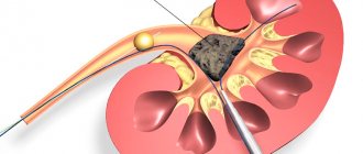
More often, struvite is diagnosed in the fairer sex. Difficult to treat with medication and require surgical intervention. In this case, shock wave lithotripsy is used.
Phosphates
Concretions of phosphate origin are soft, with slight roughness, of various shapes, light shades. Of all the existing varieties, they are the most dangerous - they quickly reach large sizes and are able to fill the entire bud. The process of growth to the size of a kidney takes several weeks.
Difficulties arise during diagnosis - phosphates can only be determined using x-ray diagnostics. Unlike struvite stones, they are not found in other organs and systems. During the growth process, neighboring organs are not damaged, which does not expose the patient to great risk. Contains calcium and phosphoric acid.
The main causes of formation are intestinal infections that penetrate from the anus to the genitourinary organs. Another reason is a violation of metabolic processes in the body and excessive love for dairy products.
With timely diagnosis, phosphates can be crushed and eliminated with the help of a special diet, medications and diuretics. It is important to drink more than 3 liters of liquid during treatment, which helps flush out deposits.
Phosphates can lead to multiple complications: wrinkling of the organ, expansion of the pyelocaliceal system, development of infections as a result of stagnation of urine, removal of the kidney.
Cystine stones
Cystine stones are composed of amino acids, making them rarely diagnosed. Occurs in patients with congenital genetic abnormalities. They are formed when the transport and absorption of cystine is impaired.
External characteristics are as follows: soft, smooth, round, yellow in color. Diagnosis is carried out using the ultrasound method. Often found in childhood and adolescence.
Treatment of cystine kidney stones is carried out with the help of special medications that help dissolve and remove the stones. Prescribed medications are aimed at changing the acidity of urine. It is also necessary to adjust your diet by adding foods and water high in sodium. If cystine stones are larger than 1 cm in diameter, surgical intervention is required.
Xanthine stones
Xanthine stones are formed as a result of genetic failures that lead to disruption of the synthesis and absorption of xanthine. Xanthine is excreted from the body in its pure form and when it is retained in the kidneys, ureter or bladder, a stone of xanthine origin is formed. It is possible to determine a calculus only by ultrasound. Younger and older children and teenagers are susceptible to this type of pathology.
Xanthine kidney stones vary in size and regardless of this, surgery is required - treatment with medications is impossible. For this purpose, shock wave exposure, endoscopic and open surgery are used.
Protein and cholesterol stones
Urolithiasis occurs with stones of various chemical and organic composition. The most rare type of protein stones is kidney stones. They contain fibrin, bacteria and salts.
Cholesterol stones contain exclusively cholesterol, so their structure is soft and crumbly, which is very dangerous. If crushed in the kidneys, bladder or ureter, damage to other organs may occur.
Having learned what types of kidney stones there are, you should not refuse to study the chemical and organic composition of the stone. As was previously thought, these studies are not required, but today the doctor needs this information to choose the correct therapy. Failure to investigate puts the patient at even greater risk.
tvoyapochka.ru
Types of stones
Before determining what kidney stones are, let's understand their types. Depending on the number of formations, stones can be single, multiple, two-stone and three-stone. In addition, kidney stones are classified depending on their location and can be unilateral or bilateral.
The stones also vary in shape and size. They come in small, medium and large sizes. As for the form, they are classified into the following varieties:
- with edges;
- with spikes;
- flat;
- round;
- coral-shaped.
I also distinguish kidney stones by their chemical composition, as well as by their organic component. According to the chemical composition, kidney stones are:
- phosphates;
- struvites;
- urates;
- carbonates;
- oxalates.
Types of stones
And in terms of the organic component, they differ and are:
- cholesterol;
- protein;
- cystine;
- xanthine
Depending on which stones are identified in the patient, the method and method of treatment is selected, and medications are selected. As for the method of getting rid of stones, it is chosen only after all the procedures and tests have been completed. Since depending on the size and composition of the stones, it depends on whether an operation will be performed to remove them, or whether it will be possible to soften the stones and remove them from the body using tablets.
Urate stones
If the patient is diagnosed with smooth and hard stones, which are yellow-orange in color and more like rock formations, they are classified as urate stones. In order to identify these types of stones, it is necessary to conduct an ultrasound. You should not try to identify such formations using regular x-rays or tests, as this will not give any results. These types of stones are mainly formed in patients in middle age. Basically, these formations appear for the following reasons:
- due to excess uric acid;
- from a sedentary lifestyle;
- for diseases of the digestive system;
- lack of vitamin B;
- in the presence of an acidic urine reaction;
- gout;
- when drinking low-quality water, as well as sour and salty foods.
In order to get rid of such stones, conservative treatment is necessary. In most cases, doctors prefer to prescribe a special diet and alkaline drink.
Oxalate stones
When specialists find dense stones in a patient’s kidneys that have sharp edges or spines, they are called oxalate stones. Basically, such stones are characterized by black or dark brown color. In order to diagnose these particular formations, it is enough to take a picture of the kidneys and undergo the appropriate tests. Basically, this type of stone is formed due to the following factors:
- for diabetes mellitus;
- pyelonephritis;
- deficiency of vitamin B and magnesium;
- metabolic disorders;
- Crohn's disease.
Oxalate stones
When a patient is diagnosed with such formations, the only treatment is surgery, since their composition is completely resistant to any influence of drugs and therefore such stones do not dissolve. Even after stones are removed from a patient through surgery, very often this subsequently leads to relapse. Therefore, during the recovery period, it is necessary to adhere to a strict diet, and also include in your diet foods and a vitamin complex that contain magnesium and vitamin B.
Struvite stones
When the formation of stones occurs due to exposure to various bacteria and infections in the human body, struvite stones appear. In appearance, they are more like smooth gray formations. Such stones pose a huge danger to the human body, as they have the ability to grow and form thorns on their surface.
Such stones appear as a result of the alkaline reaction of urine, during the development of bacteria and infectious diseases.
Struvite stones
Depending on the size of such a stone, the method of getting rid of it is chosen. This mainly occurs during the operation. The only exceptions are those cases when the stones are small in size. In this case, preference is given to lithotripsy or percutaneous lithotomy.
Phosphate stones
The main component of phosphate stones is phosphate acids. These stones are distinguished by having a soft, smooth surface. They occur in completely different shapes, but despite this they never damage internal organs. Diagnosis occurs through x-rays. Such formations are formed when infections enter the human body, through the abuse of dairy products and the wrong amount of substances.
High levels of phosphate in the urine will sooner or later lead to the formation of phosphate stones.
The main feature of such formations is that in most cases, surgical intervention is not even required to get rid of them. It will be enough just to adhere to the diet prescribed by specialists, as well as strict use of medications. The use of traditional medicine and mineral water is excellent for treating such stones.
Protein and cholesterol stones
Kidney stones such as protein and cholesterol stones are rarely found in patients. Outwardly, they most resemble soft and flat white formations.
Protein and cholesterol stones
Protein stones consist of fibrin, bacteria and salts, and cholesterol stones, respectively, consist of cholesterol. This type of formation is considered dangerous to human health. This is mainly due to their predisposition to coloring. Therefore, at the first signs, you must immediately seek medical help. Since only the doctor will tell you how to determine the composition of kidney stones. After tests and x-rays, the doctor will prescribe appropriate medication treatment and also select an appropriate diet.
Cystine stones
Cystine formations are considered one of the rarest types of stones. They mainly occur in children. Such formations can only be diagnosed using ultrasound. The fundamental component of these formations is amino acid. In appearance, they resemble a circle with a soft and perfectly smooth surface. In people with such formations, the symptoms are very pronounced and manifest themselves in the form of severe and acute pain.
Cystine stones
In order to get rid of such formations, patients are prescribed treatment with special medications, and an appropriate diet is selected. Among other things, in some situations such stones are removed through surgery. This mainly happens if the stones are of significant size.
Xanthine formations
Also, some patients may be diagnosed with xanthine stones during their lifetime, which are formed as a result of genetic predisposition. To diagnose such formations, ultrasound is performed, since other methods are completely useless. In order to get rid of this type of formation, the doctor, depending on each individual case, chooses a specific method, which may include:
- in removing stones through surgery;
- during shock wave lithotripsy.
Removing xanthine stones using medications does not bring any results.
Note! In addition to certain types of stones in nature, there are also mixed ones that contain various acids.
Causes of stone formation
If acute pain occurs in the lower abdomen and back, accompanied by severe nausea, you should consult a urologist or nephrologist. These are the first symptoms of kidney stones.
Several factors contribute to this:
- A metabolic disorder that causes an excess of salt crystals to appear in the urine.
- Poor urination due to insufficient water intake.
- Infectious infection of the genitourinary tract.
- Insufficient content in the body of special substances responsible for keeping salts in a soluble state.
- Regular use of diets that promote irregular and unhealthy eating.
Depending on the type of neoplasm, the reasons for their appearance may be different. Therefore, in order to establish what exactly influenced the deterioration of health, you need to know how to determine the type of kidney stones.
Treatment with folk remedies
Flax seed and milk
- 1 cup flax seeds
- 3 glasses of milk
Grind the flax seed, this can be done using a blender. It is important to use freshly ground seeds, otherwise they will lose their properties. Pour milk and boil until the mass is reduced to one glass. Take 1 glass per day. The course of treatment is 5 days. Repeat after 10 days. If you feel pain, this is a sign that the stones or sand are dissolving. After a year, repeat the course of treatment. Eliminate fried and spicy foods from your diet.
Disclaimer: The information provided here is for informational purposes only. Everyone's body may react differently, so please consult your doctor before using these products.
Classification of kidney stones
By number of stones:
- single;
- two- or three-concrete;
- multiple.
By location:
- one-sided;
- bilateral.
By form:
- coral-shaped;
- flat;
- with spikes;
- round;
- with edges.
By location:
- in the kidneys;
- in the ureter;
- in the bladder.
To size:
- small (about the size of the eye of a needle);
- average;
- large (sometimes reaching the size of the entire kidney).
By chemical composition:
- struvites;
- oxalates;
- urates;
- phosphates;
- carbonates.
According to the organic component:
- protein;
- xanthine;
- cystine;
- cholesterol
Pressure or pain in the lower back
Sometimes a stone gets stuck in the ureter, thereby preventing the healthy flow of urine into the bladder. It remains in the kidney, causing a feeling of pressure or pain in the lower back - on the right or left, depending on which kidney is affected.
Pain is considered the most common first sign of urolithiasis. Sometimes the symptoms are minor, but gradually increase, and sometimes they attack suddenly, without a declaration of war.
The pain can be so severe, sharp and stabbing that it leads to nausea and vomiting, and changing position or rest does not relieve it.
Urate stones
Urate stones are hard, smooth, yellow-orange stones that can appear in a variety of places in the genitourinary system. Their peculiarity is that to determine it is necessary to undergo an ultrasound. Standard tests and x-rays will not show the presence of pathology in the body. This disease occurs in patients aged 20 to 55 years. Moreover, urate formations will appear in the kidneys and ureter in middle-aged people. But in children and pensioners they are localized in the bladder.
Reasons for appearance:
- excess uric acid;
- sedentary lifestyle;
- lack of vitamin B;
- acidic urine reaction;
- diseases of the digestive system;
- gout;
- diet with excess purines;
- poor water quality;
- excess of sour and salty foods in the diet.
This disease is treated conservatively. Most often, doctors prescribe plenty of alkaline drinks and a special diet. In this case, surgery is not necessary.
Oxalate stones
Oxalate stones are dense formations with sharp edges and spines, predominantly black or dark brown in color. Sometimes there are layered types. The characteristics of oxolate stones make it possible to detect them using a urine test or an image of the kidneys. Experts say that the harbinger of this disease is oxalic acid, which reacts with calcium, causing small crystals to appear.
Oxalates are also formed due to the following factors:
- deficiency of magnesium and vitamin B in the body;
- diabetes;
- metabolic disease;
- pyelonephritis;
- Crohn's disease.
These stones differ in that they cannot be dissolved. To eliminate them you will have to have surgery. To prevent relapse, which happens quite often, it is necessary to adhere to a diet for a long time, consume vitamin B6 and magnesium.
Calcium stones
They are found in 80% of cases of urolithiasis, and are practically impossible to dissolve, since they are very hard.
Among calcium stones, there are two types: oxalates and phosphates.
Oxalates are the hardest and densest. They are gray or black in color and the surface is not smooth.
Difficulties arise when diagnosing black stones because it is difficult to recognize their chemical composition.
Due to the prickly surface, oxalates strongly scratch the kidney mucosa, which causes pain. Urine becomes red in color.
The reason for the formation of oxalates are oxalic acid salts, which predominate in the urine. As a rule, this happens due to poor nutrition, eating large amounts of parsley, baked goods, chocolate and sweets. Excess vitamin C in the body also leads to the formation of this type of stones.
Oxalates cannot be dissolved, so they can only be removed surgically. If the stone is small in size, it can be expelled with the help of medications.
The best prevention of oxalate formation is a healthy lifestyle, without drinking alcohol, and proper nutrition.
Phosphate kidney stones are slightly softer in composition than oxalates. They are also poorly soluble, but can be crushed.
They have a smooth surface, so they do not cause pain or irritation of the mucous membrane. They can be different in shape, but their color is light gray.
The cause of stones is an increase in urine alkalinity. This may happen due to metabolic disorders.
Phosphate stones grow very quickly in the kidneys, so the earlier they are diagnosed, the greater the chance of avoiding surgery.
The first sign of the appearance of this type of stones is light, loose flakes in the urine.
The friability of stones is an advantage in treatment. They can be dissolved with mineral waters, for example, Truskavetskaya or cranberry and apple juices.
Struvite stones
Struvite stones appear due to exposure to infections and bacteria. Outwardly, they are smooth gray formations, soft in condition, reminiscent of coffin lids. These stones are especially dangerous for humans, as they quickly increase in size and contribute to the appearance of the coral subspecies with spines. Bacteria react with urea, forming precipitation of ammonium, phosphates, magnesium and carbonate.
Main reasons:
- alkaline urine reaction;
- the presence of infectious diseases in the genitourinary tract;
- development of bacteria.
Phosphate stones
Phosphate acids are the main constituent of phosphate kidney stones. Can be of various shapes. Soft to the touch, smooth or slightly rough, white or light gray in color. They are dangerous because they grow very quickly, filling the entire kidney. However, due to the structure, they do not damage internal organs. A tumor can only be detected using an x-ray. Description of types and causes of occurrence:
- infection from the intestines into the genitourinary tract;
- abuse of dairy products;
- improper metabolism.
If phosphate stones are detected in time, you can even get rid of them through surgery. Fragmentation occurs by changing the acidity of urine. To do this, you need to adhere to a diet, drink special mineral water and medications prescribed by a doctor. As folk methods, you can try infusions of rose hips, barberry and grape roots.
Protein and cholesterol stones
In appearance, protein kidney stones are flat, soft, and white. They consist of fibrin, with the presence of bacteria and salts. They are very rare. Cholesterol stones are composed exclusively of cholesterol. They also look soft and black. They are dangerous because they crumble and can damage internal organs. When diagnosing these types of stones, you should consult a doctor. He will prescribe medications for crushing and withdrawal, as well as a diet. It is imperative to know what types of kidney stones are in order to take the right measures to eliminate them.
Cystine stones
The main component of cystine stone is amino acid. A rather rare species, characteristic of young people and children, due to a genetic pathological disease - cystinuria. Externally yellow in color and round in shape, with a perfectly smooth soft surface. Diagnosis can be made using ultrasound. The formation is accompanied by severe pain in the abdominal area.
Treatment of the pathology consists of changing the acidity of urine with the help of medications and a diet using sodium products. In extreme cases, if the size reaches 1.5-2 cm, surgical intervention is possible.
Xanthine stones
Some types of stones, such as xanthine stones, are a genetic defect. The neoplasm appears due to the fact that xanthine is excreted from the kidney in its original form, without being converted into uric acid. The diagnosis can be made by undergoing an ultrasound. But X-ray will not show their presence.
Removal of xanthine stones is possible only with the help of:
- operable open type accommodation;
- shock wave lithotripsy;
- laparoscopic surgery;
- endoscopic surgery.
Removing these stones on your own will not lead to a positive result.
If you feel persistent pain in the lower back, which intensifies with physical activity and sudden turns of the body, you should definitely consult a doctor. Renal colic is the next stage in the manifestation of the disease. This means that the pebbles have already entered the ureter.
Modern diagnostic methods, such as computed tomography, ultrasound, CT urography, retrograde and excretory urography, will help you quickly find out what the composition and size of the stones are in your kidneys. Thus, the treatment will take place as quickly and effectively as possible.
2pochki.com
general information
In the body, the kidneys perform the function of filtration and cleanse it of unnecessary substances. But, like any filter, they can become dirty, and the entire urinary system suffers. If there is an excess content of salts and acids in the urine, the functioning of the kidneys is disrupted - they are not able to pass large amounts of these substances.
There is an accumulation of elements that form stone deposits. The period of formation of stones is long - it can last for months or years, which depends on the water-salt balance and the chemical composition of the patient’s blood.
There are different types of kidney stones. Each type of sediment has its own characteristics. They differ in composition, density, size, shape, color and reason for formation.
Classifying kidney stones allows you to predict how large they may grow and what complications may arise.
The following types of kidney stones are distinguished according to their chemical composition:
- Urats.
- Oxalate stones.
- Phosphates.
- Carbonates.
- Struvite.
- Cystine stones.
- Protein-based deposits.
- Lipoid.
- Xanthines.
The most common deposits are inorganic stones: oxalates, phosphates, or carbonates. They account for more than 2/3 of the cases of nephrolithiasis. Uric acid stones occupy the second place in terms of frequency of formation. The rarest are stones consisting of fibrin, cholesterol, or arising from hereditary diseases.
Xanthine and cystine stones occur only in genetic kidney diseases, so metabolic disorders do not affect their formation.
How can you determine which stones are in the kidneys? To do this, you should take a urine test to determine its acidity and identify the presence of salts. Grains of sand or small fragments of stones may come out along with the urine, which makes it possible to study the chemical composition of the stones.
To determine the size, number and density of stones, the following methods are used:
- Examination of organs using ultrasound.
- Carrying out ultrasound hysterography.
- Performing an x-ray of the kidneys.
Calcium kidney stones - oxalates - are best visible in the pictures. X-ray-negative stones - urates, cystines and xanthines - appear unclearly or as shadows. But they can be seen using ultrasound or computed tomography.
Types of character
It is quite difficult to classify character. There are 4 types of characters based on the establishment of the individual and what surrounds him:
- Relations in society;
- Attitude to activity (work, work, hobby);
- Attitude towards yourself;
- Attitude towards inanimate objects.
In accordance with this classification, it is possible to create a psychological image of a person and select means of correcting character defects.
Scientist and psychologist E. Kretschmer created his own classification of people belonging to a certain type of character, which is based on the structure of the human body:
- Athletics (people of a trained type of appearance) have broad shoulders, well-developed muscles, tall stature;
- Asthenics (thin and tall) weak muscles, thin bones, elongated facial features;
- Picnics (small and overweight) tendency to obesity, overweight, short stature.
Kretschmer believed that character traits are directly related to the constitution of the body and are an innate property that cannot be trained.
Comparative characteristics
In case of urolithiasis, it is sometimes possible to determine what substances the stones are made of even before the first tests. If the stone came out and was preserved, you can determine its type by its appearance. This table shows the comparative characteristics of kidney stones and the main causes of their formation (Table 1).
Table 1 - Types of stones and their characteristics
| View | Compound | Description | Options | Causes |
| OXALATE | Ammonium oxalate and calcium salts | Available in dark gray and brown | Dense structure of stones with uneven edges. In some cases, spines grow. They have different sizes; grains of sand can grow into stones larger than 4 cm | Formed due to metabolic disorders. The following diseases may be provoking factors:
Their appearance is influenced by excessive consumption of foods rich in vitamin C and oxalic acid. |
| URATE | Consist of uric acid and its salts - potassium and sodium | Yellow to dark brown | Round or oval shape without roughness. Loose, loose structure. Dimensions can reach several centimeters | Most often diagnosed in men who prefer protein foods. They can form due to increased acidity of urine. Also affected by a sedentary lifestyle, poor nutrition, lack of B vitamins, as well as long-term use of analgesics |
| PHOSPHATE | Calcium salts of phosphoric acid | White or light gray | Smooth stones, loose in structure. The loose composition allows the formations to quickly dissolve. Grow quickly to large sizes | This species is formed only under alkaline conditions. The main reason for their formation is the presence of a bacterial infection in the kidneys, the causative agents of which are: staphylococci, E. coli, Proteus and other microorganisms. Often accompany chronic pyelonephritis |
| CARBONATE | Calcium carbonate | Most often white. May be light shades of gray and yellow | Very soft stones, smooth to the touch. Can be of different shapes and sizes | Can form in an alkaline environment. The main reason is increased calcium levels in the urine, which can be caused by:
This type of stone is quite rare |
| STRUVITE | Ammonium magnesium phosphate and carbonate apatite | Whitish or grayish | Struvite is usually smooth, but may also have slight roughness. The structure is soft. They grow very quickly and can fill most of the bud. | They are formed only in an alkaline environment under the influence of infections. Several reasons can be identified: paralysis, paresis, diabetes mellitus, neurogenic bladder, impaired urine outflow. This type is the most difficult to treat conservatively. Most patients with such stones are women |
| CYSTINE | Sulfur-containing amino acid | Pale yellow color | The density of cystine stones in the kidney is insignificant. The shape is usually round, smooth surface. Rarely reaches large sizes | They are formed only due to a genetic disease - cystinuria, in which the absorption of the cystine amino acid is impaired. Cystine stones are diagnosed at an early age |
| PROTEIN | Fibrin protein, leukocytes, bacteria and mineral salts | Whitish or grayish | Flat shape, a few millimeters in size, soft to the touch | Formed during bacterial infections of the urinary system, such as cystitis, pyelonephritis, kidney colds and others. Most often found in women due to the anatomical structure of the urethra |
| LIPOID | Low density lipoproteins | There are only black ones | Shapes - varied, consistency - soft, easy to crumble, small (several millimeters) | An extremely rare form. Occurs as a result of cholesterol metabolism disorders |
| XANTINES | Xanthine, sometimes mixed with apatite | Pink-red color | Very soft structure. Flat, can reach large sizes | They are formed due to a genetic defect - deficiency of the enzyme xanthioxidase. Diagnosed at an early age |
Having found out what types of kidney stones there are, you should remember that there are also mixed types of stones, formed from various acids and salts, and also containing fibrins, leukocytes and epithelial particles.
Consequences of ICD depending on the type of stone
With nephrolithiasis, there is a risk of developing various complications not only in the kidneys, but also in other organs. This is due to the changed composition of urine and blood, and each type of stone has its own characteristics.
Thus, a mixture of oxalic acid and calcium salts, from which oxalates are composed, can settle not only in the kidneys, but also in other organs, damaging their mucous membrane. But still, oxalate stones cause the greatest harm to the kidneys. Their dense structure does not allow large deposits to be dissolved with medicinal and folk remedies; surgical intervention is often required.
Urate stones are dangerous for children. If left unattended, uric acid salts begin to deposit in the joints and under the skin. This leads to asthmatic attacks, allergic skin reactions and constipation. In adults, exposure to acidic precipitation develops gout.
Phosphate and carbonate complications are dangerous due to their rapid growth, which is why they are extremely rarely diagnosed in the early stages. Rapid growth leads to the development of renal failure, which often becomes chronic. Other complications:
- Shrinking of the kidney.
- Expansion of the calyces and pelvis.
- With the addition of infection, the following develop: sepsis, carbuncle, pyonephrosis.
- Removal of the affected kidney.
Struvite stones grow very quickly, filling the internal cavity of the kidneys, which provokes impaired renal function. In addition, with struvite stones, there is a risk of accumulation of such deposits throughout the excretory system.
Cholesterol, xanthine and cystine kidney stones most often lead to arterial nephrotic hypertension. Whatever types of stones are formed during urolithiasis, they cause poor urine output up to absolute anuria.
Kidney stones can cause renal coma, abscess and purulent inflammation of the perinephric tissue.
Important! Regardless of the type, urinary stones without appropriate treatment can cause atrophy of the renal parenchyma, inflammatory processes in the kidneys, renal colic and many other complications.
To avoid them, you need to strengthen the body and adhere to the principles of a healthy lifestyle, which will not allow stones to form.
vsepropechen.ru
Causes of kidney stones and their types
At least 9% of the adult population is at risk of developing urolithiasis. And it looks like this risk will increase due to global warming. With climate warming, the human body is more susceptible to dehydration (dehydration). This increases the likelihood of kidney stones forming.
The following types of kidney stones are distinguished: calcium (oxalates, phosphates), urate, struvite and cystine.
One of the risk factors for the formation of kidney stones, regardless of their type, is dehydration. Everyone prone to urolithiasis must maintain an optimal drinking regime.
Drinking 2 liters of water daily reduces the risk of kidney stones by 50%. The American Urological Association recommends drinking at least 2.5 liters of water daily if you have urolithiasis.
If you have symptoms of urolithiasis, you should consult a urologist. Diagnostics involves: blood, urine, and visual studies.
Let's consider the specific causes and risk factors, as well as the features of treatment of urolithiasis for each type of kidney stones.
What types of stones are there?
Many people wonder what kidney stones are and how to get rid of them. We need to take a closer look at the answers to these questions. Stones are composed of mineral and organic substances.
Depending on the content of a particular element, stones are divided into four main groups:
- phosphates and oxalates . Formed from inorganic calcium salts. This type is often found in people who have been diagnosed with urolithiasis;
- infectious magnesium . Occurs due to abnormalities in the urinary tract. Excess fluid remains in the kidneys, infection sets in, and the normal functioning of the system stops;
- urates . Occurs after disruption of the digestive system and excessive secretion of uric acid;
- cystine and xanthine . They are extremely rare. Small formations arise due to heredity, congenital pathology and genetic disorders.
According to other parameters, stones are also divided into several types:
- by quantity: single, binary and multiple formations;
- by type of location: unilateral and bilateral;
- by differences in shape: circles, flat formations, stones with clear edges, with spikes and coral appearance;
- by size: the size of neoplasms can vary from the eye of a needle to the entire territory of the kidneys;
- by type of location: stones can be found in the kidneys, urinary tract and in the area of the ureter.
To find out exactly what type of formations you have, why kidney stones are dangerous, and how to get rid of them faster, you need to visit a doctor on time.
Causes
A couple of years ago, all scientists claimed that kidney stones are formed due to poor quality of drinking water, chlorine content and scale. But now there is a different way of thinking.
Uralithiasis occurs when the correct content of salt and colloids in urine is disrupted in the human body. It is because of this that the remnants of substances begin to accumulate in the organs and cause a lot of inconvenience to a person.
For what reasons does each type of stone occur?
- oxalates. Occur when the reaction of oxalic acid and calcium occurs. The first element is often found in vegetables and fruits, where there is a lot of vitamin C. Metabolic disorders, lack of B vitamins, diabetes mellitus, chronic kidney disease, Crohn's disease and others also lead to the formation of oxalite stones;
- urates . Appear due to the intake of poor-quality water, poor ecology, sedentary lifestyle, metabolic disorders, poor nutrition, lack of B vitamins and other reasons;
- phosphalite formations are the interaction of calcium salts with phosphoric acid. The causes are infections that enter the urinary tract and infect organs. Because of this, the acidic environment turns into an alkaline liquid, the function of the kidneys and bladder is impaired; Formations of this type have a loose structure, so they do not violate the integrity of the organs. But they grow very quickly and cover the entire kidney cavity;
- struvites. They arise due to a change in the acidic environment of urine into an alkaline environment, and the presence of bacteria in the urinary tract. Such formations have a habit of growing quickly and covering the entire kidney area, so treatment should not be delayed;
- cystine stones. They arise due to genetic pathologies, namely the disease cystinuria. Appears in children and adolescents. These formations consist of amino acids.
To find out what kidney stones look like, you need to decide on their types. After all, each formation has its own shape and size.
Calcium stones
Calcium oxalate crystals
A calcium oxalate crystal forms when calcium combines with oxalic acid. Sometimes this kidney stone occurs due to a systemic cause such as bowel disease, primary hyperparathyroidism, or primary hyperoxaluria. Only doctors can determine what disease is causing such stones.
A calcium oxalate crystal forms when calcium combines with oxalic acid
Most often, this type of stone occurs simply due to the interaction of hereditary factors, diet, and aspects of daily life. Although doctors discover the connection between daily life and stone production and choose those changes that can prevent the formation of new stones, it is up to patients themselves to create and maintain these changes.
About a million units of nephrons make up a normal adult kidney. The calcium oxalate stone does not grow in the nephron tubules, but “outside” them, on the surface of the renal pelvis, where the final urine collects and drains through the ureter into the bladder.
Calcium Phosphate Crystals
Calcium phosphate crystals form when calcium atoms combine with phosphoric acid rather than oxalic acid to produce a calcium phosphate kidney stone.
Primary hyperparathyroidism and renal tubular acidosis increase the average alkalinity of urine (increased urine pH) and stimulate the formation of calcium kidney stones. Many unusual genetic diseases do the same.
High levels of phosphate in the urine will sooner or later lead to the formation of phosphate stones.
Calcium phosphate stones can also occur because urine contains much more phosphate than oxalate. They form stones more frequently and larger than calcium oxalates. Often stones appear as crystalline plugs at the ends of the kidney tubules. More crystals are deposited at the end of the channels open to urine. Crystal plugs damage the cells that line the tubes and cause local scarring.
Symptoms
There are several symptoms that will allow patients to notice the disease in time and consult a doctor for help. Only an experienced specialist will determine what kidney stones are and how to get rid of them. The main thing is not to endure pain and discomfort, but to immediately go to a specialist. It is much easier to cure the early stage of the disease than the complicated form.
There is a list of symptoms that occur in patients with stones in organs:
- colic in the kidney area;
- a stabbing and sharp pain appears in the lower back or sides, which then disappears;
- lower abdomen tingles and hurts;
- the patient begins to feel sick and vomit;
- pain and burning appears during urination;
- pebbles or sand come out with urine;
- body temperature rises;
- the urinary system fails, trips to the toilet become difficult and frequent;
- the patient sweats, cold sweat comes out;
- the intestines are swollen and the stomach hurts;
- blood pressure rises;
- blood appears in the urine.
The frequency of manifestation of these symptoms is individual for each person, because the body reacts differently to the presence of stones, and the formations are of a different nature. The pain may occur every week or once a year. Duration is one or two hours. The main thing is to pay due attention to them and go to the doctor. Only he knows what kidney stones are and how to get rid of them.
Symptoms of a urinary tract infection
Sometimes a person with a kidney stone experiences symptoms similar to a urinary tract infection. These include:
- increased urination or frequent urge
- pain or discomfort when urinating
- colorless urine
- strong unpleasant odor of urine
- blood in urine
- heat
If you have any of these symptoms, do not self-medicate, but visit a doctor and get tested to determine the infection. If the causative agent of the disease is not found, this may be a sign of urolithiasis. However, sometimes kidney stones are complicated by a secondary infectious process, and this is a serious problem that requires emergency intervention.
Treatment
Only doctors know how to treat stones in the kidneys and urinary tract, so it is important to contact them immediately.
The first thing you will have to do is get tested to determine what kind of deposits you have and where they are located, for example, uric acid kidney stones.
Only after the specialist checks the tests and makes an accurate diagnosis will he prescribe the appropriate treatment. For different forms of the disease and stages of stone development, suitable methods of treatment are used.
First, a diagnosis is made. The patient undergoes an ultrasound examination of the kidneys and urinary tract, takes blood and urine tests, goes for urography, doctors do computed tomography, nephroscintigraphy and determine sensitivity to antibiotics. Only after this the doctor makes an accurate diagnosis and determines the type of stones that the client has. Treatment occurs in several ways.
The medicinal method allows you to remove stones using special drugs.
Thanks to this method, the formations are broken down into small particles and eliminated from the body naturally.
There are many medications that fight certain types of stones, so the doctor himself gives recommendations.
Surgical intervention is necessary when the disease is severe and there is no other way out but to remove the stones, and sometimes even the kidneys themselves. The operation can be open or using the endourethral technique.
In the first option, stones are removed by cutting the kidney or bladder. The second method is more reliable and safe and involves the use of ultrasound therapy or a laser, which is applied to the source of infection.
Diet therapy can help only when the number of formations does not reach a critical limit. By eating certain foods and eliminating others, you can maintain your health and start living without pain and colic.
Therapeutic exercise sets the body in motion. And one of the reasons for the formation of stones is a sedentary or sedentary lifestyle. Therefore, more walks and exercise will not hurt.
Herbal medicine allows you to get rid of stones using various procedures. The process is long but safe.
Rest in sanatoriums and resort centers has a beneficial effect on the body as a whole. A warm climate and fresh air will help get rid of formations in the kidneys and bladder.
Traditional methods are also widespread. They are aimed at natural removal of stones from the body. To do this, you can make a solution of rosehip root and drink it with meals or drink tea from apple peels or with milk.
To ensure that the effect is noticeable after the first weeks, you can combine several treatment methods at once. Thus, the formations will be removed from the body, and the person will begin to live actively and painlessly again.
mkb.guru
Steps
Part 1
Conducting experiments
- 1
First, let's understand the difference between minerals and regular stones.
A mineral is a natural combination of chemical elements that forms a specific structure. And, despite the fact that you can find the same mineral in different shapes and colors, it will still show the same properties when tested. In contrast, stones may be made up of a combination of minerals and do not have a crystal lattice. They are not always easy to distinguish, however, if the experiment produces different results from different sides of the object, then the object is most likely a stone.
- You can try to determine what kind of stone it is, or at least determine which of the three types of rock it belongs to.
- 2
Learn to navigate the classification of minerals.
There is a place for thousands of minerals on our planet, but many of them are classified as rare or lie too deep underground. Sometimes a couple of experiments are enough, and you are left in no doubt that this is one of the common minerals from the list in the next section. If your mineral doesn't fit any of the descriptions above, try checking your region's mineral classifier. If you have conducted many experiments, but have not been able to reduce the number of options to two or three, search the Internet. Look at photos of each mineral that is similar to yours and look for any tips you can on how to tell the difference between those minerals.
- It is best to include at least one experiment that requires exposure to the mineral, such as a hardness test or a streak test. Experiments that involve only looking and describing may be biased, since different people describe the same minerals in different ways.
- 3
Study the shape and surface of the mineral.
The collection of forms of each mineral and the characteristic features of a group of minerals is called the “common form”. [1] Geologists have many technical terms to describe these characteristics, but usually a general description is sufficient. For example, is your mineral lumpy, rough, or smooth? Is it a mixture of rectangular crystals, or is your specimen bristling with sharp crystalline peaks?
- 4
Take a closer look at how your mineral shines.
Luster refers to the way a mineral reflects light, and although it is not a scientific test, it can be useful for description. Most minerals have a “glassy” (“glossy”) or metallic luster. However, you can describe the luster as “greasy,” “pearly” (a whitish sheen), “matte” (dull, like unglazed ceramic), or any other definition that seems accurate to you. [2] Use multiple adjectives if you need to.
- 5
Pay attention to the color of the mineral.
Most people do not see any difficulties in this, but, meanwhile, this experience may turn out to be useless. Small foreign inclusions can cause color changes, which is why you can find the same mineral in different colors. However, if the mineral is an unusual color, say purple, this can significantly narrow the search area.
- When describing minerals, avoid fancy color names like “salmon” or “pussy.” Try to stick with just red, black and green.
- 6
Try the stroke experiment.
This is a useful and simple test, provided you have a piece of white unglazed porcelain. The back side of tiles from a bath or kitchen is perfect; maybe you can buy something suitable at a home improvement store. Having become the owner of a treasured piece of porcelain, simply rub the mineral on the tile and see what color streak it leaves. Often the color of the streak will be different from the base color of the mineral.
- Glaze gives porcelain and other types of ceramics a glassy (glossy) shine.
Remember that some minerals do not leave a streak, especially hard minerals (as they are harder than the streak plate).
- 7
Assess the hardness of the material.
To quickly determine the hardness of a material, geologists use the Mohs hardness scale, named after its creator. If the result fits the hardness coefficient “4”, but does not reach “5”, it means that the coefficient of your mineral is between “4” and “5”, you can stop the experiment. Try scratching your mineral using the common items mentioned below (or the minerals from the hardness test kit); start at the bottom and, if the test is positive, move up the scale to the top:[3]
- 1 - Easy to scratch with a fingernail, oily and soft to the touch (corresponds to a stearite cut)
2 - Can be scratched with a fingernail (plaster)
- 3 - Can be easily cut with a knife or nail, scratched with a coin (calcite, lime spar)
- 4 - Easy to scratch with a knife (fluorspar)
- 5 - Can be scratched with difficulty with a knife, can be scratched with a piece of glass (apatite)
- 6— You can scratch it with a file; with force, it can scratch the glass (orthoclase)
- 7— Can scratch steel for files, easily scratches glass (quartz)
- 8 – Scratch quartz (topaz)
- 9 — Scratches almost anything, cuts glass (corundum)
- 10 - Scratches or cuts almost anything (diamond)
- 8
Break the mineral and study what pieces it breaks down into.
Due to the fact that each mineral has a certain structure, it must break down into parts in a certain way.
If you observe more flat surfaces in faults of the same rock, then we are dealing with cleavage
. If there are no flat surfaces, but continuous chaotic bends and bulges are observed, then there is a fracture in the mineral. [4]- Cleavage is described in more detail using the number of planes obtained during the fault (usually from one to four); of a perfect
(smooth) or
imperfect
is also taken into account .
There are several types of fractures. They are described as splintered ( fibrous
), sharp and jagged (
hooked
), cup-shaped (
shell-shaped
,
snail-shaped
), or none of the above (
ragged
). - Cleavage is described in more detail using the number of planes obtained during the fault (usually from one to four); of a perfect
- 9
If you still have not identified your mineral, you can conduct additional experiments.
Geologists have many other tests at their disposal to classify minerals. However, many are simply not useful for identifying the most common species, and many will require special equipment or hazardous materials. Here is a brief description of several experiments that may be necessary:
- If your mineral is attracted to a magnet, then most likely it is magnetite or magnetic iron ore, the only ferromagnet among a number of common minerals. If the attraction is weak or does not meet the definition of magnetite, then your specimen may be pyrrhotite (or magnetic pyrite), franklinite, and ilmenite (or titanium ironstone). [5]
Some minerals melt easily in the flame of a candle or lighter, while others will not melt even in the flame of a jet engine. Minerals that are easier to melt have a higher fusibility rating than those that are more difficult to melt.
- If your mineral has a distinctive odor, try to describe it and look for minerals with a similar odor online. Strong-smelling minerals are not common, but the presence of the bright yellow mineral sulfur can cause the familiar rotten egg smell.
Part 2
Determination of essential minerals
- 1
If you do not understand any of the following descriptions, please refer to the previous section.
The descriptions below contain terms and numbers from the traditional classification of minerals, such as shape, hardness, appearance when broken, or other definitions. If you are not sure what they mean, refer to the previous section on conducting experiments.
- 2
Crystalline minerals are most often represented by quartz.
Quartz is extremely widespread. The bright shine and beautiful appearance of the crystal attract many collectors. On the Mohs scale, quartz has a hardness rating of 7, and if you break it, you can see all kinds of fractures, but never the flat surface characteristic of cleavage. [6] It leaves no mark on white porcelain. Its luster is characterized as glassy. [7]
- '''Milk quartz is a translucent mineral, rose quartz is pink, and amethyst is purple.
- 3
A hard glassy mineral without crystals can be another variety of quartz, flint or hornstone.
Absolutely all quartz has a crystalline structure, however, some varieties, called “cryptocrystalline,” consist of microscopic crystals that are invisible to the eye. [8][9] If you have a mineral with a hardness factor of 7, with a fracture and a glassy sheen, then it is quite possible that this is a type of quartz called flint. The most common flint is brown or gray in color. [10]
- One of the varieties of flint is chalcedony flint or, as it is also called, chalcedony variety of quartz. However, it is classified in different ways. [11] For example, some would consider any black flint to be chalcedony flint, while others would accept the definition of chalcedony flint only if it has the right luster and if it is found in a particular rock.
- 4
Striped minerals are usually chalcedony.
Chalcedony is a mixture of quartz and another mineral, morganite. There are many beautiful varieties with stripes of different colors. Here are the two most common:
- Onyx is a type of chalcedony with parallel stripes. Most often it is black or white, but onyxes are also found in other colors.
Agate has stripes that are more likely to curve or form swirls, and agates come in all sorts of colors. Agate is formed from quartz, chalcedony or similar minerals.
- 5
Check to see if your mineral matches the characteristics of feldspar.
Feldspar is the second most widespread after all varieties of quartz. The hardness index of this mineral is 6, it leaves a white streak; Feldspar can be found in a variety of colors and with different luster. When broken, it forms two flat cleavages, the smooth surfaces of which are located almost at right angles to each other. [12]
- 6
If the mineral comes off in layers when rubbed, it is probably mica.
This mineral is easy to identify, because if you scratch it with a fingernail or even just a finger, it separates into thin plates.
Potassium" (or white) mica is
pale brown or colorless, while
magnesia" (or black) mica is dark brown or black, with gray-brown veins. [13][14] - 7
Now let's understand the difference between gold and “cat” gold.
Pyrite
, also known as “cat” gold, also looks like a shiny yellow metal, but a couple of experiments are enough for the difference to become obvious. Pyrite has a hardness rating of up to, and sometimes exceeds, 6; gold, in turn, is much softer, ranging between 2 and 3. [15] It leaves a greenish-black streak and can crumble under sufficient pressure.- Marcasite (or radiant pyrite) is another common mineral closely related to pyrite. But, if pyrite crystals have a cubic shape, then marcasite forms needles.[16]
- 8
Green or blue minerals are most often malachite or azurite.
In addition to other minerals, they contain copper. It is this that gives malachite its rich green color, and makes azurite its bright blue. Both minerals often occur together, and both have a hardness coefficient between 3 and 4.[17]
- 9
Use a mineral classifier or website to identify other types.
A mineral classifier for your region will give you all the information you need about the minerals found in your area. If you still have difficulty identifying your specimen, several online resources such as minerals.net will allow you to compare the results of experiments with existing standard indicators and, thus, select the appropriate mineral.

