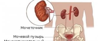
Kidney stone disease affects about 3-4% of the world's population. Moreover, about 15% of all cases are asymptomatic until the stone grows to a large size or until the patient undergoes a routine medical examination with a general urine and blood test. A stone in the renal pelvis also does not bother its “owner” until it begins to move due to provoking factors. We will discuss below what the renal pelvis is, how stones form in it and how they manifest themselves.
Potential danger of kidney stones
It is worth knowing that urolithiasis can cause a lot of trouble to its “owner”.
It is worth knowing that urolithiasis can cause a lot of trouble to its “owner”. Their severity and course depend entirely on the type of stone formed in the pelvis. Thus, the simplest and most potentially harmless are urate stones, which dissolve well on their own if you follow a certain diet and drinking regimen. In the future, such sand simply comes out with urine. All other stones dissolve more difficultly or cannot be dissolved at all. In this case, the danger is that the stone can grow and eventually fill the entire cavity of the pelvis. With this pathology, hydronephrosis occurs (accumulation of excess urine in the kidney cavity and its subsequent rupture). It is worth knowing that fixed stones lead to hydronephrosis. But there is one peculiarity here: it is the stationary stones that grow very slowly. Therefore, they are classified as conditionally favorable.
It's a different matter when it comes to moving stones. Here the patient may periodically experience renal colic, which occurs when a pebble moves. The size of movable stones is much smaller than immobile ones. However, the very shape of the stones poses a threat to the patient. So, if the stone has sharp edges or spikes, when moving, such a formation can injure the inner surface of the urinary tract. And with their high sensitivity, the patient will feel sharp pain. In the worst case, the stone, when moved, can rupture the ureter, causing urine to leak into the abdominal cavity. This will lead to additional problems, surgery and possible complications.
Treatment
There are several treatment methods, it all depends on the severity of the disease.
In mild forms, drug therapy is prescribed; in more complex cases, surgery to remove a renal pelvis stone may be required.
Drug renal treatment - the patient is prescribed drugs that promote the dissolution and subsequent removal of stones from the renal pelvis. Painkillers and anti-inflammatory drugs are also prescribed.
In cases where crushing stones in the renal pelvis is impossible due to their large size, location, and also if there are purulent processes, they resort to surgical removal of the stone from the pelvis. Abdominal surgery is usually performed under general anesthesia. The rehabilitation period after such treatment is much longer compared to the laparoscopic method. To prevent complications and restore kidney function, conservative therapy is prescribed.
At the same time, in addition to open operations, new technologies are increasingly being used in the treatment of stones in the renal pelvis: lithotomy (stone cutting is performed), contact litholapaxy (suction of fragments of a destroyed tumor), lithoextraction (instrumental removal without its destruction and section of the organ, most often from the lower third of the ureter).
An additional therapy measure is a diet that can help reduce the acidity of urine and help soften stone formations.
When doing diet therapy, it is necessary to take into account that women and men are treated differently for stones in the renal pelvis. Therefore, the menu is developed individually in each case.
Symptoms of stones in the renal pelvis
Symptoms of stones in the renal pelvis - pain in the lumbar region
As a rule, the process of formation of a kidney stone does not reveal itself in any way. The only possible symptom of the formation of stones in the renal pelvis is a change in the color of the urine. In this case, the urine may become darker in color, almost orange. But this symptom is unreliable. Clinical symptoms of kidney stones appear only when the stone has grown to a significant size. Signs of the disease will be:
Recommended reading:
How to find a kidney recipient and who can become a donor?
- Frequent urination. This is due to the fact that the kidney cavity becomes increasingly occupied by the volume of the stone. Thus, urine has nowhere to accumulate and it moves in small portions through the ureter into the bladder. Here the walls of the bladder are irritated, which leads to the urge to urinate. If the patient experiences anuria (urinary retention), you should immediately consult a doctor. In this case, prolonged urinary retention may develop, which will lead to complex toxic poisoning of the body and possibly coma.
- Pain in the lumbar region. Sometimes it can be nagging and occur periodically. As a rule, antispasmodics and heat on the lower back relieve pain. But over time the pain returns. If the stone begins to move, the patient will be bothered by renal colic. This type of pain syndrome is sharp. Sometimes it can throw the patient into shock. As a rule, with renal colic, the patient cannot find a place for himself. The patient tries to take the only correct position that relieves pain. But with renal colic this is basically impossible. In this case, it is necessary to urgently call an ambulance. Renal colic may be accompanied by an increase in temperature up to 39°, as well as nausea and vomiting.
Important: It is worth remembering that taking antispasmodics for renal colic is not contraindicated, while taking analgesics is highly discouraged.
- Against the background of severe pain, blood, mucus or pus may appear in the urine. The presence of blood in the urine indicates that the stone has injured the urinary tract.
- Among other things, the patient may experience a sharp increase in blood pressure. This symptom indicates that an infection is mixed in with the pathology.
Main symptoms
Quite often, urolithiasis may not appear for quite a long time, and sometimes throughout life. Doctors identify several main symptoms that may indicate that the body is affected by urolithiasis. These include:
Stone formation in the renal pelvis
The process of formation of a kidney stone directly depends on the speed of metabolic processes in the body and their quality.
The process of formation of a kidney stone directly depends on the speed of metabolic processes in the body and their quality. And in the first place here is an excess of salts of insoluble compounds such as phosphates, urates, oxalates, carbonates, lime salt, etc. It is worth noting that an increased concentration of these substances occurs from excessive consumption of plant and meat foods. As soon as the concentration of these salts becomes higher than is permissible for the normal functioning of the body, they settle in the renal pelvis, since the salts are not absorbed back into the blood. Here, salts settle in the pelvis first in the form of sand, and then, if clots of mucus and epithelium are present in the urine, they grow on the sand. As a result, stones form in the renal pelvis.
Recommended reading:
Apaan Mudra for the treatment of diseases and restoration of the kidneys
Important: in some cases (rarely), stones in the pelvis can form due to its abnormal structure. In this case, the accumulating urine gets into a kind of vortex, which leads to the sedimentation of salts in the pelvis. Here you can add impaired function of the epithelium of the pelvis (increased amount of mucus).
Types of stones
The stone is a mixture of minerals and organic matter. Depending on the organic and chemical origin, stones in the pelvis are of several types. All subsequent treatment will depend on their chemical composition. Oxalates are formed due to calcium salts of oxalic acid and are the most common, strong stones. Oxalate stones are the densest, they are very clearly identified using an x-ray.
Urate stones in the kidneys are formed due to the deposition of uric acid and its salts on the walls. They consist of sodium and potassium, which are not completely eliminated from the body. Less common are carbonates, white, soft formations of calcium salts.
Struvite containing magnesium ammonium phosphates. Struvite stones grow rapidly and can occupy a fairly large area of the kidney.
Depending on the shape, stones can be simple or complex. The first are formed from one element, they are smooth, with a simple structure. The second type includes coral-shaped stones, which are formed from several elements and have a complex structure and shape (often with spikes and sharp corners). Coral stones are very dangerous, as during movement they injure the mucous membranes of internal organs.
Diagnostic features
The disease can be detected at an early stage using diagnostic measures. It happens that salt stones are found unexpectedly. However, in most cases they become known when complaints already appear. If sand or stones in the urinary tract are suspected, the following examinations are prescribed:
- laboratory urine analysis;
- biochemical blood test;
- Ultrasound;
- urography;
- computed tomography.
When examining urine, you can identify the type of stone deposits and the presence of an inflammatory process. In healthy people, urine is yellow and transparent. Its shades are affected by the use of coloring foods and antibiotics. Urine becomes cloudy if there is pus in it, and when blood is mixed in, the urine becomes reddish in color. If sediments form in the urine in the form of small crystals, this indirectly indicates stone deposits in the kidneys.
In a biochemical blood test, attention is paid to the amount of creatinine and urea. An elevated level of these indicators indicates renal failure, which is caused by urate deposits. A general blood test shows an increase in the number of leukocytes, indicating an inflammatory process.
Ultrasound is an informative method for detecting stone deposits. With its help, doctors find out the location and size of stones. But in patients with excess weight and bloating, it is more difficult to find formations, so other research methods are used.
- How to remove urinary stone from toilet
X-rays using contrast agents give an idea of the structure of the kidney, allowing you to see abnormalities in the development of the organ, fluid and salt formations.
The most informative diagnostic method is computed tomography. It is performed without the use of contrast, since the stones are much denser than the parenchyma. In this case, dyes only interfere with seeing the full picture. CT allows you to determine the chemical composition of stones and prescribe the necessary treatment.
Methods of surgical treatment of urolithiasis.
Extracorporeal shock wave lithotripsy (ESWL) or extracorporeal shock wave lithotripsy (ESWL) is a non-invasive treatment for kidney and ureteral stones. The method was developed in the early 1980s in Germany, and became widely used with the introduction of the first lithotripter into clinical practice in 1983. Within a few years, ESWL became the standard treatment for small kidney and ureteral stones.
A lithotripter destroys stone using focused, high-intensity acoustic pulses. X-ray imaging or ultrasound imaging systems are used for guidance. Typically, treatment begins at the lowest power level of the equipment, with long intervals between pulses, in order to adapt the patient's tissues and reduce the likelihood of bruising. The pulse frequency and power are then gradually increased so as to crush the stone more effectively. The final power level usually depends on the patient's pain threshold or the location of the stone (with kidney stones, maximum crushing modes often lead to renal hematomas and bleeding).
The criterion for effectiveness is the fragmentation of the stone into smaller pieces, which can then easily pass through the ureter and urethra. The crushing process takes about an hour. Placement of a ureteral stent may be used at the discretion of the urologist. The stent facilitates the passage of stone fragments, relieving swelling and expanding the ureter.
Percutaneous nephrolithotomy
Drawing. Nephroscopy.
Drug therapy
If stones are detected in the renal pelvis, treatment should be prescribed by a doctor. The choice of the optimal method depends on the severity of the disease, the general condition of the patient and the diagnostic results. Treatment can be conservative or surgical. The conservative method is used for stones up to 1 mm and includes:
- mandatory diet with the exclusion of meat and offal;
- restoration of water balance;
- taking medicinal mineral waters;
- physical therapy;
- use of antimicrobial agents;
- the use of herbal infusions;
- physiotherapy room;
- if possible, sanatorium-resort treatment.
During treatment, antispasmodics and antibiotics prescribed by a doctor are used. Herbal medicines for kidney treatment are used in many cases.
- “Fitolit” removes small stones and is used for prevention.
- "Blemaren" is used to alkalize urine and effectively fights mixed formations.
- "Cyston" is a mild diuretic that dissolves stones.
- Canephron is the most popular drug.
Why are kidney stones dangerous?
There are two types of kidney stones: dynamic and immobile. The former can dissolve and be excreted in urine. The latter grow for a long time, are able to take the shape of an organ and are not removed from the kidney on their own. Both are dangerous to human health.
Small moving stones. Their surface is smooth or jagged. Mobile deposits with sharp edges when leaving the urinary tract can damage the mucous membrane and cause bleeding and pain.
Fixed stones are larger and have a smooth surface. Gradually growing, they are able to block the renal pelvis and disrupt the outflow of urine.
- Kidney stone 5 mm what to do
Stones in the renal pelvis lead to the development of the following pathologies:
- Hydronephrosis (acute stagnation of urine in the upper ureter).
- Chronic inflammatory diseases of the urinary system (cystitis, pyelonephritis).
- Kidney failure.
- Urosepsis (acute inflammatory process of the urinary tract, which, if left untreated, leads to death).
Both types of stone deposits pose a threat to the health and life of the patient. Therefore, symptoms cannot be ignored.
Shock wave therapy
Removal of stones from the renal pelvis is possible using the shock wave method. The advantage is that there are no incisions on the body. Thanks to this, the recovery period is reduced. The sand is broken down by shock waves and removed from the body along with urine.
But this method does not break down all types of stones, so it is necessary to find out what type of tumors are in the body. The method also has some contraindications. After this procedure, antibiotics and diuretics are prescribed to prevent bacterial complications.
Causes of kidney stones
There is no clear evidence about the causes of stones in the pelvis or calyx of the kidneys. Those prerequisites that lead to the formation of deposits in one person do not cause disease in another.
Factors that cause kidney stones include the patient’s health status and external causes:
- Chronic inflammatory diseases and other pathologies in the urinary system associated with complications in the outflow of urine from the kidneys.
- Genetic abnormalities leading to insufficient production of kidney enzymes.
- Disorders of the thyroid gland. Hyperparaterosis - excessive production of parathyroid hormone, leads to stagnation of calcium in the body and, as a result, hypercalcemia.
- Poor nutrition. The diet of people suffering from kidney stones contains foods with a lot of salt. Alcohol, soy products, carbonated drinks with artificial sweeteners, dyes and preservatives are harmful for pelvic stones. Regular consumption of sorrel, spinach, sardines and red meat provokes the accumulation of purines, oxalic and uric acid in the kidneys.
- Due to a sedentary lifestyle, blood supply deteriorates and blood stagnates in the pelvis.
- The use of sulfonamide pharmaceuticals for a long time leads to the deposition of stones in the kidneys.
- Overweight.
In addition, the cause of kidney stones can be the environmental situation in the place of residence, the quality of water consumed, and a lack of vitamins A and D.
Sources used:
- https://1urolog.ru/questions/urolithiasis.html
- https://lecheniepochki.ru/kamni-v-pochkax/kamen-v-loxanke-pochki.html
- https://pochki5.ru/other/kamen-v-chashechke-pochki.html
Causes
Under the influence of unfavorable environmental factors, as well as internal pathological processes, there is a gradual accumulation of salts and sand in the renal collecting system, which will eventually unite into conglomerates, forming stones.
- Kidney stone: symptoms in men, causes, prevention, diagnosis and treatment
Environmental factors include unfavorable environmental and climatic living conditions. More often, the pathological process is diagnosed in people living in hot climates and polluted regions.
Radiation exposure, abuse of bad habits, and uncontrolled use of medications can cause the formation of stones.
The likelihood of nephrolithiasis increases in patients with a hereditary predisposition, congenital abnormal structure and location of the organs of the urinary system, which disrupts the normal discharge of urine.
The process of accumulation of salts in the calyx of the kidney is affected by poor nutrition with excessive consumption of salt, smoked foods, sweets and canned foods. Lack of fluid in the body and low physical activity make it impossible to wash out accumulated salts and sand, which gradually form a calculus.
The formation of renal stone pathology often occurs against the background of infectious and inflammatory processes, which are accompanied by disruption of the passage of urine and a significant accumulation of salts, waste, toxins and other harmful compounds in the kidney.


