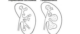
What is it and how is it different from hydronephrosis?
Pyeelectasia is a kidney pathology characterized by dilation of the renal pelvis, which obstructs the outflow of urine.
The right kidney is predominantly affected. This is due to the anatomy of the human body, because... the kidney on the right is located 2-3 cm lower than on the left. Right-sided kidney damage is more often observed in men and newborns.
According to ICD 10, the pathology was assigned code Q62.
According to the location of the lesion, it is divided into unilateral and bilateral. With unilateral pyelectasis, for example, left-sided, one renal pelvis is affected, with bilateral pyelectasis, respectively, both - left and right.
Bilateral damage is extremely dangerous, as it can cause atrophy of the kidney tissue.
Based on the severity of the course, they are classified as moderate, moderate and severe. At the moderate stage, the symptoms of the disease do not manifest themselves at all. There is no pain, urination is not impaired. Usually a moderate degree does not need adjustment and disappears over time.
If dilation of the renal pelvis is added to the expansion of the calyceal region, then this condition is called hydronephrosis. In other words, it is progressive pyelectasis. This is their difference.
Some researchers draw a parallel between this disease and Down syndrome. According to studies, pyeelectasis occurs in 25% of fetuses with Down syndrome, and in children without chromosomal abnormalities it is diagnosed in only 2.8%. The disease can be diagnosed in the fetus as early as the second trimester of pregnancy.
Treatment
The necessary treatment should be prescribed by an appropriate specialist and only after a thorough preliminary diagnosis. The main goal of therapy is to eliminate the root cause of pathological enlargement.
If the pathology is caused by dynamic factors, then drug treatment tactics are chosen. For nervous disorders, the use of sedatives and sedatives is indicated; for infectious lesions, antibiotic therapy is recommended.
If pyeelectasis is characterized by congenital etiology, then treatment is possible only with surgical methods:
- Urolithiasis - a calculus that impedes urination is removed surgically or conservatively, which is determined by the size of the stone and its sensitivity to stone-dissolving drugs.
- Narrowing of the ureter - a frame rim is implanted at the site of pathological narrowing, which in the professional language of doctors is called stenting.
After treatment, it is necessary to take all possible measures to prevent recurrence of pathological enlargement of the renal pelvis.
To do this, it is necessary to maintain an optimal drinking regime, avoid unauthorized and uncontrolled use of diuretics, and do not abuse foods with a diuretic effect, as well as salty, fatty and fried foods. If pyeloectasia is detected and treated in a timely manner, the risk of developing characteristic pathological complications will be practically minimal, and the renal structures will function uninterruptedly. Treatment tactics for renal pyelectasis
How does ICD 10 pyeloectasia occur?
Each kidney has a central area where urine collects called the renal pelvis. The ureters are tubes that drain urine from the renal pelvis and direct it to the bladder. If the ureters are blocked or compressed, they cannot drain urine effectively from the kidneys.
The accumulation of urine in the renal pelvis causes it to dilate, which usually leads to decreased urine output and urinary tract infections. This condition is called pyelectasis.
Differential diagnosis
Considering that pyeelectasia and hydronephrosis in young children have similar symptoms, during the examination it is necessary to carry out a differential diagnosis, which will allow an accurate diagnosis and prescribe adequate treatment.
First of all, an external examination and palpation is carried out to determine the areas of localization of pain, and if the pain syndrome extends to the kidney area, the following procedures are performed:
- urography (excretory and general);
- pneumopyelography;
- ultrasonography (to exclude or confirm the presence of nephrolithiasis).
Pyeelectasis is much easier to detect, and in most cases, the presence of such a disease can be seen already during a routine ultrasound scan in pregnant women in the period from 16 to 22 weeks of pregnancy.
Sometimes the disease is complicated by an associated infection, which leads to more severe symptoms. In such cases, a full examination is prescribed, including urography, cystography and radioisotope scanning.
Read all about kidney urography here.
During such procedures, it is possible to establish the exact cause of the development of the pathology and see which urinary tract disorders led to the disease.
Main symptoms and signs
Pyeelectasia is a kidney pathology associated with dilation of the renal pelvis due to impaired urine output. The disease can be unilateral (when one kidney is affected) or bilateral (two kidneys).
Mild forms do not require special treatment, but severe forms of pathology require surgical therapy.
According to the International Classification of Diseases (ICD-10), the expansion of the renal collecting system is assigned codes N00-N99.
What it is?
The kidneys are a paired organ located in the posterior part of the peritoneum and covered with a protective membrane (capsule). The organ consists of small cups in which urine accumulates. The collection of these cups is called the pelvis.
Kidney structure
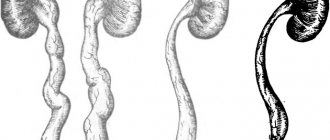
Initially, urinary fluid accumulates in the renal pelvis, where it is processed. Then it enters the bladder through the ureter and is excreted from the body through the urethral canal. When the outflow of urine is disrupted, urine accumulates in the kidneys, which causes renal hydronephrosis and deformation of the organ.
In children, pyelectasia is more often congenital and is mainly found in boys. When urinary outflow is impaired, pathological changes in the structure of the kidneys occur - they are compressed, stretched and lose their functionality. The main cause of pathology in newborns is unfavorable factors that affected the mother’s body during gestation.
There are 3 stages of development of pathology of the pyelocaliceal system:
- The initial stage with a mild degree of expansion, which does not require special therapy - it is enough to register the patient with a therapist who will monitor his condition.
- The second stage with a moderate form of expansion requires regular monitoring and observation via ultrasound. The causes of the disease are treated.
- The third stage is the most severe - there is a risk of complete blockage of the urinary canal and the development of acute renal failure. Treatment is surgery.
Signs
As a rule, the pathology itself does not have any specific signs and is more often a consequence of other diseases of the urinary system - inflammatory processes and infectious lesions, congenital anomalies.
Important! The disease may not manifest itself for a long time, especially when only one kidney is affected and the second one is working with double load.
Children with congenital lesions of the pelvis experience the following symptoms:
- temperature increase;
- frequent urination;
- pain in the lumbar region;
- atrophic and sclerotic changes in the organ (reduction in kidney size and proliferation of fibrous tissue).
In adults, in addition to the above symptoms, constant weakness, fatigue and decreased physical activity occur.
As noted above, pyeelectasia is not an independent disease, but a consequence of any pathological processes occurring in the body.
Experts identify common causes of the disease:
- abnormalities of intrauterine development or genetic predisposition;
- reverse flow of urine from the bladder to the kidneys (reflux);
- formation of stones in the urinary ducts;
- tumors of various etiologies;
- infections;
- neurogenic disorders;
- diseases of endocrine organs.
Dilatation of the pelvis in newborns can occur under the influence of the following factors:
- hereditary diseases;
- improper formation of organs in the fetus, which leads to compression of the urinary canals;
- abnormal structure of the valve between the pelvis and the ureter, accompanied by its curvature;
- premature birth;
- disruption of neural conduction, resulting in rare, copious urine discharge;
- kidney disease in a pregnant woman;
- various complications during the period of bearing a child in the form of swelling, surges in blood pressure, difficulty removing proteins from the body;
- weak muscular system in a newborn baby (hypotonicity of the bladder muscles);
- rarely visiting the toilet to empty a full bladder.
Important! If, with rare urination in a child over 3 years old, the size of the renal pelvis is within normal limits, then there is no reason for concern and no treatment is required.
Causes of impaired urine flow in adults:
- drinking large amounts of liquid;
- narrowing of the walls of the ureter or blockage with particles of pus and mucus released during inflammatory processes in the kidneys;
- blockage of the urinary ducts with stones;
- urethral torsion due to renal anomaly (prolapse);
- signals from the brain do not enter the bladder and do not cause the urge to empty;
- pregnancy.
Complications
Basically, stagnation of urine in the renal system can cause the development of renal sclerosis, when the functioning tissues of the organ are replaced by fibrous ones, which will subsequently lead to renal failure.
The main complications of pyelectasis:
- an increase in the size of the ureter in the area of its connection with the bladder, accompanied by a decrease in its opening (urethrocele);
- prolapse of the ureter into the urethra or vagina (ectopia);
- reverse flow of urine from the bladder to the kidneys (vesicoureteral reflux).
- an increase in the size of the ureter due to pressure in the bladder;
- pyelonephritis;
- sclerosis of the renal system;
- kidney failure.
Treatment
As a rule, therapy is aimed at treating the cause of the renal abnormality. If the cause of the disease is congenital organ pathologies, then doctors resort to surgical treatment methods aimed at restoring the normal outflow of urine from the kidneys.
For example, with a narrow ureter, a special stent is inserted into the organ to prevent further narrowing of the duct. If the urinary tract is blocked by stones or mucus, stones are removed.
In children, in most cases, doctors simply monitor the progress of the disease for some time, since most often pyelectasis in infants goes away without treatment. If changes in the pelvis and ureter are minor and urine output is not impaired, then the child is under medical supervision until the anomaly completely disappears.
Medication
If a violation of urinary outflow provokes the development of inflammation in the kidneys, then antibiotics and anti-inflammatory drugs are prescribed:
- fluoroquinolones (Norfloxacin, Pefloxacin, Ciprofloxacin);
- sulfonamides (Biseptol, Groseptol);
- nitrofurans (Furamag, Furadonin, Canephron);
- anti-inflammatory (Nimesil, Paracetamol).
At the same time, drugs are prescribed to improve intestinal microflora after taking antibiotics (Bifidumbacterin, Lacto G). To facilitate urine output, diuretics are prescribed - Furosemide, Diacarb, Veroshpiron, Uregit.
Folk remedies
Alternative medicine methods help patients completely relieve or alleviate the symptoms of pathology.
- Decoction of corn silk To prepare the drink: 1 tbsp. dry herbs are poured into 250 ml of boiling water, settled and filtered. Drink 2 times a day as a diuretic.
- Yarrow infusion 1 tbsp. herbs pour 1 tbsp. boiling water and let stand. Drink 3-4 times a day to relieve inflammation and drain urine.
- Herbal collection Pour a pinch of adonis, nettle leaves, horsetail and bearberry into a thermos and pour 500 ml of boiling water, leave overnight. Drink 0.25 cups 4 times a day.
The main goal of the diet for pyeloectasia is to reduce protein intake (no more than 60 grams per day) and increase the intake of fats and carbohydrates. If possible, food should be prepared with a minimum amount of salt; spicy foods, preservatives, and dairy products should be limited in the diet.
Exclude:
- various cereals and porridges;
- flour and pasta products;
- mushrooms and marinades;
- chocolate products.
Recommended:
- lean varieties of meat and fish, boiled or steamed;
- fresh vegetables and fruits.
To relieve the load on the organ, especially if both kidneys are damaged, you need to reduce fluid intake - 1.5-2 liters of water per day, including first courses, teas, decoctions and other liquids.
The medical term “pyelectasia” is translated from Greek as pyelos - pelvis, ectasia - expansion. The disease indicates that the outflow of urine from the renal pelvis is impaired due to an incorrect anatomical structure or a previous infection. According to the ICD-10 code classification, it is assigned class Q 62.
The kidneys are a paired organ that is located behind the peritoneum and is protected from damage by the capsule. They are a system of cups connected to form a pelvis. Pyeelectasis is an enlargement of the renal pelvis that prevents the full outflow of urine. There are other names for this process and its varieties:
- Dilatation of the pelvicalyceal system (pyelocalyceal system);
- Pyeloureterectasia;
- Pyeloureterectasia;
- Pyelocalicoureterectasia;
- Calicopyelectasia;
- Ureteropyelectasia;
- Ureterocalicopyelectasia;
- Ureteropyelocalicoectasia.
The pathological change is not a separate disease, but only accompanies other inflammatory processes. There are different reasons why the disease occurs:
- oncology;
- congenital malformations;
- urolithiasis disease;
- reflux of urine into the pelvis or ureteral reflux.
Based on etiology, doctors distinguish four types of disease:
- Dynamic congenital occurs due to the fact that the ureter narrows, the process of urination becomes difficult. It is a consequence of congenital phimosis.
- Dynamic acquired - when compression of areas of the urethra occurs after a prostate tumor, compression of the ureter, pyelonephritis.
- Organic acquired - narrowing and curvature of the ureter occurs after external or internal trauma, inflammation, ingestion of copious amounts of fluid, hormonal disorders, prolapse of the kidney.
- Organic congenital - with an intrauterine malformation of the urinary function, which arose as a result of renal inflammation.
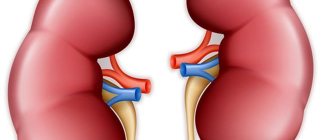
According to the observation of nephrologists, patients more often have an enlarged renal pelvis on the right. This is due to the structure of the body. Pyeloectasia of the right kidney is a type of unilateral dilatation of the renal pelvis.
Adult men and newborn children are predominantly affected. In severe cases, inflammation spreads to the ureters and calyces.
A complication of the disease is pyelocalicoectasia of the right kidney - the formation of stones in the cavity of the pelvis.
Pyeelectasis of the left kidney, like the right one, is unnoticed and asymptomatic, although on the left the disease occurs much less frequently. Obvious signs of left-sided enlargement in adults begin to appear only at the last stage, so you need to undergo regular examinations in order to prevent the disease in time and choose the right treatment.
Pyeelectasia :: Symptoms, causes, treatment and code according to ICD-10
Title: Pyeelectasia.
Pyeelectasis
This is an expansion of the renal pelvis due to the accumulation of urine. This is considered an independent (physiological) or concomitant condition in many urological diseases with impaired urodynamics. Complaints are often absent, but they can be represented by symptoms of the underlying pathology.
Diagnosis is based on ultrasound examination of the kidneys, excretory urography and cystography, pyelography, MRI or CT scan with contrast.
Treatment is necessary as enlargement occurs; surgery to restore adequate urination is performed in 25-45% of cases, which helps prevent end-stage hydronephrosis and chronic renal failure.
The term “pyelectasia” comes from the Greek words “pyelos” - “pelvis” and “ektasis” - “expansion”. Changes are sometimes detected by ultrasound of the fetus in pregnant women in the second trimester of pregnancy; in boys - 3-4 times more often than in girls.
Pyelectasia is often diagnosed in women aged 30-35 years during pregnancy, this condition is considered physiological in the absence of changes in the urine, and goes away on its own 4-8 weeks after birth.
In older men, pelvic enlargement is associated with prostate adenoma, which causes obstruction of the lower urinary tract. Urologists often consider pyelectasia as the initial symptom of hydronephrosis.
Pyeelectasis
There are two main causes of this condition: obstruction of urine flow (obstruction) and backflow of urine (reflux). The renal pelvis may enlarge due to excessive simultaneous intake of fluid, which is not considered a pathology; the situation resolves spontaneously. There are a large number of conditions accompanied by pyelectasis.
There are the following reasons leading to impaired urine outflow: A stone, a blood or salt clot, a tumor of the bladder, or the prostate gland can cause pyelectasia.
Narrowing of the pelvicalyceal segment, stricture of the ureter and urethra, prostate adenoma is also considered as obstructive uropathy; with these pathologies, vesicoureteral-pelvic reflux is often formed with an expansion of the renal cavity system.
Any part of the ureter can be compressed by external tumors located in the uterus, ovaries, and colon. In advanced forms of pelvic cancer, the lumen of the ureter narrows due to metastatic lesions.
Inflammatory processes in the retroperitoneal space, for example, Ormond's disease, hip lipomatosis, can have a compressive effect. • Developmental anomalies.
Horseshoe kidney, pelvic dystopia, nephroptosis are urological disorders, which in mild cases manifest themselves as pyeloectasia, and in advanced cases – hydronephrotic transformation. Similar changes were noted with ectopia, torsion, severe ureteral protrusion, accessory vessel, and flexure of the ureter.
Posterior urethral valves are the most common cause of pyelectasis in newborn boys. • Neurogenic disorders.
The neurogenic bladder is caused by impaired innervation and constant stagnation of urine in the bladder after urination.
Long-term condition leads to the formation of vesicoureteral reflux, which is often accompanied by recurrent urinary tract infections and pyelectasis. Predisposing factors include endocrine disorders associated with increased urine production, previous urological surgeries and radiation therapy. Teratogenic effects are significant for the development of intrauterine pyelectasia: irradiation, the use of certain drugs, viral diseases transmitted at a critical time for organogenesis. A certain role belongs to hereditary predisposition to urological or nephrological pathology.
When urine stagnates, compensatory-adaptive reactions begin, which lead to atrophy of the renal structures. When the secondary microbial flora is attacked, an inflammatory process occurs, which aggravates morphological changes.
Their severity is related to the degree of ureteral occlusion, the stage of the disease, the age of the patient, the involvement of contralateral organs in the process and the compensatory capabilities of the body.
In children, pyelectasis sometimes resolves spontaneously due to changes in the position of organs relative to each other, maturation of the structures of the urinary tract, when pressure in the urinary system increases and is redistributed.
Anomalies and malformations (stenosis, valves, vascular malformations that have a compressive effect on the ureter) significantly complicate the prognosis and, without treatment, lead to hydronephotic transformation of the kidneys.
Clinical manifestations of the pathology are usually absent; some patients note traction pain in the lower back, worsening in the morning or after drinking large amounts of liquid. Fever, weakness, and dysuric disorders indicate the development of concomitant kidney inflammation.
Often the symptoms are not caused by pyelectasis itself, but by the underlying pathological process.
Thus, in diseases accompanied by obstruction of the lower urinary tract, the patient is bothered by urination without effort, weak blood flow, frequent impulses (BPH, edema, narrowing of the urethra), periodic renal colic with the release of stones or sand (nephrolithiasis), leakage of urine from the ureter vagina (ectopia) etc.
Because pelvic dilatation remains asymptomatic for a long period of time and the kidneys work under significant strain, there is a progression of pyeloectasia to pyelocalcectasia and hydronephrosis, in which normally functioning tissue is replaced by connective tissue with loss of organ function.
Standing urine during pyeloectasia is a favorable environment for persistent pathogenic microflora, which leads to recurrent urinary tract infections. Another complication is the development of drug-resistant nephrogenic hypertension.
Nephrologists see increased blood pressure and hydronephrotic transformation of the kidneys as a harbinger of chronic renal failure.
The patient is managed by a urologist or nephrologist. If a neoplasm of the uterus or ovaries is suspected, consultation with a gynecologist or oncologist is necessary. In adults, a single detection of pyelectasis is not considered a pathology; in these cases, dynamic ultrasound is mandatory.
Testing for pyelectasis is aimed at eliminating organic or functional causes, and in addition to ultrasound, may include: • Laboratory tests.
In the compensated form, there are no changes in urine tests; leukocyturia, proteinuria, bacteriuria are characteristics of the inflammatory process.
Salt precipitation is characteristic of dysmetabolic nephropathy or urolithiasis. A blood test for creatinine and urea is justified by bilateral damage; an increase in the level indicates renal failure. If bacteria are found in urine, a study of the biomaterial in the flora is prescribed. • Instrumental diagnostics.
If ultrasound findings are unclear, excretion urography is performed using cystography, CT or MRI of the kidneys with contrast agent, nephroscintigraphy and angiography. If bladder cancer is suspected, cystoscopy and TRB are performed to confirm prostate cancer.
In the fetus, pyeloectasia is determined using ultrasound scanning in the 2nd trimester of pregnancy (25%) after birth. Depending on the indications, one or another method is selected for additional diagnostics. There are physiological and pathological forms of pyelectasia.
With the first observed pelvic expansion without any other changes, it is impossible to predict whether the condition will progress, so the patient's dynamics are monitored. The main task of the clinician is to determine the root cause of pyelectoasis.
Concomitant confirmed pyelitis, pyelonephritis or cystitis requires the prescription of drugs to stop the infectious and inflammatory process. This applies to: • antibiotics; • uroseptics; • immunomodulators; • drugs that improve blood circulation; • multivitamin complexes; • Litholytic drugs, the action of which prevents the formation and loss of crystals (for urolithiasis). At the same time, a diet that takes into account the composition of salt is recommended. During the treatment of pyeloectasia, saturated meat, chicken, mushrooms, fish broths, chocolate, strong tea and coffee, all alcoholic beverages, sausages and marinades are excluded from the diet. This is shown in the progression of pyelectasis. The size of the operation depends on the reason that led to the expansion of the intracellular structures of the kidneys. The intervention can be laparoscopic, open or endourological, aimed at restoring normal urodynamics. The following methods are used: • Plastic surgery of the ureteral segment with removal of the distended pelvic mucosa and suturing of the ureter to the kidneys, vascularization, balloon expansion, endotomy using a laser scalpel or electric current. • Removal of dental plaque using one of the methods (lithotripsy with contact or at a distance, endoscopic nephrolitholapaxis, open surgery). • Palliative operations and manipulations to normalize the urinary tract in cases of acute inflammation: the use of epicystostomy, nephrostomy, catheterization of the bladder, placement of a catheter stent through the ureter in the pelvis, etc. • Removal of tumors that negatively affect urodynamics.
• Nephrectomy with loss of kidney function and destruction of its parenchyma to eliminate the source of infection in the body. In children, if at least 10% of normal tissue is preserved, organ-conserving surgery is not performed due to its high regenerative capacity.
If the dilation of the renal pelvis does not progress, active therapeutic measures are not needed. For preventive purposes, decoctions of diuretic herbs and herbal uroseptics are prescribed. It is not recommended that the patient drink a lot of liquid at the same time. To reduce the load on your kidneys, you need to urinate at night. 1. Precision surgery of hydronephrosis / Alyaev Yu. G., Grigoryan V. A., Adamyan R. T., Enikeev M. E., Chinenov D. V. // Annals of plastic, reconstructive and aesthetic surgery - 2008 - No. 1. 2. Clinical significance of ultrasound examinations in monitoring children with pyeelectasis. Abstract of the dissertation / Mavricheva I. S. – 2002. 3. Comparative assessment of modern research methods for hydronephrosis. Abstract of the dissertation / Kasiteridi I. G. - 2005.
4. New endourological technologies in the diagnosis and treatment of diseases of the kidneys and upper urinary tract / Martov A. G., Ergakov D. V. // Minimally invasive technologies in the treatment of urological diseases. Thematic collection. – 2006.
42a96bb5c8a2ac07fc866444b97bf1 Content moderator: Vasin A.S.
Source: https://kiberis.ru/?p=280366
Symptoms: can the disease be recognized at an early stage?
In case of a bacterial infection, a full examination is prescribed:
- Detailed blood and urine analysis.
- Excretory urography.
- Cystoscopy. A probe with a camera is inserted into the bladder through the urethra, and the walls of the bladder are examined with its help.
- Radioisotope scan of the kidneys. Used to detect tumors. A special radioisotope substance is injected into the blood and collects near the tumor. The camera scans and determines the location of the tumor.
This is an x-ray with the introduction of contrast agents. If the urinary system is functioning normally, the contrast agent injected into the blood will be in the urine within 5 minutes. Carry out no more than once a year.
This study is prohibited for children.
Pregnant women are allowed only ultrasound diagnostics.
Extended diagnostics allows us to identify diseases that accompany the severe stage of pyelectasis:
- vesicoureteral reflux, i.e. throwing urine from the bladder into the kidney;
- megaureter - abnormal dilatation of the ureter;
- A ureterocele is a swollen ureter.
After the examination, a decision is made on treatment methods for identified concomitant pathologies.
Congenital malformations
- dystopia (abnormal location) of the kidney or ureter;
- megaureter;
- ureteral strictures at any level.
Acquired kidney pathologies
- urolithiasis disease;
- benign and malignant formations;
- chronic infections of the urinary system;
- injuries;
- bladder neoplasms;
- pregnancy;
- prostate adenoma in men.
Violation of the physiological outflow of urine from the urinary tract through the ureters into the bladder causes its accumulation in the pelvis and calyces, an increase in their volume and overstretching of the walls. Subsequently, these structures put pressure on the functional apparatus of the kidney, the nephrons undergo atrophy and cannot perform their main functions - the formation and excretion of urine. If the pathology is not treated, the patient will eventually develop chronic renal failure.
The clinical manifestations of calicoectasia of the right or left kidney are not significantly different. Due to scant symptoms, a person may not be aware of a urinary organ disease for years. Compensated calicoectasia often becomes an incidental finding on ultrasound during examination for another disease.
With a significant increase in the volume of the calyces, increased renal pressure and overstretching of the organ capsule, the following symptoms may appear:
- Pain in the lower back on the affected side, radiating to the lower abdomen, groin, external genitalia.
- A sharp increase in body temperature, chills, flushing of the skin of the face and body.
- Nausea and occasional vomiting;
- Changes in the amount, color and transparency of urine excreted: darkening and turbidity of the urine is usually observed;
- Frequent urination in portions of 50-100 ml;
- The appearance of blood in the urine (if the vascular wall is damaged by a growing tumor or calculus).
If left untreated, the syndrome may be complicated by calicopyeloureterectasia. And what is it? This syndrome is a consequence of the expansion of not only the renal calyces, but also the pelvis and ureters. It is characterized by a clear clinical picture and increasing signs of urinary organ failure. In addition, with calicopyelectasia, the following often develop:
- secondary pyelonephritis;
- urolithiasis;
- hydronephrosis – persistent progressive dilatation of the calyces and pelvis, in which the renal parenchyma is critically thinned;
- chronic renal failure.
- Unilateral renal pyelectasis.
- Bilateral renal pyelectasis.
In the first case, the anomaly affects only one kidney, while the second remains healthy.
The second option is much worse - both kidneys are affected by anomalies.
Additional facts
The term "pyelectasia" comes from the Greek words "pyelos" - "pelvis" and "ektasis" - "expansion". Changes are sometimes detected during fetal ultrasound in pregnant women in the second trimester of pregnancy; in boys - 3-4 times more often than in girls. Pyelectasia is often diagnosed in women aged 30-35 years during pregnancy, this condition is considered physiological in the absence of changes in the urine, and goes away on its own 4-8 weeks after birth. In older men, pelvic enlargement is associated with prostate adenoma, which causes obstruction of the lower urinary tract. Urologists often consider pyelectasia as the initial symptom of hydronephrosis.
Pyeelectasis
Complications with enlarged renal pelvis
If pyelectasis is left untreated, increased pressure in the kidneys can reduce the kidney's ability to filter blood, remove waste and produce urine, and regulate electrolytes in the body. This can lead to kidney infection and, in some cases, complete failure of kidney function or death.
If the enlargement of the renal pelvis turns into inflammation, the patient may develop pyelonephritis.
Lack of timely treatment can lead to the development of a number of complications:
- chronic renal failure;
- chronic pyelonephritis, pyelitis;
- urosepsis;
- hydronephrosis.
The disease cannot be started, as this is fraught with serious consequences
Prevention measures
There is no specific method for preventing the disease. Doctors recommend monitoring the condition of the genitourinary system, paying attention to discomfort in the kidney area, lower abdomen, and problems with urine excretion.
Additional measures:
- prevention of inflammatory processes;
- timely emptying of the bladder: stagnant liquid is a suitable environment for the proliferation of dangerous microbes;
- treatment of urethritis, pyelonephritis, cystitis, vaginitis, sexually transmitted diseases, prevention of chronicity;
- following doctor’s recommendations during pregnancy, reducing various risks for the fetus;
- avoiding excess consumption of water, tea, and juices if there are problems with urine excretion.
With congenital and acquired pyeloectasia, you should not delay in starting treatment. Whether the patient takes medications or undergoes surgery is decided by the urologist based on the diagnostic results. It is important to know the cause of the deviations, the condition of the renal tissue and ureters. Untreated pyeloectasia provokes congestion, disrupts the functioning of natural filters, and leads to nephron atrophy or kidney sclerosis.
Pyeelectasia in children
The doctor will prescribe the required dose of drugs
A once-daily dose of antibiotic therapy is recommended depending on the result of a renal ultrasound done after birth. This should reduce the likelihood of a urinary tract infection.
Usually the disease is congenital in nature. According to statistics, boys are more susceptible to pyelectasis than girls. Its occurrence during the prenatal period is influenced by various unfavorable factors associated with the urinary system. When the outflow of urine is disrupted, the kidney tissues are compressed, which leads to a decrease in the functionality of the organ.
This condition is detected on ultrasound in about 1.4 percent of fetuses. This is the most common fetal abnormality and accounts for about 50 percent of all findings. Pyeloexatia in the fetus is most often bilateral, and unilateral is most likely to be on the left side. It is more common in male fetuses both in the prenatal and postnatal periods.
Pyeelectasia in infants is the result of a genetic abnormality. The most common cause is ureteral obstruction, which occurs when the ureter narrows as it drains from the kidneys. Another cause is reflux of urine as it returns to the kidney. Reflux is often caused by problems with the valves in the ureters that control the flow of urine.
To diagnose abnormal urine flow from the bladder into the upper urinary tract, voiding cystourethrography (MCUG) may be performed, which requires placement of a catheter in the bladder. This condition occurs in 5-25% of children with pyeelectasis.
Some signs that a child may have pyelectasis include:
- elevated temperature,
- poor appetite
- irritability or fatigue and drowsiness,
- abdominal pain.
What is renal pyelectasis?
Pyeelectasis is represented by deformation of the structure of the kidney outlet element. The dimensional parameters of the renal pelvis in this disease differ significantly from the parameters corresponding to a healthy organ. Depending on the degree of expansion of the pelvis, several stages of the disease are distinguished: moderate, mild, severe. Pyeelectasis can be left-sided, right-sided, or bilateral (less commonly). This is due to the localization of deformation. When the pelvis of the right kidney is enlarged, a diagnosis of right-sided pyeelectasis is made, and when the pelvis is deformed on the left, a diagnosis of left-sided pyelectasia is made. If deformation of the pelvis is diagnosed on both kidneys, the patient is diagnosed with bilateral pyloecstasy.
The pathology can be acquired or congenital. Often the disease is diagnosed in utero during a screening ultrasound examination at 16-24, 32-36 weeks.
In male patients, this disease is diagnosed 3 times more often than in women. Approximately 35% of diagnoses occur in boys aged 0-10 years. Congenital pyelectasis is often detected in children. But cases of acquired disease are also not uncommon.
Treatment of pyelectasis in children and adults
Pyeelectasia (Q62) is treated by eliminating the factors that caused it, that is, mainly congenital factors.
There are cases that the disease can eliminate itself, but this was observed only in children (as a result of the final formation of their genitourinary system, congenital defects may be mitigated). But such cases are very rare and occur only in mild stages of the disease.
More severe stages, namely moderate and severe, may even require surgical intervention, but usually everything is managed only with conservation and drug therapy. The doctor prescribes antibiotics and anti-inflammatory drugs for such therapy.
If calculi, or more simply stones, form in the kidneys, then two treatment methods can be taken:
- An attempt to dissolve the stones.
- Remove stones through surgery.
In addition, junk food (fast food, soda, alcohol...) is completely excluded from the patient’s diet and vegetables, fruits and nuts are recommended for consumption, but not all of them either, since some types can contribute to the formation of stones.
As the pelvis swells, pressure rises in the kidneys, in the pelvis itself, in the bladder and in the calyxes. In this situation, antispasmodic myotropic drugs are prescribed. It can be:
- “Scopolamine”;
- “Platifillin”;
- “Atropine sulfate”;
- “Hyoscyamine.”
They relax the muscles, which helps relieve the pressure a little, but these drugs do not completely solve the problem.
If you have a pathology of the genitourinary system, then with a probability of 0.1 you will have surgery. This probability is quite high. In other words, every tenth person undergoes surgery.
"{amp}gt;
Surgical intervention can be performed from an open approach (abdominal surgery) or endoscopically using special equipment through two punctures in the abdominal wall. The choice of surgical intervention method depends on the cause that caused the development of the urinary outflow disorder.
The prognosis of the disease also depends on the cause of calicopyelectasia: if the development of the disease is caused by the formation of stones, then their elimination will lead to a complete recovery; the congenital malformation can be eliminated surgically. If the cause of calicopyelectasia is a neoplasm, the prognosis largely depends on its type and the possibility of removal.
- anatomical abnormalities,
- compression of masses or expansion of surrounding tissues, such as an enlarged prostate, pregnancy or inflammation,
- functional abnormalities, when parts of the urinary tract do not work properly,
- mechanical obstruction by uric acid crystals or blood clots.
The obstruction causes urine to accumulate in the renal pelvis. When fluid accumulates in the pelvis, a kidney cyst can often develop.
In most cases, pyeelectasis in adults is discovered during other examinations that were carried out to determine other diseases. In this condition, the following conditions are observed on ultrasound in adults:
- narrowing of the lower part of the ureter, due to which a cyst may form,
- drainage of the ureter into the vagina or urethra,
- reverse flow of urine through the ureter,
- dilatation of the ureter.
What it is?
Kidney structure
Signs
Diagnostics
The patient is managed by a urologist or nephrologist. If a neoplasm of the uterus or ovaries is suspected, consultation with a gynecologist or oncologist is necessary. In adults, a single detection of pyelectasis is not considered a pathology. In these cases, dynamic ultrasound is mandatory. Testing for pyelectasis is aimed at eliminating organic or functional causes, and in addition to ultrasound, may include: • Laboratory tests.
In the compensated form, there are no changes in urine tests; leukocyturia, proteinuria, bacteriuria are characteristics of the inflammatory process.
Salt precipitation is characteristic of dysmetabolic nephropathy or urolithiasis. A blood test for creatinine and urea is justified by bilateral damage; an increase in the level indicates renal failure. If bacteria are found in urine, a study of the biomaterial in the flora is prescribed. • Instrumental diagnostics.
If ultrasound findings are unclear, excretion urography is performed using cystography, CT or MRI of the kidneys with contrast agent, nephroscintigraphy and angiography. If bladder cancer is suspected, cystoscopy and TRB are performed to confirm prostate cancer. In the fetus, pyeloectasia is determined using ultrasound scanning in the 2nd trimester of pregnancy (25%) after birth. Depending on the indications, one or another method is selected for additional diagnostics. There are physiological and pathological forms of pyelectasia. With the first observed pelvic expansion without any other changes, it is impossible to predict whether the condition will progress, so the patient's dynamics are monitored. The main task of the clinician is to determine the root cause of pyelectoasis.
Pyeelectasis of the kidneys in a child, an adult and during pregnancy - causes, symptoms and treatment
What it is?
Kidney structure
Signs
Treatment with folk remedies is also a fairly effective way to combat diseases of the collecting system. This article presents the most popular recipes that are sure to give positive results.
- To prepare the infusion, we need ginseng root and eleutherococcus in equal proportions. Pour vodka over all this and place in a cool, dark place for 7 days.
- Doctors recommend drinking fresh milk until the swelling goes away.
- Take dried rosehip root, remove shavings (12 pieces) approximately 15 cm long. Pour all this with 1 liter of purified water and place in a cool, dark place. In the morning, strain the contents. Take throughout the day. The duration of treatment is 1 month.
Nutrition and diet for kidney caliectasis must also be correct and balanced. Nutritionists recommend adhering to dietary table No. 7.
It is necessary to exclude fried, fatty foods, confectionery products, and alcoholic beverages. To improve the effectiveness of treatment, you can add the following products to your diet:
- Who would have thought that stewed pumpkin would help get rid of kidney problems?
- Regular consumption of watermelon, red currant and raspberry juice has an excellent effect on the condition of the genitourinary system.
Symptoms such as chills and low-grade fever may occur.
How to find a kidney recipient and who can become a donor?
- chills, fever up to low-grade levels;
- change in diuresis (decrease in fluid volume against the background of an increased urge to urinate);
- red blood cells are found in the urine; if they appear in multiples (macrohematuria), the liquid becomes slightly pink;
- pain from the affected organ (calicoectasia of the left kidney or pathology of the organ located on the right) in the lumbar region, increasing with deep palpation or tapping (positive Pasternatsky’s symptom);
- with partial impairment of renal function, nausea may be felt, accompanied by isolated attacks of vomiting.
It is characteristic that the right kidneys are more susceptible to the expansion of the calyces, as well as the development of hydronephrotic conditions, which is associated with some anatomical features of the location of the organs themselves, their circulatory and urinary systems. Calicoectasia of the right kidney is detected approximately twice as often as a similar pathology on the left, which helps to suspect this particular disease during the initial collection of diagnostic data.
Often the symptoms of hydronephrotic conditions, accompanied by expansion of the calyces, are erased. This happens when the outflow of urine is partially impaired and the fluid pressure in the pyelocaliceal complex is not so significant as to lead to rapid progression of the disease. If the lumen of the ureter is not completely obstructed, pathological changes in the renal cavities may be insignificant or absent for a long time.
But still, the pathology begins to manifest itself over time, especially when, due to insufficient urinary excretion, an infection occurs with the development of pyelitis or pyelonephritis, and the formation of stones in the pelvis begins. The influence of additional pathological factors provokes a further deterioration in the outflow of urine, against the background of which calicopyelectasia of the right kidney or similar abnormal changes in the left organ quickly develops.
Sometimes right- or left-sided caliectasis is mistaken for diseases of the liver, intestines or pancreas.
As it progresses, the patient may experience the following symptoms:
- severe increase in body temperature (up to 40 degrees);
- nausea and vomiting;
- frequent urge to urinate (as a rule, most of them are false);
- pain in one of the sides or groin area (in men, pain symptoms are much stronger than in women or children);
- change in the color and smell of urine;
- severe chills.
We must remember: the sooner treatment begins, the more effective it will be. Therefore, when the first unpleasant symptoms appear, you should definitely consult a doctor for diagnosis.
In infants, this disease can appear due to disruption of many blood vessels in the kidneys. There are no symptoms and caliectasis is diagnosed during one of the examinations with a pediatrician. As the disease progresses, the temperature may rise, severe pain may appear, and changes in the color and nature of urination may occur.
- Severe pain in the lower back.
- Body temperature up to 39 ºС and above.
- Severe chills, fever.
- Nausea, vomiting.
- Blood in the urine (hematuria).
- Frequent and painful urination.
- Small amounts of urine.
When the disease occurs, the patient begins to experience severe chills, which is accompanied by an elevated body temperature reaching 40 degrees. In this case, the temperature rises when urine is not removed from the body in time.
Characteristic is the appearance of nausea and vomiting, which does not bring relief to the patient. With caliectasis of the kidneys, a person feels severe pain that spreads to other areas, in particular to the groin area.
Pain appears when the patient moves or touches the lumbar area. Pain in men becomes more severe if the patient develops calicopyeloureterectasia.
Calicopyeloureterectasia is understood as the addition of inflammatory processes in the urethra to the disease. Calicopyeloureterectasia leads to mild bleeding due to stones damaging the walls of the canal through which urine passes.
Symptoms
Clinical manifestations of the pathology are usually absent; some patients note traction pain in the lower back, worsening in the morning or after drinking large amounts of liquid. Fever, weakness, and dysuric disorders indicate the development of concomitant kidney inflammation. Often the symptoms are not caused by pyelectasis itself, but by the underlying pathological process. Thus, in diseases accompanied by obstruction of the lower urinary tract, the patient is bothered by urination without effort, weak blood flow, frequent impulses (BPH, edema, narrowing of the urethra), periodic renal colic with the release of stones or sand (nephrolithiasis), leakage of urine from the ureter vagina (ectopia) etc.
Pathology treatment options
Treatment tactics for pyeloectasia of the right kidney will depend on the underlying cause of the condition and the degree of its manifestation. If the disease is detected in newborns in mild or moderate severity, then over time it goes away on its own or becomes less pronounced. No one can say exactly how the pathology will manifest itself after the birth of a child.
Find out what toxic kidney nephropathy is and how to treat the disease.
The rules and methods of using bearberry for the treatment of cystitis are described in this article.
Diagnostic methods
The pathology can be detected by the doctor even at the time of the next ultrasound examination of the fetus, that is, before the birth of the child.
The minimum period for detecting an anomaly is 16 weeks of gestation. The diagnosis of “renal pyeloectasia” can be established if the size of the pelvis exceeds the age norm.
For the second trimester, the indicator should not be more than 5 mm, for the third - 2 mm more. A deviation of 1 mm allows us to hope that the kidneys will be able to return to normal during the process of extrauterine ripening. An increase in size of 2-3 mm should be a serious concern.
When hydronephrosis (the second name of the anomaly) is detected, the child’s genitourinary system is dynamically monitored using an ultrasound diagnostic device. There is no other way to monitor changes in the baby's kidneys at the prenatal stage.
If there were no positive changes before birth or they remained insignificant, the newly born baby, if necessary, may additionally be prescribed other tests.
Often this is cysto-urography with the introduction of contrast through venous access.
These methods are used infrequently, in extreme cases, when for some reason ultrasound turns out to be uninformative. But in most cases, an ultrasound examination is sufficient to visualize the overall picture of the disease.
Ultrasounds of the baby’s kidneys are performed regularly throughout the first year of his life. The examination is carried out every 2-3 months to monitor the pathology over time.
Routine examinations cannot be skipped, because changes sometimes develop very quickly, and time may be lost for effective treatment and prevention of complications.
In a number of cases, such a waiting tactic is completely justified and the dilation of the pelvis, caused by physiological reasons, regresses on its own by the period of introducing complementary foods.
Nutrition for pyeelectasis
If pyeelectasis is detected in children or adults, the specialist prescribes a diet that must be followed. The main principle of dietary nutrition for this disease is to reduce the amount of protein consumed (no more than 60 grams per day), as well as increase the consumption of fats and carbohydrates.
Also, with pyelectasis, control over the amount of fluid consumed is necessary. The daily dose of liquid is calculated by the formula: 30 ml per 1 kilogram of the patient’s weight.
Symptomatic manifestations and diagnosis
With a milder course of the disease in children, the first signs are noticed several months or years after the dilatation of the renal pelvis. Severe pyeelectasis in children of different ages is characterized by the following manifestations:
- increase in abdominal volume due to enlargement of the kidneys;
- dysuric disorders, especially with the symptom of painful urination;
- positive effleurage syndrome (painful reaction to tapping from the back);
- laboratory signs of inflammation;
- symptoms of chronic renal failure.
Main symptoms and signs
What it is?
Kidney structure
Signs
The expansion can affect not only the pelvis, but also the calyces and ureter. This is especially common with long-term progression of the disease. Pyeelectasia develops when urine is not removed from the pelvis and flows back into the kidney. The disease can be congenital or acquired. These forms are organic and dynamic, based on reasons.
- ureteral strictures, causing stagnation of urine in the collecting system;
- chronic inflammation of the bladder;
- excessive fluid intake;
- infectious lesions of the genitourinary system (pyelonephritis, glomerulonephritis) in which the ureter is blocked by clots of dead tissue and pus;
- obstruction of the ureter due to the passage of a stone during urolithiasis;
- prolapse of the kidneys. as a result of which twisting of the ureter occurs;
- neurogenic bladder. as a result of which the pressure inside the organ increases;
- intrauterine pathologies associated with compression of the organs of the urinary system by vessels;
If a child or adult has already had diseases of the kidneys or ureter and bladder, you should pay close attention to these organs in the future: treat the diseases in a timely manner, and undergo regular examinations by medical specialists. It is recommended to ensure that the pelvic area and lower back are not overcooled, and to perform moderate-intensity physical exercise to prevent the occurrence of congestion in the pelvic organs. During pregnancy, you need to get tested regularly and monitor the disease.
Forms of severity and consequences
There are several types of renal pyelectasis: right-sided, left-sided or bilateral. Based on severity, there are mild, moderate or severe forms of pyelectasis. The severity of the pathological process is determined by the preservation of organ functions, while concomitant and complicating diseases are taken into account.
In general, the dilation of the pelvis itself is asymptomatic, and the appearance of pathological signs is caused by a concomitant, provoking pathology or the development of complications.
Most often, renal pyelectasis is accompanied by complications such as:
- Vesicoureteral reflux – most typical for children; against the background of ureteral atony, urine is thrown back from the vesical cavity into the renal pelvis;
- Ureterocele - when at the junction of the bladder and the ureter there is expansion, i.e. swelling of the ureter;
- Ureteral prolapse - characteristic of severe forms of pyelectasis, in which the ureter grows into the urethra in men or the vaginal wall in women;
- Impaired valve functionality in the urethra, which is typical for male patients;
- Megaureter is a pathological condition characterized by enlargement of the ureters against the background of impaired urinary outflow from the lower urinary tract or from the bladder structures;
- Atrophy of renal tissue - when a pathological decrease in the size of the organ occurs;
- Acute pyelonephritis - an inflammatory process in the kidney tissues;
- Decreased renal functionality;
- Death of kidney tissue (renal sclerosis).
In a child, renal pyelectasis is dangerous because in the future it can cause disruption of the normal functioning of the kidneys. Urinary outflow is hampered, kidney tissue is compressed, which leads to renal atrophy and deterioration in the functioning of the organ. Poor urine excretion leads to infectious processes and the formation of pyelonephritis, which leads to a decrease in renal function and the development of sclerosis.
Severity of renal pyelectasis
Etiology and forms of development of the disease
At the first stage of its onset, renal pyeloectasia has no visible signs and resolves spontaneously over time. When complications occur, the pathology undergoes three phases of development: mild (moderate), moderate and severe. Each stage is determined by the degree of functionality of the filtration organs. The reasons for the stretching of the renal calyces depend on the possible appearance of obstacles to the outflow of urine in the form of narrowing of the urinary passages or the occurrence of ureteral reflux (reverse reflux of urine from the bladder).
Boys are more likely to experience problems with the genitourinary system than girls. In children, renal pyeelectasis in the vast majority of cases is of a congenital organic or dynamic nature and is distinguished by the following factors:
Kidney pyeelectasis in adults is acquired in nature and is associated with organic and dynamic disorders when:
- 1. Partial narrowing or complete blockage of the diameter of the ureter with a calculus (stone) occurs in renal colic.
- 2. Organ tissue becomes inflamed with pyelonephritis.
- 3. The kidney prolapses, followed by blocking of the ureter with a mass of pus.
- 4. Excessive fluid intake.
- 5. Infection of the urinary system with bacterial forms;
- 6. Increased pressure in the bladder cavity.
- 7. Decreased ureteral motility in elderly people or bedridden patients.
Medical practice indicates the development of pyeloectasia of the right and left kidneys and simultaneously of two organs - bilateral pathology.
We recommend
What is renal hydronephrosis and how is it treated?
The essence of pathology
In the ICD-10 catalog, pyeelectasis is designated by code Q62. The disease is diagnosed in cases where, when there is difficulty in the outflow of urine, increased pressure is exerted on the kidneys from the fluid accumulated in the pelvis.
The disorder also develops in cases of problems with the urinary tract, which are narrowed due to infectious diseases or injuries.
There are three types of disease in the medical history, depending on which organs are affected: pyeelectasis of the left, right or both kidneys. In the first two cases, the disease spreads to only one of the organs; bilateral pyelectasis leads to the spread of the disease to the pelvis of both organs.
In any form, pathology can be classified according to the degree of development:
- Easy. The pelvis enlarges slightly and is mainly a temporary phenomenon, which is diagnosed immediately after birth and over time, when the process of formation of kidney tissue is completed, the condition of the organs returns to normal.
In this case, no special treatment is required, but until the child is three months old, ultrasound scans are regularly performed.
Average. There is a noticeable increase in the affected organs, which requires moderate conservative therapy and constant monitoring. Treatment is prescribed on an individual basis and depends on the reasons that caused such disorders. Heavy. Enlargement of the kidneys leads to disruption of their functions. In most cases, at this stage, surgical intervention is necessary, in the absence of which sclerotic processes or hydronephrosis may develop as complications.
Statistically, pyeloectasia is more common in boys than in girls. This is due to the anatomical features of the kidney structure.
Forms and types
Overdistension of the renal pelvis can be of three degrees. For mild to moderate cases, systematic observation by a specialist and ultrasound examination of the urinary tract are recommended. Severe severity requires surgical treatment.
Depending on the time of occurrence, congenital and acquired pyeloectasis are distinguished. In this case, the process can be unilateral, in which one kidney is affected, or bilateral, with dilation of the pelvis observed in both kidneys.
Treatment tactics
Treatment is based on the clinical picture and research data
After diagnostic measures and determination of the severity of pyeloectasia, therapy is prescribed. The treatment regimen varies greatly and depends on many factors: the causes of urinary stagnation, concomitant diseases and complications, and the clinical picture. Drug therapy is based on the following groups of drugs:
- anti-inflammatory and antibacterial agents;
- uroantiseptics;
- anti-stone formation drugs;
- means for reducing creatinine, urea, restoring the electrolyte composition of blood plasma;
- diuretics.
For hematuric syndrome, iron supplements are prescribed. Along with drug therapy, a therapeutic diet low in protein and salt is indicated. For patients with edema, the volume of daily fluid is limited.
Surgical treatment is prescribed in severe cases, especially if stagnation of urine is caused by abnormalities in the development of the organs of the genitourinary system, in a bilateral condition. After the operation, the child is given a long rehabilitation period.
Some clinicians believe that moderate renal pyelectasis in a child goes away on its own without special intervention. All that is required is observation of the dynamics of the pathological process. During a child's growing up, there are three main periods of development of renal structures that can affect the development of pyelectasia: up to one year, the period of intense stretching and growth of internal organs at 6–7 years, puberty.
Causes
Obstructions in the urethra and bladder:
- Stones, tumors, diverticula, foreign bodies of the urethra and bladder.
- Valves, strictures of the urethra.
- BPH.
- Neurogenic bladder disorders.
Compression of the ureter from the outside:
- Cystitis.
- Enlarged lymph nodes.
- Inflammatory lesions of the retroperitoneal space.
- Intestinal diseases.
- An accessory renal vessel that can compress the ureter.
Abnormal position and course of the ureter:
- Bend, curvature of the ureter.
- Narrowing of the ureteropelvic segment and ureter.
- Decreased ureteral tone.
Prevention and prognosis
There is no specific prevention of pyeloectasia, but the risk of developing pathology can be reduced even at the stage of pregnancy. Women are advised to control the volume of daily fluid, avoid exposure to harmful factors, and monitor the condition of the kidneys in case of a complicated nephrourological history.
The prognosis for renal pyeloectasis in a child varies greatly, depending on the causes of the pathology, combination with other diseases, and symptoms. In case of persistent functional disorders of the kidneys, appropriate treatment is carried out. With the development of chronic renal failure, an adequate diet, long-term drug therapy to prevent or treat complications, and a kidney transplant for end-stage chronic renal failure are required.
Renal pyeloectasia is a pathological expansion of the renal cavities due to dysfunction of urine output into the ureters and bladder. This disorder is not an independent disease and indicates the beginning of an abnormal process. Accumulating in the cavities of the kidneys, the fluid contributes to the formation of obstruction of the renal calyces, followed by hydronephrosis. According to statistics from the International Classification of Diseases, renal pyeloectasia belongs to class Q, the ICD-10 code is Q 62.