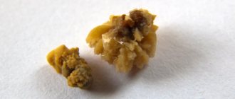
Calcification is the abnormal accumulation of calcium salts in body tissues: a rare but sometimes life-threatening syndrome caused by the crystallization and deposition of calcium salts in small blood vessels and soft tissues of the body. With nephrocalcinosis, diffuse calcification of the renal parenchyma occurs due to the deposition of calcium salts in the area of the renal medulla.
In most cases, the cause of the disease is a metabolic disorder. Nephrocalcinosis is common in premature newborns due to the fact that they are born with an immature kidney.
Causes and symptoms of the disease
In the kidneys, there are primary and secondary calcifications. Nephrocalcinosis is classified in a similar way - a condition in which formations appear in the organ.
Primary calcifications appear as a result of congenital diseases and various disorders in the development of the urinary system. This process is called primary nephrocalcinosis. It affects the renal parenchyma.
Nephrocalcinosis is caused by:
- increased levels of calcium in the body;
- loss of calcium from the skeletal system;
- excess vitamin D content, which regulates the concentration of calcium in the blood.
Secondary calcifications appear after inflammatory diseases, especially after kidney tuberculosis, thyroid diseases and other endocrine disorders.
Untreated pyelonephritis also leads to stone formation. With secondary nephrocalcinosis, scarred tissue of the organ is damaged.
Secondary nephrocalcinosis occurs when:
- improper flow of blood into the cortical layer of the kidney;
- mercury poisoning;
- radiation exposure;
- abuse of diuretics;
- disorders of the acid-base balance of the blood;
- kidney tuberculosis;
- pathological changes in the endocrine system.
The disease is classified according to the location of the formations in the organ. Cortical nephrocalcinosis is manifested by changes in the cortical layer, medullary - by damage to areas of the renal pyramids.
In the early stages, nephrocalcinosis does not make itself felt, especially if the formation appears in one kidney. It is difficult to identify, because the healthy kidney takes over the function of the diseased one, thus creating the appearance of complete well-being.
The danger of formations lies in disruption of the functioning of the affected organ. Therefore, when calcifications are detected, it is important to undergo a comprehensive comprehensive examination.
Poor nutrition often causes the development of nephrocalcinosis.
https://youtu.be/https://www.youtube.com/watch?v=Tgqsvu6ozD4
_
Cyst with calcifications: how dangerous is it?
A cystic formation forms when salts accumulate in the renal parenchyma and healthy cells die. In this case, the tubules become clogged and the connective tissue grows, replacing the parenchyma of the organ. With a cyst with calcifications, an inflammatory reaction and infection occurs, causing failure of the urinary system. On average, the size of the cyst is no more than 0.5 cm. If the patient is not operated on in time and the cyst is not removed, nephrosclerosis will manifest itself.
Signs of illness
If the stones do not lead to disruption of the urinary system, then it is quite difficult to detect them. Most often, calcifications are diagnosed accidentally as a result of an ultrasound examination.
Symptoms of the disease:
- frequent urination;
- the appearance of protein in the urine;
- constant thirst;
- smell of acetone from the mouth;
- change in skin color;
- swelling of the limbs;
- pain in the lumbar region;
- high blood pressure.
Patients complain of poor appetite, weakness and decreased performance.
Large formations block the lumen of the ureter, causing severe pain and the appearance of blood in the urine. Renal colic requires urgent hospitalization.
Nephrocalcinosis can be a sign of cancer. But if the formations are isolated, then there is no need to worry about the likelihood of cancer.
How to get rid of calcifications. Treatment
To avoid complications, even single calcium tumors should be treated. Recovery may take a long time.
The main factor in successful treatment is identifying the main source of the disease. Depending on what laboratory tests show, antiparasitic, antiviral, anti-inflammatory or anticancer treatment is prescribed.
For large calcified lesions, surgical intervention is indicated. Microcalcification in the soft tissues of the lung is observed over time and preventive measures are taken to prevent the development of tuberculosis and cancer.
Diagnosis, treatment and prevention
The disease often occurs without any symptoms. A number of tests and studies are carried out for diagnosis.
Sometimes formations are discovered during the diagnosis of other diseases during an ultrasound examination. X-ray shows a very advanced stage of nephrocalcinosis. In some cases, a puncture biopsy of organ tissue is prescribed.
Calcification of the parenchyma of the right kidney
If calcification is detected in the kidney, treatment pays a lot of attention to eliminating the causes of metabolic disorders in the body. Removing formations surgically is ineffective. Sometimes the problem is accompanied by infectious and inflammatory processes in the urinary system. Then therapy is aimed at treating these manifestations.
If the composition of urine changes and there are no other manifestations, the patient is recommended to limit treatment to a special diet and taking vitamins.
You should exclude from your diet foods rich in calcium, such as cheese, as well as parsley, legumes, wheat, condensed milk, black bread, and cabbage.
In advanced cases of the disease, treatment includes taking painkillers and medications to improve the functioning of the organ.
The therapeutic course is aimed at treating dysfunctions such as pyelonephritis and arterial hypertension.
Children and adults are prescribed the same treatment.
In case of imbalance of calcium and magnesium, intravenous administration of saline solutions is prescribed. The patient's advanced condition requires hemodialysis or an organ transplant.
Physical activity will help improve the flow of urine, along with which unnecessary formations will be removed. You should avoid taking medications that have a negative effect on the organ, after consulting with your doctor.
The prognosis of the disease is very favorable. But if the disease is diagnosed too late, it can lead to serious kidney problems.
If calcifications are detected, work in hazardous industries and hot shops is prohibited.
Traditional medicine in the treatment of kidney tumors
Birch sap is a medicine that has no contraindications for the treatment of kidney diseases.
It removes salts perfectly. The juice is preserved with honey and citric acid so that it can be consumed all year round.
Birch buds have the strongest diuretic effect. Five grams are poured into one glass of boiling water, infused and drunk one-third of a glass throughout the day. Bear's ear herb also helps with salts.
To prepare the infusion, take one part of the herb and forty parts of boiling water. Take the infusion three times a day, twenty milliliters.
View on topic
Ultrasound of early nephrocalcinosis of the kidneys:
So, if calcifications are diagnosed early, they can be easily cured with folk remedies and diet. But the asymptomatic course of the disease leads to a malfunction of not only the kidneys, but also the entire urinary system: the development of renal failure and uremia. If a large number of formations are diagnosed, this may indicate the presence of cancer. Prevention of the disease consists of regular examinations by a nephrologist, a balanced diet and adherence to healthy lifestyle standards.
Calcifications in the kidneys, or nephrocalcinosis, is the deposition of calcium salts in the parenchyma of the paired organ. This pathology is diffuse (widespread) in nature, accompanied by inflammatory and sclerotic processes, which if left untreated leads to chronic renal failure.
What treatment is prescribed for nephrocalcinosis?
Treatment is aimed at eliminating factors that contribute to the development of renal calcification. The following treatment measures are carried out:
- a course of intravenous drips with solutions that should change the balance towards an alkaline or acidic environment;
- taking vitamin B;
- treatment of concomitant pathologies present in the body, for example, pyelonephritis, urolithiasis, renal failure and problems with the cardiovascular system;
- course of treatment with hormonal drugs;
- to normalize the concentration of potassium in the blood, a course of intravenous injections of solutions of magnesium sulfate and sodium phosphate is carried out;
- in emergency, severe cases, a hemodialysis procedure is performed, in which the blood is purified by bypassing the kidneys;
- If the kidneys fail, an organ transplant is required.
The doctor will prescribe the necessary treatment
After normalizing the level of calcium in the body, you should maintain the achieved results with the help of diet and proper nutrition. This will help relieve excess stress on the kidneys, improve the patient’s well-being and avoid relapse of the disease.
What to do if calcifications are found in the kidneys
First of all, it is necessary to identify and eliminate the cause that led to such a pathological condition.
Depending on its type, calcinosis is classified into primary, which develops in healthy tissues, and secondary, which forms in the affected and pathologically altered organ.
Primary nephrocalcinosis
This pathology is not an independent disease. Rather, we can talk about it as a symptom of a disease that is accompanied by a violation of calcium-phosphorus metabolism with the development of hypercalcemia (excessively high levels of calcium in the blood) and hypercalciuria (active excretion of calcium along with urine).
Often the causes of the primary form are hidden in the following pathologies:
- Too much intake of a substance into the body, for example, with a diet enriched with this element, or taking similar medications;
- Damage to bone tissue with the release of calcium salts from them into the blood (eg osteoporosis, bone tumors, metastases in the bones);
- Malignant neoplasms capable of producing parahormone;
- Disturbances in the removal of this element from the body (eg kidney pathologies, hormonal diseases);
- Diseases of the paired organ, accompanied by dysfunction of the renal tubules responsible for the release of calcium ions into urine (eg congenital or acquired tubulopathies);
- Excess vitamin D, which leads to hypercalcemia;
- Sarcoidosis;
- Hyperparathyroidism is the excessive production of parathyroid hormone by the parathyroid glands. Basically, this pathology develops due to a tumor of the gland.
The secondary form occurs with necrosis of kidney tissue, circulatory disorders (eg thrombosis, atherosclerosis, embolism of the renal arteries), radiation damage, intoxication with mercury compounds, taking phenacetin, amphotericin B, sulfonamide, thiazide, anthranil and ethacrine drugs.
How calcium salts are deposited
Three substances are responsible for its metabolism: vitamin D, parathyroid hormone, calcitonin. It is stored in the bones and, when necessary, enters the blood.
Vitamin D enters the body with food, and is also formed in the layers of the skin under the influence of ultraviolet radiation. It increases the concentration of calcium in the blood in several ways: by activating the activity of its absorption by the intestines, increasing the reabsorption of ions in the kidneys, and increasing resorption from bones. If there is too much of it, then calcification occurs.
Parathyroid hormone is produced by the parathyroid glands. Its production is regulated by calcium - with a high content of the latter, the synthesis of the hormone decreases and, accordingly, vice versa.
Parathyroid hormone leads to calcification in the following ways: by washing out the element from the bones; increased reabsorption in the kidneys; activation of vitamin D synthesis; increased absorption in the intestine. That is, with an increase in the concentration of parathyroid hormone, hypercalcemia and nephrocalcinosis develop. Calcitonin is a thyroid hormone. It reduces the concentration of the element, suppressing the process of resorption in bone tissue; inhibiting the reabsorption of ions, which leads to their excretion in the urine.
Kidney cyst formed with calcifications
Due to the influence of one of the above reasons, the flow of calcium to the kidneys is activated. The paired organ cannot constantly withstand such an increased load, which ultimately leads to the accumulation of the latter in the renal parenchyma. When there is too much of it inside the epithelial cells lining the renal tubules, degenerative processes develop, cells die, and deposits appear inside the tubules themselves.
Such pathological processes lead to the formation of peculiar cylinders that completely clog the tubules, and therefore the latter cease to function. Deposits provoke the growth of connective tissue, which replaces the functioning parenchyma of the organ.
As a result, the cyst leads to tissue shrinkage, insufficiency, and nephrosclerosis. And against the background of the listed pathologies, inflammatory and infectious diseases develop (eg pyelonephritis, urolithiasis), which further aggravates the health condition and leads to the progression of insufficiency.
Symptoms of the presence of pathology
The clinical picture of this condition is combined with signs of the underlying disease and includes the following manifestations:
- General weakness, drowsiness, fatigue, poor concentration, depression;
- Muscle weakness, joint, bone and muscle pain;
- Lack of appetite, nausea, vomiting, constipation, pancreatitis, abdominal cramps;
- Thirst and constant dry mouth;
- Arrhythmia, heart pain, hypertension;
- Manifestations of urolithiasis, pyelonephritis, lower back pain, signs of failure and other kidney diseases;
- With an irreversible pathological process - edema, high blood pressure, proteinuria.
Establishing diagnosis
The earlier pathology is detected, the higher the chances of preserving organ function. In the early stages, the most effective diagnostic method is a puncture biopsy, because pathological changes are not yet visible on ultrasound and x-ray.
X-ray can only show an advanced disease, when the parenchyma has already been sufficiently damaged. Sometimes the disease can be suspected using ultrasound, but in this case it is necessary to differentiate the diagnosis from a spongy kidney.
In addition, it is mandatory to do a blood test for calcium concentration, as well as a similar urine test. A study on the level of parathyroid hormone and vitamin D will also be required.
Of course, the diagnostic complex also includes a general/biochemical analysis of blood and urine. The doctor may prescribe additional tests if the cause of the pathology cannot be determined using the methods listed above.
Treatment of calcifications found in the kidneys
Therapy is primarily aimed at eliminating the root cause of the disease.
To normalize calcium levels, resort to the following measures:
- Application of sodium bicarbonate and citrate solution;
- For acidosis, administration of potassium citrate/aspartate (balance shift to the acidic side) or ammonium/sodium chloride for alkalosis (balance shift to the alkaline side);
- Taking B vitamins;
- A diet that involves limiting the intake of its ions into the body;
- Hemodialysis in the event of a crisis and the appearance of a threat of cardiac arrest;
- Treatment of concomitant pathologies (pyelonephritis, urolithiasis, insufficiency, blood pressure);
- When the process is started, program hemodialysis or an organ transplant is required.
Diet prescribed for kidney calcifications
To reduce the intake of this substance into the body, you need to limit the following products in the diet: poppy seeds, sesame seeds, sunflower seeds, hard cheese, wheat bran, halva, processed cheese, feta cheese, tea, yeast, condensed milk, almonds, mustard seeds, wheat grits, sago, nutmeg and walnuts, pistachios, parsley, dill, chickpeas, garlic, milk, beans, cottage cheese, sour cream, oatmeal, peas, cream, oatmeal, cabbage, black bread. Recovery depends on the stage of the pathology and treatment methods.
As a rule, in the initial stages of development, treatment is very effective in coping with the disease.
But with progression and development of insufficiency, there is a high probability of developing severe complications, which without hemodialysis and transplantation lead to death.
Renal nephrocalcinosis is a metabolic syndrome based on the death of the renal glomeruli and the deposition of calcium salts in areas of necrotic tissue. By their consistency, calcifications imitate stones that occur with urolithiasis, but unlike them, they are located directly in the parenchyma of the urinary organ. Although this pathology is more common in older patients, it is diagnosed in people of all ages. Why calcifications appear in the kidneys, signs of what disease they can become, and how to treat such a metabolic disorder in the body: let’s try to figure it out.
Symptoms of the presence of pathology
The clinical picture of this condition is combined with signs of the underlying disease and includes the following manifestations:
- General weakness, drowsiness, fatigue, poor concentration, depression;
- Muscle weakness, joint, bone and muscle pain;
- Lack of appetite, nausea, vomiting, constipation, pancreatitis, abdominal cramps;
- Thirst and constant dry mouth;
- Arrhythmia, heart pain, hypertension;
- Manifestations of urolithiasis, pyelonephritis, lower back pain, signs of failure and other kidney diseases;
- With an irreversible pathological process - edema, high blood pressure, proteinuria.
Causes of calcifications in the kidneys
There are several reasons for the development of nephrocalcinosis. They are divided into primary and secondary. Primary ones are associated with diseases of the urinary organs, accompanied by impaired filtration in the renal glomeruli. Secondary nephrocalcinosis is a consequence of ischemic necrosis or sclerosis of renal tissue, metabolic disorders in the body, and vascular diseases.
Most often, calcifications in the kidneys develop when:
- infectious and inflammatory processes in the kidneys (pyelonephritis, glomerulonephritis);
- chronic renal failure;
- tubulopathies;
- malignant neoplasms;
- poisoning with certain toxic substances (for example, inhalation of mercury vapor);
- intrauterine infections;
- disorders of placental circulation in the mother-child system;
- Graves' disease - diffuse toxic goiter;
- hypovitaminosis D;
- excess protein in the diet;
- pregnancy.
In the pathogenesis of the development of the syndrome, there are three main points associated with increased reabsorption (reabsorption) of calcium in the kidneys, leaching of the macroelement from the bones and its active absorption in the intestine.
Clinical manifestations: how to recognize the disease at an early stage
At the initial stage, when calcifications do not yet reduce the filtration capabilities of the organ and do not cause complete or partial blockage of the ureter, nephrocalcinosis is asymptomatic.
Later, patients develop the following symptoms:
- deterioration of health, weakness, loss of strength;
- decreased performance;
- dizziness;
- lack of appetite;
- insomnia;
- joint pain;
- skin itching;
- the appearance of clear mucus in the urine;
- dyspepsia caused by disruption of the gastrointestinal tract.
An increase in the number and size of calcifications leads to a progressive deterioration of the condition. Complaints come to the fore:
- nagging, aching pain in the lower back;
- thirsty;
- pallor, yellowness of the skin;
- frequent urge to urinate;
- increase in the volume of urine excreted per day;
- increased blood pressure;
- swelling localized on the arms and legs;
- the appearance of an unpleasant, “acetone” odor from the mouth.
The danger of nephrocalcinosis lies in its effect on the functions of the urinary organs. The deposition of calcium salts in the kidney tissue causes severe disturbances in the water-salt balance in the body. Often, calcification migrating through the urinary tract causes disruption of the physiological outflow of urine. In addition, multiple lesions are a sign of kidney malignancy.
Diagnostic methods
Renal calcifications can be diagnosed based on the characteristic clinical picture, as well as laboratory and instrumental data. The standard patient examination plan includes:
- Collection of complaints and medical history.
- General medical examination, palpation of the abdominal cavity and kidneys, determination of the symptom of effleurage.
- Blood pressure measurement.
- Laboratory tests – OAC, OAM, biochemical blood test.
- Instrumental tests - ultrasound of the kidneys, general X-ray examination and urography with contrast agent, CT, MRI, kidney biopsy (if indicated).
How to treat calcifications in the kidneys? Therapy for this metabolic disorder should be comprehensive, aimed at one of the main causes - high levels of calcium in the blood.
Therapeutic nutrition and lifestyle
All patients with nephrocalcinosis should adhere to treatment table No. 7. The diet involves excluding foods rich in vitamin D from the diet (improves calcium absorption):
- cabbage;
- sunflower seeds;
- sesame;
- Walnut;
- almond;
- halva;
- black and white bread;
- legumes;
- milk and dairy products.
Lifestyle recommendations include giving up bad habits, physical activity, and physical therapy. These measures will improve the outflow of processed fluid through the urinary tract and reduce the risk of the formation of new calcifications.
The action of toxic substances plays a major role in the formation of the disease, so if possible, you should avoid working in hazardous industries.
What does official medicine offer?
All patients with nephrocalcinosis are treated in the clinic at their place of residence. The following medications are usually prescribed:
- sodium chloride - to increase the volume of bcc and remove excess calcium from the body;
- sodium bicarbonate/citrate – to normalize the alkaline environment;
- potassium citrate – to normalize the acidic environment.
Traditional medicine recipes
Treatment with folk remedies can be used as additional therapy. Recipes based on:
- bearberry;
- birch buds;
- motherwort;
- oak bark;
- bay leaf.
Unfortunately, active measures to prevent nephrocalcinosis have not been developed to date. It is recommended to monitor the health of the kidneys and the body as a whole, and undergo timely treatment for metabolic diseases. A balanced diet and sufficient physical activity play an important role in preventing the disease.
The kidneys are a very vulnerable organ to various damage and infections. The normal functioning of the entire body depends on the stability of their work. Thanks to the kidneys, excess substances and chemical compounds are filtered and eliminated.
When metabolic processes are disrupted, the excretory and filtration function of the kidneys deteriorates. Various salts, including calcium salts, begin to settle in the parenchyma of the organ. They are the most common formations that form in the area of infiltration inflammation; they represent a symbiosis of dead kidney tissue and calcium salts. Calcifications can be detected in both adults and children.
Mechanism of formation of calcifications
Salts are excreted from the body in urine. When metabolism is disturbed, they begin to accumulate in the kidneys. If their formation is not eliminated at the initial stage of formation, stones will gradually form from salts. The deposition of calcium salts causes the formation of calcifications and the development of nephrocalcinosis.
Three components are responsible for calcium metabolism:
Calcium is found in the bones and, if necessary, enters the bloodstream. Vitamin D can be obtained through food, as well as under the influence of ultraviolet rays from the sun, which stimulate its synthesis in the layers of the skin. It is thanks to vitamin D that the concentration of calcium in the blood increases, its resorption from bones increases, and absorption by the intestines increases. If calcium is supplied in excess, calcification develops.
Parathyroid hormone is produced by the parathyroid glands. This process is regulated by calcium. If it becomes in excess, the synthesis of parathyroid hormone decreases, and if there is not enough, it increases. That is, an increase in the concentration of this hormone causes hypercalcemia and nephrocalcinosis.
Calcitonin is a hormone that is synthesized by the thyroid gland. It affects the decrease in calcium concentration, suppressing its resorption in the bones, inhibiting the reabsorption of ions that are excreted in the urine.
Learn about the symptoms of cervical cystitis in women and treatment options for the disease.
A list of drinks and foods with a diuretic effect can be seen in this article.
Preventive measures
There are no basic principles for the prevention of calcinosis, since there are many reasons for the appearance of the pathological process.
But doctors advise timely and comprehensive treatment of inflammatory and infectious diseases.
You need to monitor your diet by consuming high-quality foods and monitoring the composition of drinking water. You also need to lead an active lifestyle.
The recovery period depends on the stage of the pathological process.
Basically, in the initial stages, therapy becomes effective, but as renal failure develops and progresses, severe complications can develop with the appearance of uremia, which without surgery can lead to death.
Classification
The formation of calcifications in the kidneys can be:
- Primary – observed in congenital diseases of the urinary organs with damage to the renal tubules. Calcium falls out in the area of the papillae, which causes a decrease in the filtration function of the kidneys. Primary nephrocalcinosis develops.
- Secondary - kidney stones form against the background of other diseases (kidney tuberculosis, thyroid disorders, tumor formations). Sometimes secondary nephrocalcinosis develops due to mercury poisoning or drug overdose. Calcium salts can be deposited in all parts of the nephron.
Causes
Various factors can cause kidney calcification. The presence of stones signals pathological processes in the body.
Reasons for the formation of calcifications:
- excessive intake of calcium from food and medications;
- lesions of the skeletal system, in which calcium salts are released from the bones into the blood (osteoporosis, tumors);
- neoplasms that cause increased synthesis of parathyroid hormone;
- violation of calcium excretion from the body;
- hypercalcemia due to excess vitamin D;
- pathologies of the renal tubules that prevent the excretion of calcium ions;
- kidney diseases (pyelonephritis, glomerulonephritis, tuberculosis);
- diseases of the endocrine system;
- intoxication with chemicals, drugs;
- thrombosis, atherosclerosis, causing impaired blood flow.
Formation of a cyst with calcifications
When exposed to favorable factors, the flow of calcium to the kidneys is activated. The organ cannot constantly be in such an intense operating mode and bear the load. Therefore, calcium begins to constantly accumulate in the parenchyma. When its amount is very large, the renal tubules are completely lined, cell death occurs, and tissues atrophy.
In the process of these pathological phenomena, cylinders are formed that completely clog the tubules and their functionality is lost. Connective tissue grows, replacing the parenchyma. A kidney cyst forms, which causes shrinkage of the paired organ, nephrosclerosis. Against this background, infections and inflammation develop, which worsens health and subsequently leads to kidney failure.
Preventive measures
Timely examination and diagnosis will help prevent many problems with metabolic processes in the body.
Nutrition should be balanced, taking into account individual needs and medical indications. If you have to treat pathologies of the kidneys and urinary tract, you need to limit household and work loads, observe the correct work and rest schedule.
With an active lifestyle and physical exercise, the immune system and general condition of the body are strengthened, and congestion in the urinary system is less likely to occur.
The kidneys are a vulnerable organ that is susceptible to various infections, injuries and colds.
Therefore, it is necessary to constantly monitor their health and immediately engage in treatment if any problems arise.
The kidneys remove all excess substances from the body with urine, cleansing it of unfavorable compounds, so the stability of their work is important.
Experts recommend periodically undergoing routine tests and ultrasound examinations of the kidneys.
Symptoms
At the very beginning of the development of nephrocalcinosis, the presence of calcifications may not be manifested by external symptoms, especially with a unilateral pathological process. If calcium deposits do not affect the functioning of the urinary organs, then they are difficult to diagnose. Calcifications are usually discovered randomly during ultrasound of the kidneys.
Symptoms of nephrocalcinosis gradually begin to appear:
- frequent and copious urination;
- protein in urine;
- hematuria;
- nagging and aching pain in the lumbar region;
- weakness;
- drowsiness;
- fast fatiguability;
- poor appetite;
- disruptions in the gastrointestinal tract (flatulence, nausea, vomiting);
- dizziness;
- swelling of the limbs;
- arterial hypertension;
- thirst.
Due to obstruction of the ureter, an attack of renal colic may occur.
The presence of calcifications in the kidneys is dangerous because they affect the functionality of the organ. The stones themselves are not dangerous, but when they become large and begin to migrate through the urinary tract, they can cause various problems. The balance of water and salts in the body is disrupted.
Calcium deposits in the mammary gland
It is impossible to detect these formations in the mammary gland by palpation, but they are clearly visible during a study such as mammography. The presence of calcifications is not always a suspicion of a malignant tumor, but rather the opposite - in 80% of all cases, these formations indicate the presence of a benign tumor process.
If this is so, then these areas themselves are not treated in any way, and treatment is carried out only for the identified tumor formation. However, it also happens that diagnosed single calcifications are not a sign of a breast tumor, which is simply not found during further diagnosis.
In some cases, diseases can be diagnosed that lead to calcium deposition in soft tissues, most often fibrocystic mastopathy and various adenoses. The calcifications themselves are never removed through surgery, but it is worth remembering that such formations can also appear in the area of other organs.
Diagnostics
The presence of calcium salts is easy to detect in a general urine test. It is always prescribed for suspected kidney stones. Additionally, the doctor prescribes a blood test to check the concentration of vitamin D and parathyroid hormone.
To clarify the diagnosis, instrumental studies are performed:
- Ultrasound of the kidney;
- plain radiography;
- MRI;
- biopsy.
X-ray makes it possible to visualize calcifications due to their similarity in structure to bone. They stand out clearly against the background of the parenchyma. Ultrasound does not always provide comprehensive information about stones. Small formations may remain undetected. MRI and CT provide a more detailed picture.
Establishing diagnosis
- The earlier pathology is detected, the higher the chances of preserving organ function. In the early stages, the most effective diagnostic method is a puncture biopsy, because pathological changes are not yet visible on ultrasound and x-ray.
- X-ray can only show an advanced disease, when the parenchyma has already been sufficiently damaged. Sometimes the disease can be suspected using ultrasound, but in this case it is necessary to differentiate the diagnosis from a spongy kidney.
- In addition, it is mandatory to do a blood test for calcium concentration, as well as a similar urine test. A study on the level of parathyroid hormone and vitamin D will also be required.
- Of course, the diagnostic complex also includes a general/biochemical analysis of blood and urine. The doctor may prescribe additional tests if the cause of the pathology cannot be determined using the methods listed above.
General rules and methods of treatment
Treatment tactics for nephrocalcinosis depend on the clinical picture, the degree of kidney damage, and the stage of the pathological process. First of all, it is necessary to reduce the concentration of calcium in the blood, which becomes the root cause of the stone formation process. If calcifications are detected at an early stage of their formation, it is enough just to adjust your lifestyle and diet to stop the pathological process. If nephrocalcinosis occurs against the background of gastric, endocrine, renal and other pathologies, measures must be taken to treat them. Consultation with other specialists (gastroenterologist, endocrinologist) may be required.
Learn about the benefits of chamomile for the kidneys and how to use the herb.
The relative density of urine is increased: what does this mean and how to correct the indicators? Read the answer at this address.
Go to https://vseopochkah.com/bolezni/pielonefrit/ostryj.html and read the information about nutrition and diet rules for acute pyelonephritis.
Diet and nutrition rules
Proper nutrition is of paramount importance for calcifications. Its task is to reduce the consumption of foods rich in calcium and vitamin D.
It is necessary to exclude from the diet:
- sunflower seeds and products containing them;
- cabbage;
- beans;
- sesame;
- walnuts;
- almond;
- milk;
- dill.
It is recommended to enrich the menu with foods high in magnesium. For calcifications, treatment table No. 7 is usually prescribed.
Medications
To normalize calcium concentrations, you need to use drug therapy, which includes:
- sodium citrate and bicarbonate;
- NaCl to change the balance towards alkalization;
- potassium aspartate to normalize the balance towards oxidation;
- B vitamins.
- when there is a critical increase in blood Ca, magnesium sulfate and sodium phosphate are administered intravenously.
If the calcification process is started and renal failure progresses, extrarenal blood purification (hemodialysis) may be prescribed. As a last resort, they resort to nephrectomy and kidney transplantation.
Folk remedies and recipes
At the stage of initial formation of calcifications, you can resort to traditional medicine recipes to cleanse the kidneys of salts. The use of any remedy must be agreed with a doctor.
Proven recipes:
- Pour 1 spoon of flaxseeds into a glass of water. Boil and drink 50 ml every 2 hours for 2 days. You can add a little lemon juice to the broth.
- Pour 1 spoon of rosehip into 200 ml of boiling water. Let it sit, drink instead of tea three times a day after meals.
- Take 2 teaspoons of corn silk and birch leaves, 1 teaspoon of burdock root and steelhead. Pour a glass of boiling water and leave for 1 hour. Take 1 spoon three times a day.
What can and should be done?
The main goal of treatment is to eliminate the factors that provoke kidney calcification.
To normalize calcium volume, the following methods are used:
- sodium citrate and bicarbonate are introduced;
- for alkalosis, sodium chloride (ammonium) is prescribed (to change the balance to an alkaline environment); for acidosis, potassium aspartate (citrate) is administered (to change the balance to an acidic environment);
- B vitamins;
- diet with limited intake of calcium ions into the body.
Prevention of salt deposits
Since calcium salts can be deposited in the kidneys for various reasons, there is no single preventive scheme to prevent the pathological process.
To reduce the risk of stone formation, the following recommendations must be followed:
- balance nutrition;
- treat infectious and inflammatory diseases in a timely manner;
- drink clean water;
- avoid stagnation of processes in the urinary organs, move more.
Calcified kidney stones: how to avoid their occurrence? Video - expert recommendations and important rules that everyone should know:
Features of diet therapy
For calcifications in the kidneys, therapeutic diet No. 7 is prescribed, in which it is necessary to strictly control nutrition.
It is recommended to consume the following products: low-fat broths, boiled or baked meat and fish, foods with a high magnesium content, which are beneficial for metabolism. You should limit your salt intake to 3-4 grams per day.
It is necessary to exclude alcohol, coffee, foods with a high content of vitamin D (fatty, fried and smoked foods, baked goods, hard cheeses, legumes). You need to eat little and often so as not to burden the digestive tract and urinary system.

