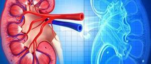
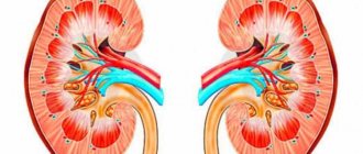
Urine tests characterize the functioning of the kidneys, their ability to filter or retain certain substances. Indirectly, they can be used to judge the state of the cardiovascular, endocrine system, and the patient’s severity.
A separate line in the test form contains such an indicator as urine osmolarity. This is the concentration per liter of various dissolved substances. It should not be confused with specific gravity. Its change is influenced by bacteria, protein, and leukocytes present in the urine. Only absolutely pure urine can be compared in terms of osmolarity and specific gravity.
What is it made up of?
Osmosis is the one-way movement of liquid through a semi-permeable membrane that separates two solutions with different concentrations of substances. Water moves to the side with more dissolved substances.
Osmotically active ions in the human body are sodium and chlorine ions, glucose, proteins, urea, and bicarbonate. They are able to actively attract water. If we talk about the concentration of these substances in 1 liter of urine, then this is osmolarity. When it comes to the amount per 1 kg of liquid, this is osmolality. They are measured in mOsm/l and mOsm/kg, respectively.
The concept of osmotic window is used - this is the difference between the measurement results and theoretical calculations. For the last indicator, it is necessary to determine the concentration of active substances.
What does osmolarity mean?
This concept refers to the movement of water in the direction with a higher concentration of minerals, ensured by the movement of substances with high osmotic activity. These substances include ions of chlorine, sodium, proteins, urea, glucose and bicarbonate. A violation of the osmolarity indicator indicates a malfunction of the endocrine gland, diseases of the cardiovascular system and indicates a decrease in the ability of the kidneys to retain certain substances from water. And since the concentration of sodium ions in the body is controlled by the kidneys through signals coming from the nervous system, a change in this indicator leads to a general decrease in immunity and the body’s resistance to disease. The physical meaning of the concept means the concentration of active components in a liter of urine, expressed in milliosmoles per liter of solution mOsm/l.
Determination methods
For analysis, special devices are used - osmometers. Their work is based on certain laws of physics.
- Steam pressure. To operate the device, a minimum amount of urine is required, measured in microliters. But they also give the biggest error. The action is based on lowering the vapor pressure of the solvent above the solution. More often used in pediatric practice for newborns.
- Increasing the boiling point (the higher the osmolarity, the later the solution boils). Devices of this type are not widespread in medicine; they are used for non-biological fluids.
- Lowering the freezing point (the higher the osmolarity, the longer it takes to freeze). Cryoscopic osmometers are most widely used.
- Membrane osmometers pass the test liquid through an artificial or natural membrane and at this moment performs the necessary measurements. Blood is most often used for analysis.
LEUKOCYTES IN URINE.
Normal: men 0-3 in the field of view, women 0-5 in the field of view.
Pathology options:
5-20 in sight.
Interpretation of the results: pyelonephritis or lower urinary tract infection without severe exacerbation, glomerulonephritis, any nephropathy.
more than 20 in sight.
Interpretation of results: pyelonephritis, cystitis, urethritis, prostatitis in the acute stage.
Active Steinheimer-Malbinn leukocytes.
Interpretation of the results: pyelonephritis, cystitis, urethritis, prostatitis in the acute stage, diagnostic value is not high.
Preparing for the study
The day before the test you need to eat right. Sometimes it is recommended not to drink for 12 hours before donating urine. The results of the analysis may be affected by certain medications (dextrose), or by performing an X-ray examination with a contrast suspension the day before.
It is very important to obtain bacteria-free urine. To do this, women and men wash their genitals. Women are advised to cover their vagina with a cotton swab. A few drops of urine are released into the toilet, then a sterile jar is placed and the rest is collected.
BACTERIURIA
Norm: absent.
Pathology options:
Rods (cocci).
Interpretation of results: Escherichia coli, streptococcus (microscopy with staining required).
Fungi.
Interpretation of results: yeast fungi (use of antibiotics).
Undifferentiated bacteria.
Interpretation of results: microscopy with staining is required.
Decoding indicators
Normal values are considered to be 800-1200 mOsm/l. Changes may be as follows:
- 600-800 mOsm/l – primary renal dysfunction;
- 400-600 mOsm/l – moderate decrease in renal function;
- less than 400 mOsm/l – significant violations.
A hyperosmolar condition can be caused by:
- dehydration;
- congestive heart failure;
- glucosuria;
- renal artery stenosis;
- shock.
Pyelonephritis is a condition in which hypoosmolar urine is observed.
Hypoosmolarity is observed when:
- excess fluid;
- renal failure;
- pyelonephritis;
- necrosis of renal tubules.
Sometimes hypoosmolarity is a symptom of diabetes insipidus.
Osmolarity norms for cerebrospinal fluid, blood, urine and the whole body
The normal values of osmolarity of biological fluids such as blood, or rather its serum (plasma), as well as cerebrospinal fluid (CSF), differ little, which cannot be said about urine, in which the norms for this parameter are 2 to 4 times higher.
Table 1. Normal values of osmolarity of various biological environments of the body
Biological environmentNormal limits
| Blood plasma (serum) | 280 – 300 mOsm/l |
| Cerebrospinal fluid (CSF) | 270 – 290 mOsm/l |
| Urine (urine) | 600 – 1200 mOsm/l |
| IO (osmolarity index) | 2,0 – 3,5 |
| SWR (free water clearance) | (-1.2) – (-3.0) ml/min |
Numerical indicators of blood osmolarity in children, although not as significant, are still different from those in adults (Table 2). OSK (norm) in children begins to change starting from 9 months of age. By the age of one year it reaches 280 – 300 mOsm/l (the norm for an adult), remaining within these limits, regardless of the person’s age, until the end of life.
Table 2. Normal plasma osmolarity in children
Child's ageNormal, mosm/l
| Newborns up to 1 week of life | 275 – 300 |
| Newborns from 1 week to 1 month of life | 276 – 305 |
| Children from 1 month to 1 year of life | 274 – 305 |
| Children from one year and older | 280 – 300 |
It should be noted that the above standards for adults and children may differ from those in other laboratories. In this regard, patients should first of all focus on the limits of normal values indicated in the analysis form of a particular laboratory.
When the analysis is justified
What does a general urine test reveal?
It is indicative to determine osmolarity if the development of renal failure is suspected. The concentration of urea and creatinine changes only when more than 50% of the nephrons are affected.
This is observed on the 3-4th day of decreased urine production (oliguria). This is not indicative for early diagnosis of acute renal failure. Therefore, a urine osmolarity of 400-350 mOsm/L precedes the development of acute kidney failure.
It is recommended to conduct research in arterial hypertension, diabetes mellitus, to evaluate the treatment of hyperosmolar comas, the effectiveness of infusion therapy, and the diagnosis of intracranial hypertension.
What does the analysis show?
How to understand the tests you receive? This is probably possible if you try to follow the guidelines below:
- It is known that changes in blood plasma osmolarity parallel fluctuations in the content of sodium cations in it. Consequently, an increase in Na+ concentration (hypernatremia) and an increase in TSC (more than 290 mOsm/l) will lead to an increase in the activity of the drinking center, the person will not leave the feeling of thirst, and stimulation of vasopressin synthesis will begin to prevent the removal of water resources from the body. An increase in blood plasma osmolarity by 50–60 mOsm/L is a dangerous sign, since in this situation the patient may die from cerebral edema.
- And, conversely, a decrease in Na+ levels (hyponatremia) and a decrease in TSC (below 280 mOsm/l), inhibiting the production of vasopressin, promotes increased release of water from the body through the kidneys.
CYLINDERS.
Normal: Single in the preparation.
Pathology options:
Hyaline (high content).
Interpretation of results: “witnesses” of proteinuria.
Granular (high content).
Interpretation of the results: damage to the kidney parenchyma of any origin.
Waxy (high content).
Interpretation of the results: damage to the kidney parenchyma of any origin.
Epithelial (high content).
Interpretation of the results: damage to the kidney parenchyma of any origin.
Erythrocytes (increased content).
Interpretation of the results: damage to the kidney parenchyma of any origin.
Pigment (high content).
Interpretation of the results: damage to the kidney parenchyma of any origin.
Leukocyte (increased content).
Interpretation of the results: damage to the kidney parenchyma of any origin.
SALT.
Norm: none.
Pathology options:
Uric acid.
Interpretation of results: hypovolemia (diarrhea, vomiting, excessive sweating), severe pneumonia, leukemia when taking cystostatic drugs.
Urats.
Interpretation of results: hypovolemia (diarrhea, vomiting, excessive sweating), severe pneumonia, leukemia when taking cystostatic drugs.
Acid ammonium urate.
Interpretation of results: inflammation of the urinary tract (pyelonephritis, cystitis, etc.).
Calcium phosphate.
Interpretation of results: rheumatism, anemia.
Calcium sulfate.
Interpretation of results: has no diagnostic value, may occur when using sulfur mineral waters.
Hippuric acid.
Interpretation of the results: diabetes mellitus, consumption of lingonberries, blueberries, intake of salicylic and benzoic acids.
Ammonia - magnesium phosphate.
Interpretation of results: intake of plant foods, cystitis.
Magnesium phosphate neutral.
Interpretation of the results: repeated vomiting, frequent gastric lavage.
Amorphous phosphates.
Interpretation of results: no diagnostic value.
Calcium oxalate.
Interpretation of the results: eating large amounts of tomatoes, spinach, sorrel, apples, grapes, oranges.
Calcium carbonate.
Interpretation of results: no diagnostic value.
Cystine.
Interpretation of results: hereditary cystinosis.
Leucine.
Interpretation of results: protein decomposition products, liver diseases, B-12 deficiency anemia, leukemia.
Xanthine.
Interpretation of the results: products of the cleavage of purine bases, promotes stone formation.
Cholesterol.
Typical results: amyloidosis, renal tuberculosis, cystitis.
Bilirubin.
Interpretation of results: hyperbilirubinemia.
Hematoidin.
Interpretation of results: bleeding from the urinary tract.
Hemosiderin.
Interpretation of results: intravascular hemolysis.
Fatty acid.
Interpretation of results: fatty degeneration of organs.
Sulfanilamide crystals.
Interpretation of results: treatment with sulfonamide drugs.
Osmotic concentration and dilution of urine.
Introduction
We are starting to study the section “Physiology of the kidneys and regulation of water-salt metabolism.” Knowledge of this section is necessary to understand the pathophysiological basis of diseases of the kidneys themselves, as well as the pathogenetic mechanisms of various types of disorders of water-electrolyte homeostasis. This is important for clinical practice: competent and effective correction of water and electrolyte metabolism disorders that accompany many diseases and aggravate their course; understanding the mechanism of action of diuretics, treatment of the renal form of hypertension, edematous conditions, etc.
We are aware that it is impossible to give a complete picture of the current state of the problems of renal physiology in a small textbook. Therefore, we will consider only those questions that are necessary for you to master this material as part of the curriculum, and prepare for the study of subsequent clinical disciplines.
Given the importance of this topic for your professional education, prepare yourself to study complex theoretical material. We will assume that you have basic knowledge of the functional anatomy of the kidneys from the anatomy and histology course. You will have to remember them, because in the kidney, like in no other organ, morphology and function are closely interrelated.
When presenting the educational material, we used drawings and diagrams as much as possible; with their help, we tried to make it easier for you to perceive. Therefore, we strongly advise you to: refer to the pictures, carefully read the explanations to them, and they will help you understand the complex material.
| Mechanisms of urine formation |
The idea of the kidney only as an excretory organ in no way corresponds to modern data on the wide range of functions it performs in the body. The kidneys are a multifunctional organ.
They participate in the regulation of the volume of internal fluids, the concentration of individual ions, the total concentration of osmotically active substances, and blood pH. The kidneys provide excretion of the final products of nitrogen metabolism, foreign substances, and excess organic and inorganic substances. The production of physiologically active substances in the kidney (renin, the active form of vitamin D3, erythropoietin) and its metabolic function are important for the body. The main functions of the kidneys are presented in table 1
Table 1
Kidney functions
| Function |
| Blood volume regulation |
| Regulation of blood osmotic concentration |
| Regulation of blood ion composition |
| Regulation of the acid-base state of the blood |
| Blood pressure regulation |
| Removal of end products of nitrogen metabolism |
| Regulation of eryropoiesis |
| Regulation of blood clotting |
| Regulation of calcium metabolism |
| Regulation of protein, lipid, carbohydrate metabolism |
| Production of biologically active substances |
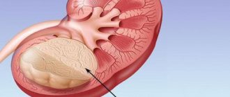
Nephron and its blood supply
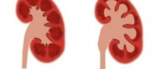
The mammalian kidney is structurally composed of two layers: the outer, cortical, and the underlying medulla, which contains the outer and inner parts.
The structural unit of the kidney is the nephron; there are about 1 million of them in the human kidney (a diagram of one of the nephrons is shown in Fig. 1). Each nephron begins with a double-walled Shumlyansky-Bowman capsule, inside which there is a glomerulus of capillaries - the glomerulus.
Between the walls of the capsule there is a cavity from which the proximal tubule (PT) begins. The part of the nephron next to the proximal tubule is the descending part of the loop of Henle; it ends with a hairpin-shaped elbow and then passes into the ascending part of the loop, located parallel to the descending one; then comes the distal tubule (DC), which returns to the capsule of its nephron and lies between the afferent and efferent arterioles, so that its border with the thick ascending loop of Henle (the area of the macula densa) is close to the afferent arteriole. Next, urine enters the collecting ducts (CT), which in transit pass through all layers of the kidney and are located parallel to the loops of Henle. Strictly speaking, CTs are not part of the nephron, since they have a different embryonic origin, but from a physiological point of view they are considered an integral part of the nephron.
Figure 1 Diagram of the structure of the nephron
Remember: the location of each part of the nephron in the kidney, as well as their relative position, is important for understanding their participation in the process of urine formation.
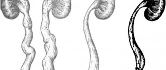
In the kidney of humans and mammals, there are several types of nephrons, differing in the location of the glomeruli: superficial, intracortical (lying inside the cortex) and juxtamedullary (their glomeruli are located at the border of the medulla cortex (Fig. 2). The difference between them lies in topography, loop length Henle and features of the blood supply. Thus, the juxtamedullary nephrons have a long loop of Henle, descending deep into the inner medulla. Due to these features, they will take part in the process of concentrating urine.
Figure 2 Types of nephrons
Blood supply to the kidney
Let us now turn to the blood supply to the kidneys. The blood supply to the kidney plays a special role, since it not only ensures cellular metabolism, but is also directly involved in urine formation.
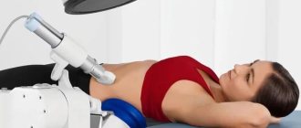
In 1 minute, about 1200 ml of blood passes through the vessels of both kidneys in a person, i.e. about 20-25% of the blood ejected by the heart into the aorta. Since the mass of the kidneys in humans is only 0.43% of body weight, an exceptionally high level of organ blood flow is obvious (Fig. 3). The amount of renal plasma flow and blood flow is determined by the PAG purification method (guidelines for laboratory work).
91-93% of the blood entering the kidney flows through the vessels of the renal cortex, the rest is supplied by the renal medulla. Blood flow in the renal cortex is normally 4-5 ml/g of tissue. An important feature of renal blood flow is a high level of self-regulation - the blood flow remains constant when blood pressure changes more than twice (for example, from 90 to 190 mm Hg).
Figure 3 Comparison of renal and coronary blood flow
The arteries of the kidney arise from the abdominal aorta, which ensures a high level of blood pressure in the afferent arterioles, through which blood enters the glomerulus, which contains a branched capillary network. Blood flows from the glomerulus through the efferent arteriole, which again breaks up into a secondary network of capillaries intertwining the proximal and distal tubules (peritubular capillaries). Further through the veins, the blood leaves the kidney and enters the inferior vena cava. From the glomeruli of the juxtamedullary nephrons, the efferent arteriole delivers blood to the medulla, where the straight vessels (vasa recta) are formed, descending deeply into it along with the loops of Henle and participating in the osmotic concentration of urine. Thus, the blood supply to the kidneys is arranged like two sequential systems of vessels with adjustable resistance.
The main stages of the urine formation process
Urine formation consists of three main processes presented in Fig. 4.
Glomerular or glomerular filtration.
Tubular reabsorption.
Tubular secretion.
Let's move on to their description.
Glomerular filtration
Urine formation in the kidney begins with ultrafiltration of blood plasma in the renal glomeruli. The fluid passes from the lumen of the blood capillaries into the cavity of the glomerular capsule through the glomerular filter.
Figure 4 The main processes responsible for the formation of urine
Let us take a closer look at the structure of this filter and the forces that ensure the filtration process.
Filter membrane . The filtering membrane consists of three layers: the endothelium of the capillaries, the basement membrane and the inner layer of the Shumlyansky-Bowman capsule, which is formed by epithelial cells - podocytes. (Fig.5).
Capillary endothelial cells have very thin peripheral areas; only the area of the cell where the nucleus is located protrudes into the lumen of the vessel. The lateral parts of the cell are pierced with rather large holes, usually covered with thin diaphragms. At normal blood flow speed, large protein molecules form a barrier layer above these pores, which serves as an obstacle to the passage of not only globulins, but also albumins through the pores.
Thus, the fenestrated endothelium of the capillaries limits the passage of formed elements and proteins through the glomerular filter, but freely passes low-molecular substances dissolved in the blood plasma.
The next barrier of the glomerular filter is the basement membrane. Its “pores” restrict the passage of molecules depending on size, shape and charge. Since the membrane has a mesh structure formed by thin filaments, the passage of molecules larger than 3.4 nm is limited. The negatively charged pore wall makes it difficult for molecules with the same charge to pass through. The pores are not round, which is also important for limiting the filtration of albumins.
Figure 5 Structure of the glomerular filter
The last barrier to filtered substances are podocytes. Their processes (“legs”) are adjacent to the basement membrane on the side of the glomerular capsule; between the podocyte legs there are spaces through which the filtered fluid flows. However, even in this case, there is a barrier to the path of filtered substances - slit membranes that block the space between the podocyte stalks. They restrict the passage of albumins and other molecules with high molecular weight. The surface of the legs of neighboring processes is covered with negatively charged sialoglycoproteins, which limit the passage of negatively charged particles. Because podocytes contain actomyosin myofibrils within their processes, they can contract and relax, acting as micropumps that pump filtrate into the capsule cavity.
Such a multilayer filter ensures the preservation of proteins in the blood and the formation of practically protein-free primary urine, which contains the majority of inorganic ions and dissolved low molecular weight organic substances in almost the same concentration as in plasma.
Let's move on to consider the forces that ensure the filtration process. The driving force for filtration is the effective filtration pressure (Pf). It is created by the difference between the hydrostatic blood pressure in the capillaries of the glomerulus (Pg) and the forces opposing it - the oncotic pressure of blood plasma proteins (Pron) and the hydrostatic pressure of the fluid in the glomerular capsule (Pk).
Accordingly, the formula for calculation is as follows:
Рф=Рг-(Ron+Рк).
Let's substitute the numerical values of the pressures and make the calculation:
RF = 70 mm Hg. – (30mmHg+20mmHg)=20mmHg.
Thus, the effective filtration pressure is 20 mmHg. As we have already said, the resulting protein-free filtrate is similar in composition to blood plasma and has the same concentration of osmotically active substances as plasma - 300 mOsm/l. In both human kidneys, 110-130 ml of ultrafiltrate is formed in 1 minute. Thus, each ml of plasma from 600 ml passing through the kidney vessels in 1 minute (the value of the renal plasma flow) loses approximately 1/5 of its volume. The volume of primary urine filtered per minute is usually called the glomerular filtration rate (GFR). The method for determining GFR and renal plasma flow is based on the principle of purification (for a detailed description of the method, see the laboratory manual). Filtration is considered a fairly stable process, but GFR can change under various physiological conditions and pathology. Regulation of renal blood flow and GFR occurs with the participation of sympathetic nerves, the renin-angiotensin system and other factors.
During the day, a huge amount of primary urine is formed - 180 liters, final urine is released only 1.5-2.0 liters. The remaining fluid is reabsorbed in the renal tubules. As a result of reabsorption, most of the water and substances dissolved in it that “failed” through the filter and are valuable to the body are returned back to the blood. The result of the complex work of the tubules, in which, as we will see later, there is a kind of “division of labor,” will be the formation of final urine, the composition and quantity of which will be determined by the water-salt balance of the body. Let us move on to a description of the processes occurring in the tubules.
Mechanisms of tubular reabsorption.
The next two stages of urine formation occur in the kidney tubules - the processes of reabsorption and secretion . Reabsorption is the process of reabsorption of substances from the lumen of the tubules into the blood, while their excretion in the urine decreases. Secretion is a process reverse to reabsorption, as a result of which products to be excreted are transported into the lumen of the tubules; at the same time, their excretion in urine increases. Localization of the most important transport processes is presented in Fig. 6.
We draw your attention to the fact that reabsorption and secretion are based on the processes of membrane transport through the walls of the tubules. They are universal, and in principle the same as those that act during the transfer of substances through other plasma membranes (during absorption in the intestine, transport in capillaries).
Figure 6 Reabsorption and secretion in the renal tubules.
The direction of the arrows indicates reabsorption and secretion
Due to the variety of transport processes, their intensity, specificity, selectivity, the kidneys can be called a unique organ. Let's move on to consider specific mechanisms of reabsorption.
Proximal reabsorption
The ultrafiltrate formed in the glomeruli then enters the proximal tubules. Epithelial cells that form the walls of the proximal tubules, like all cells capable of transporting substances, have an asymmetric structure, that is, they are characterized by the direction of processes from the apical to the basal surface of the cell. The apical membrane of the cell, facing the lumen of the tubule, has a brush border, which increases the absorption surface by almost 40 times and has a high sorption capacity. The basement membrane of cells forms folds, the space between which is called the basal labyrinth. This is where the reabsorbed fluid enters before entering the peritubular capillaries. Cells are connected to each other by so-called tight junctions or tight junctions. Throughout the rest of their length, they are separated by a fairly wide intercellular space - the basolateral labyrinth.
Figure 7 Scheme of the structure of the epithelium of the proximal tubules
Look at Fig. 7. and you will see that for the reabsorption of solutes and water from the lumen of the tubule into the basal labyrinth and further into the blood, there are two paths: number 1 shows the first path - transcellular - through the cell. In this case, the substance on its path must overcome two plasma membranes (apical and basal) and the cytoplasm of the cell. The second path of reabsorption - paracellular, between cells - is shown at number 2. It passes through the zones of tight junctions. With such transport, the mechanisms of diffusion, osmosis, and transfer of a substance together with a solvent can be used.
Consideration of reabsorption in the proximal tubule should begin with the mechanisms of Na reabsorption, since the reabsorption of other substances is directly or indirectly related to Na. The process of sodium reabsorption can be divided into 3 stages: passage through the apical membrane, movement through the cell to the basement membrane, and evacuation from the cell through the basement membrane into the intercellular space.
Let's look at them step by step.
Apical transport . The entry of Na into the cell through the apical membrane is a passive process. It occurs along an electrochemical and concentration gradient. These gradients are created due to the active transport of sodium out of the cell through the basal and basolateral membranes (which we will discuss below). The apical membrane of cells contains Na channels and Na transporters that facilitate passive Na entry. The fact is that the lipid base of the membrane is impermeable to the hydrophilic Na ion even in the presence of a large electrochemical gradient and a negative charge on the inner surface of the cell membrane. In order for Na ions to penetrate the cell membrane, it contains hydrophilic proteins - facilitators (permeases), which form channels through which Na passes. The scheme of Na transport in proximal tubule cells is shown in Fig. 8.
The next group of mechanisms for the apical supply of Na is carried out using secondary active transport. A cotransporter (carrier) can transport Na and some second substance in one direction using the symport mechanism. An example of this type of transport is the combined transfer of Na with glucose and Na with amino acids. Alternatively, a substance, such as H+, can leave the cell in exchange for the Na+ ion, which moves into the cell: this mechanism is called countertransport or antiport. Na transport can be coupled with the transport of bicarbonate and phosphates. The Na that enters the cell does not mix with the total Na of the cell, but moves to the evacuation sites through a special transport channel system without disturbing the cellular internal environment.
Basal transport. Na+ is actively transported through the basal and basolateral membranes against the electrochemical and concentration gradient using Na+–K+ pumps. In this case, the Na+ ion is exchanged for the K+ ion. The main role in the operation of the pumps belongs to the enzyme Na+/K+ - ATPase, which causes the breakdown of the ATP molecule, which provides the energy necessary for reabsorption. This type of transport is called primary active. The fact that Na+ is constantly pumped out of the cell is very important, because due to this, the concentration of Na+ in the cell remains low, which, together with the electrochemical potential, ensures the entry of new portions of sodium into the cell.
Figure 8 Scheme of sodium transport in proximal tubule cells
We looked at how Na+ is reabsorbed through the cell (transcellularly), but some Na+ can pass through tight junctions (paracellularly) along with Cl- ions.
Following the electrolytes, water flows passively along the osmotic gradient from the tubules; it is transported partly through the zones of cellular contacts, partly through the cell through special water channels. Moving, water captures and carries away in its flow substances dissolved in the tubular fluid (mainly Na, Cl and urea). This transport mechanism is called “solvent following” or “solvent transport.”
In the proximal tubule, most of the filtered Na (65-80%) and 80% of water are reabsorbed. A distinctive feature of reabsorption in the proximal tubule is that, after Na and other osmotically active substances, in equivalent quantities , so the fluid in the proximal tubule remains isosmotic to the blood plasma, and its osmotic concentration is 300 mOsm/l.
Glucose reabsorption . We have already mentioned that glucose enters through the apical membrane through the symport system with Na. The movement of glucose is mediated by the transporter and is a secondary active transport, since the energy required to transport glucose across the apical membrane is generated by Na transport pumps. Through the basement membrane, glucose leaves the cell by facilitated diffusion (Figure 9).
Figure 9 Mechanism of glucose reabsorption in the proximal tubule
At a normal blood glucose concentration (3.3-5.5 mmol/l), all filtered glucose is almost completely (100%) reabsorbed by the cells of the proximal tubules and it is absent in the final urine. When the blood glucose level increases from 5 to 10 mmol/ l glucose appears in the urine (glucosuria). In this case, the renal threshold is exceeded - the amount of filtered glucose exceeds the reabsorption capacity of the tubules, the transport systems are maximally saturated, and excess glucose is excreted in the urine. The reabsorption capacity of the tubules is determined by glucose using the clearance method (purification method, description of the method in the laboratory manual).
Like glucose, amino acids are almost completely reabsorbed in the proximal tubules. Reabsorption of amino acids is carried out by secondary active transport, together with Na. However, for amino acids in the apical membrane there is not one transporter, as for glucose, but 5-7 types of transporters specific for different groups of amino acids.
Low molecular weight proteins are reabsorbed in the proximal tubules, which in small quantities (about 1.8 g/day) are filtered and enter the proximal tubules. In fact, all filtered protein is reabsorbed, and its excretion in urine is negligible (up to 100 mg/day). With kidney disease, the amount of protein in the urine can increase to 50.0 g/day. (proteinuria). Protein reabsorption occurs by endocytosis. The protein molecule is adsorbed on the apical membrane, the membrane invaginates, forming vacuoles. These vacuoles split off from the cell membrane and merge in the cell with lysosomes, where, under the action of lysosomal enzymes, the protein is broken down into amino acids. The breakdown products then leave the cell. Bicarbonate, potassium, phosphates, and vitamins are also reabsorbed in the PC.
Na and other reabsorbed substances, passing through the wall of the PC, enter the basal and basolateral labyrinths. The fluid is then evacuated into the blood by the peritubular capillaries, but this mechanism is not entirely clear. There is a hypothesis according to which the oncotic pressure in these capillaries significantly exceeds the oncotic pressure of the blood entering the glomeruli, since during the filtration process the blood loses part of the plasma, and the blood flowing from the glomerulus becomes somewhat thickened. Another hypothesis attaches the main importance to the hydrostatic pressure that arose in the basal labyrinth due to the accumulation of a large volume of fluid in it. Perhaps both hypotheses are true.
Let's summarize: look again at Figure 6 and be surprised at the enormous work that PC cells do. They reabsorb 100% of glucose and amino acids, proteins, 80% Na, 80% water, 80% bicarbonate, vitamins and other substances. But with all the complexity and diversity of transport processes in the proximal tubules, they are a miracle of economy and efficiency.
Remember the features of proximal reabsorption:
1. Large volume (out of 120 ml of liquid filtered in 1 minute, 20 ml remains at the outlet of their PC).
2. Returns biologically valuable organic and mineral substances to the blood.
3. The leading ion in reabsorption is the Na ion, which is associated with the reabsorption of other substances.
4. Reabsorption in the proximal tubules is called isoosmotic, because. water and Na are reabsorbed interconnectedly. As a result, the chemical composition of the tubular fluid changes, but the osmotic concentration does not change (300 mOsm/l).
Figure 10 Interaction between the loop of Henle and the collecting loop
tubes when concentrated urine is formed.
In the process of osmotic concentration of urine, the following are involved: the loop of Henle, the distal tubule, the collecting duct, the vessels and the interstitium of the medulla, which function as a single rotary-countercurrent multiplying system. We have already mentioned that juxtamedullary nephrons with long loops of Henle, which, like the collecting ducts, penetrate deeply into the medulla of the kidney, are involved in the concentration of urine.
Let’s immediately “open the cards” - the process of final concentration of urine occurs in the collecting ducts, and the conditions for this are created by the work of the entire rotary-countercurrent multiplying system. This system creates hyperosmolarity of the medulla and, under the action of antidiuretic hormone (ADH), causes water to move from the collecting duct into the interstitium and then into the blood vessels of the medulla, resulting in concentrated urine. A key issue when performing urine concentration is how the interstitial fluid of the medulla becomes hyperosmolar. Let's try to answer it. Follow our reasoning in Fig. 10: from the proximal tubule, a liquid isosmotic to the blood plasma, with a concentration of 300 mOsm/l, enters the thin descending limb of the loop of Henle and, moving along it, begins to lose water; as a result, its osmotic concentration progressively increases and reaches its maximum at the bend of the loop in the papilla (1400 mOsm/l). Then it turns and flows along the ascending limb in the opposite direction (hence the name turning-countercurrent system), while it is diluted and the osmolarity decreases to 100 mOsm/l. These processes are due to different functional properties of the walls of these sections of the loop: the “flywheel” of this system is the thick ascending portion of the loop, which is completely impermeable to water, but actively reabsorbs Na and Cl. In the apical membrane of the cells of this section, Na transport occurs simultaneously with K ions and two Cl ions using the Na, K, 2Cl cotransporter . Na is actively transported through the basement membrane of cells.
NaCl enters the interstitium and causes the release of water from the descending limb of the loop, the walls of which, on the contrary, are highly permeable to water, but do not allow Na ions to pass through. Thus, the reabsorption of sodium chloride in the ascending part of the loop is “responsible” for the reabsorption of water in the descending part. Let's look at this first with a simple diagram (Fig. 11).
Let's imagine that the loop of Henle is filled with stationary fluid that came from the proximal tubule, then the osmotic concentration in any part of the loop of Henle is 300 mOsm/l. (1). Now let us assume (2) that the active transport system in the ascending limb of the loop reabsorbs sodium chloride into the interstitial space until a maximum gradient (say 200 mOsm/L) is established between the fluid in the ascending limb of the loop of Henle and the fluid in the interstitial space . Remember that the walls of this part of the loop are impermeable to water.
Note that there is now a difference in osmolarity between the fluid in the descending limb (300 mOsm/L) and the interstitial fluid surrounding the tubule (400 mOsm/L). Since the walls of the descending limb of the loop are highly permeable to water, water passively leaves its lumen into the interstitial space, in which there is a higher osmolarity resulting from the reabsorption of sodium chloride in the ascending limb, and the osmolarity of the fluid in the descending limb increases to 400 mOsm/L. (3).
Now let’s complicate the diagram: let the fluid in the loop not stand still, but continuously move, then as we move down the descending limb, more and more water leaves it, the concentration of intratubular fluid increases more and more and, since osmotic equilibrium is established, the concentration of interstitial fluid increases by the same value (200 mOsm/l). A gradient of 200 mOsm/L, the so-called transverse or horizontal gradient, is maintained at each of the “floors” of the medulla.
Figure 11 Scheme of interaction between the descending and ascending limbs in the process of concentrating the tubular fluid.
Thus, between adjacent sections of the descending and ascending limb the difference in osmotic concentration is small, but along the loop and the length of the renal papilla - vertically - this single effect increases, is summed up (multiplied), and as a result a significantly larger difference in osmotic pressure is formed - the so-called cortical -papillary vertical osmotic gradient.
300 mOsm/l bark
600 mOsm/l
900 mOsm/l
1200 mOsm/l
1400 mOsm/l papilla
Now you understand why the system is also called multiplying. Thus, the descending and ascending legs of the loop are in close contact with each other, are located in parallel, influence each other and function as a single coupled mechanism - a counterflow-rotary-multiplying system. Thanks to the work of this system, a corticopapillary osmotic gradient is created in the interstitium of the medulla.
It should be noted that the osmolarity of the interstitium is created not only by sodium chloride ions. About half of the osmolarity is due to the presence of urea . Urea has its own circulation in the kidney, in particular, from the collecting duct it passively diffuses into the interstitium of the medulla and thereby increases its osmolarity.
The straight vessels of the medulla, like the limbs of the loop of Henle, also form a rotary-countercurrent system. They are located parallel to the loops of Henle and undergo the same changes in osmolarity as in the loop. This maintains a longitudinal osmotic gradient in the medulla, preventing it from being washed out.
Let's return to our reasoning.
When starting to study the operation of the concentrating mechanism of the kidney, we set ourselves the task of finding out how a zone of hyperosmolarity is created in the interstitium of the medulla, and we solved it. But we have not yet solved the problem of concentrating urine, which was posed to the kidney: as the tubular fluid moved along the descending limb, its osmolarity increased and in the bend area reached 1400 mOsm/l, just like the osmolarity of the interstitium, but as it moved along the ascending limb it again diluted to 100 mOsm/l. Next, reabsorption of NaCl, water and other substances occurs in the distal tubule and the osmotic concentration again becomes 300 mOsm/l, but the tubular fluid is still isosmotic to the blood, i.e., concentration did not occur in the loop. Where will concentrated urine be formed?
Remember, we first “revealed our cards.” Yes, the decisive circumstance will be that from the distal tubule the liquid enters the collecting duct, where the formation of the final urine and the process of its concentration will occur. It is no coincidence that the collecting ducts are located parallel to the loops of Henle and the vasa recta. They pass through all zones of the kidney in transit and are surrounded throughout by interstitium with osmotic pressure progressively increasing in the direction from the cortex to the papilla. Note in Figure 10 that the interstitial fluid osmolarity at each level is identical to that in the descending limb and CT. In other words, around the ST on each “floor” of the medulla there is a horizontal osmotic gradient of 200 mOsm/l, and vertically there is a powerful corticopapillary osmotic gradient created by the rotary-opposite system of the loop of Henle. Thus, we can say that the loop of Henle “works” on the collecting duct, creating a zone of hyperosmia in the interstitium of the medulla. This will be the force that is capable of drawing water from the collecting duct and concentrating urine. When tubular fluid enters the collecting duct, its osmolarity is at the same level as the osmolarity of the interstitial fluid in this area of the kidney. In the area of the papilla, osmolarity reaches a maximum (in humans it is 1400 mOsm/L), therefore the maximum osmotic concentration of urine in humans can also reach 1400 mOsm/L.
Summarizing the above, we can present the following diagram of the main processes that ensure the osmotic concentration of urine.
The main elementary act in the concentrating system of the kidney is the creation of a transverse (horizontal) osmotic gradient between the ascending and descending limbs of the loop of Henle. As a result of their interaction, and due to the countercurrent movement of tubular fluid and blood, a certain value of the longitudinal (vertical) osmotic gradient is established.
But this is one part of the concentrating mechanism, now let's consider the second - the osmolarity of the final urine will depend on the permeability of the walls of the collecting ducts for water, the main regulator of which is ADH. If there is a lot of ADH, the permeability of the walls of the collecting duct for water increases and water, subject to the osmotic gradient existing in the interstitium of the medulla, is reabsorbed and enters the interstitium and then into the blood. The fluid in the collecting ducts comes into equilibrium with the surrounding hyperosmolar interstitium, and concentrated urine is released. If ADH is low, the walls of the collecting ducts become watertight, hypotonic urine is formed, and diuresis increases.
Remember: The final volume and composition of urine is determined by the function of the CT. Their role in the osmotic concentration and dilution of urine is determined both by the characteristics of their anatomical location in the kidney and by the effect of ADH on the permeability of their walls to water. In other words, what happens to the urine in the collecting duct will determine its final concentration.
Before moving on to the mechanism of action of ADH, we draw your attention to the fact that, unlike the proximal tubule, where Na and water are reabsorbed together, in the distal tubule and collecting duct, water and Na are reabsorbed independently. It is this circumstance that allows the distal nephron to produce both concentrated and dilute urine. Distal reabsorption is called facultative (optional).
Remember:
1. The loop of Henle, distal tubule, collecting duct, vessels and interstitium of the medulla take part in the process of osmotic concentration of urine. Their unification into a single concentrating apparatus of the kidney is due to their mutual arrangement and the commonality of the processes occurring in them.
2. The process of final concentration of urine occurs in the collecting duct due to facultative reabsorption of water.
3. The conditions for it are created by the cortical-papillary osmotic gradient of the medulla, created by the rotary-countercurrent-multiplying system of the loop of Henle.
4. Facultative reabsorption of water in the collecting duct is regulated by ADH.
5. In the distal nephron, sodium and water are reabsorbed independently.
Figure 12 Mechanism of action of ADH on collecting duct cells
Let us summarize the effects of ADH in the diagram:
There is a disease associated with insufficient secretion of ADH. It is called diabetes insipidus and is characterized by the release of