The kidneys are a paired excretory organ in which the processes of filtration, reabsorption and primary excretion of urine occur. Its most important structural element is, of course, the kidney parenchyma - what it is, how it is structured, and what its characteristics are considered normal we will discuss in our review and video in this article.
How does our main excretory organ work?
Structure
Parenchyma is a Greek word that combines the totality of the functioning elements of an internal organ. In other words, this is a part of it that performs specific functions.
This is where urine is formed
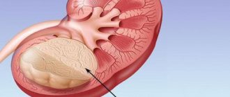
Renal parenchyma is the tissue in which nephrons, the main functional units of the kidneys, are localized.
It consists of two layers, with no clear boundary between them:
- cortical, located closer to the outer shell;
- cerebral, internal.
The main part of nephrons is located in the cortex. There are about 1,000,000 of them in total, but no more than a third of them function in a healthy person.
This is such a complex structure
This is interesting. Considering that the kidneys filter and purify about 1,700 liters of blood every day, you can understand how difficult and stressful their job is.
The structure of nephrons in the kidney parenchyma is quite complex. Each of them consists of a glomerulus and a system of tubules. The glomerulus is located in the outer (cortical) layer, and the descending ends of the tubules descend into the medulla, forming the renal pyramids.
The latter open into small calyces, of which there are from 8 to 10 in each kidney. Then they enlarge, forming 3-4 large cups, and they, in turn, flow into the renal pelvis.
Typical liver changes
By conducting a study, a specialist will be able to identify the main typical changes in the parenchyma:
- change of size;
- increased echogenicity;
- changing the clarity of the contour;
- change in the pattern of blood vessels;
- violation of the homogeneity of the structure;
- focal, local or diffuse changes in liver tissue.
Sometimes even an experienced diagnostician will not give you a completely complete picture of the condition of your organs. Minor changes can hide severe pathology. And sometimes the changes are associated with previous illnesses. For a complete clinical picture, it is necessary to undergo a comprehensive examination, an MRI, and a urine and blood test.
Changes in the parenchyma are quite dangerous and lead to diseases such as cirrhosis, hepatitis, and sclerosing cholangitis.
Functions performed
The structure of the kidney parenchyma described above ensures the following functions:
- excretory (excretory);
- concentration;
- homeostatic (ion-regulating, osmoregulating);
- endocrine;
- metabolic.
First of all, the excretory function of the organ should be noted. A large volume of blood is pumped through the kidneys every minute. Primary urine containing a large amount of liquid is formed in the glomeruli.
Then several liters of primary urine enter the tubules, where the processes of reabsorption (reabsorption) and concentration occur. The resulting secondary urine passes through the pyramids, small and large cups, pelvis and, finally, is excreted from the kidneys through the ureters into the bladder.
The photo shows the process of urine formation
This is interesting. In addition to excreting urine and maintaining a constant internal environment, the kidneys can be called a hormone-producing organ. The fact is that they produce renin, which controls blood volume, as well as erythrooetin, a stimulator of hematopoiesis.
Symptoms
If the filtration and excretory functions of an organ are disrupted, this means that pathology of the organ tissue develops. Signs of diffuse disorders can either develop rapidly or increase gradually. For the most part, the following symptoms may indicate this:
- increased body temperature, especially in acute infectious pathologies of the kidneys;
- change in skin color, it acquires a yellowish tint and becomes dry;
- weakness, fatigue, possible headaches;
- impaired urination and changes in urine volume, which may decrease or increase;
- the presence of cutting and burning sensation when emptying the bladder;
- the formation of edema in the face; in certain cases, the arms and legs may swell;
- increased blood pressure, shortness of breath;
- lower back pain, both acute and aching in nature.
If kidney problems are diagnosed during a laboratory test, the urine is diagnosed by a change in its color; most often it is cloudy and dark, with pus or blood appearing in it. Also, decreased echogenicity may indicate fluid accumulation or tumor formation.
Research methods
The structure and internal structure of the kidney parenchyma is determined quite easily.
To do this, you need to pass one or more instrumental tests:
- Ultrasound;
- computed tomography;
- MRI.
Ultrasound is the easiest way to “look” inside the human body
With the help of these modern diagnostic methods, the parenchyma of the right kidney, as well as the left, is well visualized.
Standard instructions instruct the doctor to evaluate the following parameters:
- anatomical structure;
- internal structure;
- parenchyma thickness;
- density;
- absence/presence of pathological changes.
Ultrasound results in adult women and men
Diagnosis of kidney condition does not differ between people of different genders. The norms of indicators are the same for both men and women. The normal size of a woman's kidneys is different during pregnancy. The norm is considered to be lengthening of the organ up to 2 cm; slight expansion is allowed along with the pelvis and ureters. The norm for adults when deciphering the results is as follows: thickness - 40-50 mm, length 100-120 mm, width 50-60 mm, thickness of the functional part - 15-25 mm. The sizes of the right and left kidneys differ, but not more than 2 cm. The normal ultrasound scan of the kidneys in an adult is determined by the height indicator. Using the table below, you can determine the normal size of the kidneys relative to a person's height.
To maintain normal functioning, the body needs to carry out metabolism. In order for the body to receive everything it needs from the environment, there must be a continuous cycle between the person and the external environment.
During metabolic processes, metabolic products are formed in our body, which must be excreted from the body. This includes urea, carbon dioxide, ammonia, etc.
Substances and excess water are removed, as well as mineral salts, organic substances and toxins that enter the body with food or other routes.
The elimination process occurs through the excretory system, namely the kidneys.
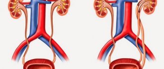
The kidney is a paired parenchymal organ, bean-shaped . The kidneys are located in the abdominal cavity, in the lumbar region, retroperitoneally.
Normal kidney values:
- length 10-12 cm,
- width – 5-6 cm,
- thickness from 3 to 4 cm;
- the weight of one kidney is 150-200 g.
The structure of the kidney also includes the main tissue - parenchyma .
Main indicators of parenchyma: norm and pathology
Thickness
The normal thickness of the kidney parenchyma is 15-25 mm. In elderly patients over 60 years of age, this figure is slightly lower - about 11 mm. If it deviates either upward or downward, it is necessary to promptly establish the cause.
To understand, you need to remember the anatomy
Table: Reasons for deviation from the norm:
| Thickening of the renal parenchyma | Thinning of the kidney parenchyma |
| Acute inflammatory processes (for example, with glomerulonephritis) | Chronic glomerulonephritis |
| Acute nephrotic syndrome | Infectious diseases |
| surge arrester | Chronic inflammatory lesions of the parenchyma |
| Cysts | Neoplasms originating from epithelial tissue |
| Tumors and other neoplasms originating from parenchymal tissue | Hydronephrosis |
| Compensatory (vicarious) kidney hypertrophy |
If we compare all parenchymal dystrophies, the most common is a decrease in the kidney parenchyma. This condition is fraught with a decrease in the number of functioning nephrons and the development of renal failure. The final stage of the pathological process is parenchymal atrophy.
Structure
When a patient undergoes an ultrasound of the kidneys, he may often find in the conclusion an indication that the echostructure of the parenchyma is changed. What does it mean?
Normally, the structure of the organ tissue is homogeneous, without pathological inclusions. With any disease, diffuse or focal changes can be observed.
This is what healthy kidneys look like on an ultrasound
Uniformly heterogeneous renal parenchyma is observed with:
- development of urolithiasis;
- inflammatory, including autoimmune diseases;
- some endocrine diseases - diabetes mellitus, hyperthyroidism;
- atherosclerosis of renal vessels.
- diffuse metabolic changes (a typical example is calcifications in the parenchyma).
Focal
Limited (focal) changes in the parenchyma are observed with:
- benign neoplasms (oncocytoma, angiomyolipoma, adenoma);
- malignant neoplasms (cancer);
- cysts.
A pathological focus can also be caused by a stone in the parenchyma.
The calculus is well visualized
Density
The echogenicity of the parenchyma is its ability to reflect or transmit ultrasonic waves.
Normal on ultrasound:
- the pyramids have low echogenicity (almost black);
- the cortex and columns are isoechoic and identical to each other (gray);
- sinuses are hyperechoic (the lightest).
Most often in medicine, increased density of the kidney parenchyma is encountered.
This may indicate development:
- diabetic nephropathy;
- chronic infectious diseases;
- consequences of arterial hypertension;
- glomerulonephritis;
- amyloidosis;
- sclerotic changes.
If the echogenicity of the renal parenchyma in the fetus is increased, this is a consequence of congenital developmental anomalies. Severe parenchymal fibrosis should alert the physician.
During pregnancy, it is very important to undergo screening ultrasound examinations
In adults, the renal parenchyma is denser more often in chronic diseases, chronic renal failure. Typically, this pathology is diagnosed in elderly patients. As a rule, parenchymal fibrosis is a consequence of the replacement of functioning nephrons with connective tissue.
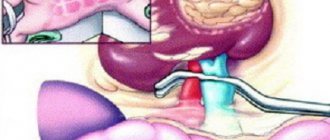
Note! Ultrasound diagnostic doctors are well acquainted with so-called pseudopathologies. For example, sometimes enlarged Bertin's columns extend quite deeply beyond the parenchyma into the renal sinus. It seems that such a parenchymal bridge of the kidney literally divides the organ in two. However, this is not a tumor or pathological tissue, but simply an individual characteristic of the patient.
Parenchyma cells are an important structural element of any organ. Kidneys are no exception. Regular preventive examinations and disease prevention ensure that they remain healthy for a long time.
Preparation
To obtain the most reliable information, you need to pay attention to preparatory activities. Before the procedure, it is important not to eat for 6 hours. For 3 days you should avoid eating foods that cause increased gas formation. It is also not recommended to smoke, suck candy, or chew gum immediately before an ultrasound.
In addition, in order to properly prepare, you should drink at least 1 liter of clean water an hour before the procedure. Filling the bladder will help improve the ultrasound and improve the quality of the examination. During pregnancy, women can have their kidneys diagnosed by ultrasound; this procedure does not have a detrimental effect on the fragile fetus.
For your information, if the transcript indicates increased pneumatosis, then this is considered a sign of increased gas formation. This circumstance is evidence that the preparation for the procedure was poorly carried out. Ultrasound is a fairly informative method for diagnosing the condition of the renal apparatus. It allows you to identify many diseases at their initial stage of manifestation.
Leave a comment 44,663
At the moment, one of the frequently prescribed diagnostic methods that determine the condition of the kidneys is ultrasound examination. The results of a kidney ultrasound will help identify possible organ diseases or pathological manifestations. Using ultrasound, the following parameters are determined: quantity, location, contours, shape and size, structure of parenchymal tissue. It is determined whether there are neoplasms, stones, inflammation and swelling. Renal blood flow is visualized.
Indications for an ultrasound are: impaired urination, the appearance of blood in the urine, pain in the lumbar region, injuries, existing inflammatory processes, poor urinalysis.
Kidney ultrasound allows you to examine the health or progress of the organ’s disease, selecting appropriate therapy based on the data obtained.
Questions for the doctor
About kidney cyst
Hello! I am 36 years old, female, and have not been ill with anything special. Recently I decided to do an ultrasound of all organs for prevention, and during an examination of my kidneys, a cyst on the right measuring 13*17 mm was discovered. What could have provoked its formation? Is it dangerous? How to treat? Thank you.
Good day! A kidney cyst is in most cases a benign tumor formation that occurs quite often. The causes of this disease, unfortunately, have not been sufficiently studied; parenchymal cysts are usually congenital. The process of cyst formation itself is associated with overflow of the renal tubule with fluid.
The choice of treatment tactics in each case is selected by the doctor individually. Since your tumor is relatively small in size, conservative treatment or even just observation may be successful. Be sure to consult a urologist.
Why is atrophy dangerous?
Dad has had kidney problems for a long time; doctors diagnosed urolithiasis. When he was last seen, the doctor said that his condition was worsening, and the ultrasound clearly showed signs of hydronephrosis on the left. I scared that soon the kidney would stop working completely, since almost all of the “working” tissue was compressed. Is there any way to prevent this?
Hello! Hydronephrosis is a very dangerous syndrome that ultimately leads to atrophy of parenchymal tissue and the development of renal failure. In the later stages, it can be treated surgically; be sure to show your dad to a urologist to decide on the need to install a nephrostomy. Be healthy!
Children's sizes
Kidney ultrasound is often performed to diagnose children. It allows you to determine the presence of congenital anomalies. Most often, the procedure is prescribed if there are complaints of pain in the lower back, lower abdomen, after injuries, or problems with urination. An examination of the newborn is carried out to exclude anomalies that are associated with heredity, in case of a difficult pregnancy, or in the serious condition of the child at the time of birth.
What is the normal parenchymal layer on the kidney?
The tissue outer membrane of the kidney is called parenchyma, which consists of the medulla and cortex. The structure of the layer is very thin, consisting of small capsules that are connected by a system of blood vessels. There are more than a million of them in the structure of the right and left kidneys, and they are responsible for the reproduction of urine. Bypassing the sinuses, which are located in the medulla of the parenchyma, the fluid enters the pelvic and calyceal sections of the kidneys.
Moderate structural changes are considered normal and develop as the human body ages. At the age of 16-18 years, the thickness of the upper layer of the kidney is 1.3-1.6 cm. After both sexes reach a certain age (16 years), the thickness of the kidney parenchyma decreases to 1-1.1 cm.
Return to contents
Causes
Quite often, diffuse changes in the renal parenchyma occur as a result of exacerbation of pathological processes in the kidneys. But this is not the only reason for this pathology; other diseases can also provoke negative changes.
The following abnormalities in the body can provoke the disease:
- inflammation of adipose tissue;
- diabetes;
- congenital vascular pathologies;
- hyperthyroidism;
- vascular diseases that increase the echogenicity of the parenchyma;
- formation of calcifications in renal tissue;
- initial stage of development of urolithiasis;
- inflammation of the tubules and parenchyma nodules;
- fat deposits;
- accumulation of cholesterol.
All of the above reasons can provoke the development of such a deviation as renal parenchyma. If a person is diagnosed with at least one of the pathologies listed above, then it is necessary to systematically undergo examinations and be observed by a doctor in order to immediately begin treatment when the first signs indicating the development of the pathology appear.
Note! Signs indicating the disease can be pronounced, or they can be completely invisible. The presence of pathology can only be determined by ultrasound results. In order to identify pathology, it is recommended to undergo annual preventive examinations.
Among other things, the following problems can provoke diffuse changes in the kidneys:
- salt deposits that arise as a result of metabolic disorders, as well as malnutrition and pathologies of certain organs. In this case, dead kidney tissue accumulates in the parenchyma, as a result of which calcium microcrystals accumulate on the surface. To prevent this situation, it is recommended to move as much as possible and avoid systematically consuming protein foods;
- development of tumors, mostly benign tumors. But the danger in this case is that in some cases the tumor can develop into kidney cancer. With this degeneration, the patient exhibits symptoms such as chills, increased blood pressure and tissue swelling;
- thinning of the parenchyma - this problem develops if a person is diagnosed with chronic renal abnormalities. In the human body, pathogenic bacteria develop, which provoke the progression of the inflammatory process. In order to avoid this, you need to carefully monitor your health and start treatment when the first symptoms appear, otherwise the infection can lead to the death of natural filters. If the disease leads to thinning of the parenchyma, then a serious disruption of the functionality of human organs occurs. Delay in such a situation, as well as self-medication, pose a serious threat to both human health and life. With such symptoms, renal failure may develop.
After an ultrasound examination, the doctor will be able to identify the following disorders and abnormalities:
- the presence of fluid in the renal pelvis;
- reduced echogenicity;
- reverse circulation in the renal arteries;
- poor visualization of the renal veins;
- echo signal from the area of the kidney sinuses;
- tumor thrombosis;
- the presence of unclear outlines of the parenchyma.
After this, the doctor will be able to prescribe effective treatment that will help the patient get rid of the disease in a fairly short period of time.
How are pathological processes treated?
Treatment of diseases of the kidney parenchyma without professional help is almost impossible.
Since diffuse changes in the renal parenchyma have different development factors, they should be treated separately. If infection is considered the basis of a pathological deviation in the parenchyma, then its source must first be eliminated. This treatment is carried out with the help of antibiotics, a dietary regimen and increased rest. Drug therapy, as well as specially selected nutrition, will help cope with poor blood flow. When none of the approaches help, doctors resort to surgery to preserve the functionality of one or both kidneys.
Self-medication is dangerous to human health and can lead to surgery.
The surface layer of the kidney plays an important role in the filtration and excretory capacity of the organ. Parenchymal abnormalities develop due to various factors and can significantly undermine human health. At the first unpleasant manifestations in the lumbar region, you should seek advice from a specialist, since self-medication can provoke a deterioration in the condition of the organ.
prourinu.ru
Video on the topic
This video clearly and simply presents the anatomy of the kidney:
It is easy to maintain the normal state of the renal parenchyma. To do this, you need to lead a healthy lifestyle, eat a healthy and balanced diet, and do not abuse table salt, spicy foods, or alcohol. Be attentive to your own health, follow your doctor’s recommendations and do not indulge in self-medication. If any pathologies are detected, carry out timely treatment under the supervision of an experienced specialist.
It happens that you have heard a word and even intuitively understand what it is about, but you cannot clearly formulate your knowledge. It seems to me that “parenchyma” is one of these words.
The uncertainty that arises can be understood, because the term does not mean something specific. Historically, the term “parenchyma” was introduced to distinguish the collection of tissues that fill the organ from its outer shell and internal bridges that extend from this shell. This term describes structures of different origin or functionality that are located in the space between the connective tissue frame of the organ, which is called the stroma. Schematically, the structure of an organ can be represented as follows: the outside of the organ is covered with a membrane of connective tissue, often containing smooth muscle fibers.
From this shell, partitions - trabeculae - extend into the thickness of the organ, through which nerves, lymphatic and blood vessels penetrate inside. The gap between these partitions is filled with the working part of the organ - parenchyma. It is different in different types of organs: the liver parenchyma is glandular tissue, in the spleen it is reticular connective tissue. Parenchyma can have a different structure within the same organ, for example, as a cortical and medulla layer. Organs rich in parenchyma are called parenchymatous.