Renal angiography is a method of studying the renal vessels and arteries using a contrast agent and x-rays. Your doctor uses it to look for an aneurysm, narrowing of blood vessels (stenosis), or blockages. He will see how blood flows to the kidneys.
For the test, a radiologist injects a contrast dye into the artery that sends blood to the kidney. The doctor uses X-ray images to watch the dye as it flows through the kidney's blood vessels.
Renal angiography is a type of X-ray. Its beams produce small amounts of radioactive waves to create images.
A renal angiogram involves fluoroscopy. It's a kind of X-ray cinema.
Types of renal angiography
Renal angiography is divided into 2 types: translumbar and transfemoral aortography.
Carrying out translumbar aortography
The patient takes a prone position on the X-ray table. The puncture site is first numbed. A catheter with a contrast agent is inserted into the lumbar region and the patient must hold his breath before insertion.
Four pictures are taken, the first two while the aorta is being filled with the drug, the third picture at the end of the procedure, and the fourth 6 minutes after completion. To prevent thrombosis, low molecular weight heparin is used, which is injected drip into the aorta.
Carrying out transfemoral aortography
First, the puncture site is anesthetized, then the femoral artery is punctured and a contrast agent is injected at maximum speed. A special probe is inserted into the aorta, through which it is filled with contrast. Two photographs are taken at the beginning and at the end of the drug administration.
Types of procedure
Depending on the physics of the process when examining internal organs, how kidney angiography is performed and how information is processed, the two most common methods are:
- CT angiography of the kidneys using X-rays;
- MRI examination on a tomograph with a radiofrequency scanner.
Additional Information. When applied to vessels, the MR imaging technique is less informative compared to CT angiography. After irradiation with radio frequencies on an MRI scanner, the doctor can only evaluate the “picture” visually. CT technique makes it possible not only to see vascular tissue, but to assess their real physical density, which is very important when
CT angiography of renal vessels. Taking into account the higher price for MRI diagnostic services (renal angiography, the price in Moscow for MRI with contrast exceeds 16 thousand rubles, while CT with contrast costs between 7-9 thousand rubles), CT technology is widely in demand in renal angiography.
When X-ray irradiation is completed, unambiguous information is provided about the location of the renal vessels and their functional activity.
Note! Computer processing of scan results, when angiography of renal vessels is taken, allows for the creation of a three-dimensional model (3D model) of the circulatory system.
What does a 3D model of arteries look like?
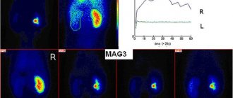
With the development of radioisotope diagnostics, radioisotope renal angiography has been introduced into medical practice, using radionuclide scanning with the introduction of a radioisotope pharmaceutical such as hippuran-131i with extremely low radioactivity.
The radioisotope scanning technique (scintigraphy) is based on studying the distribution of hippuran in the kidneys. One of the scintigraphy methods is a dynamic study of the kidneys with indirect angiography or renoscintigraphy. During indirect dynamic examination, hippuran is administered in a concentration determined by the age of the patient being examined. Indirect dynamic examination makes it possible to identify indications for kidney disease long before the onset of disturbances in their functioning.
Additional Information. The use of radionuclide scanning for indirect dynamic examination is 30-100 times safer than CT tomography. Its price is lower than prices for other methods.
Indications
- Cystic neoplasms.
- Various kidney anomalies.
- Suspicion of malignancy.
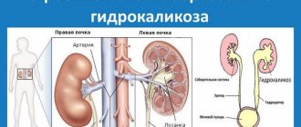
- Hydronephrosis, a condition where the kidneys are filled with fluid.
- The appearance of bloody urine to determine its cause.
- In cases where it is impossible to make a diagnosis using other methods.
- To assess blood flow in the transplanted kidney.
- Atherosclerotic stenosis of the renal arteries.
- Hypertensive chronic kidney disease.
- Fibromuscular dysplasia.
- To exclude diseases not related to the kidneys.
Why is renal artery angiography required?
The study is carried out to determine the condition of the renal arteries, i.e. main arteries that supply blood to the kidneys.
Angiography of the renal arteries may show the presence of:
- aneurysm (expansion of a vein or artery by more than 50% of the proper diameter);
- abnormal connections between an artery and a vein (fistula or fistula);
- thrombi (blood clots) in the lumen of the arteries;
- narrowing of the blood vessels in the kidneys (renal stenosis);
- benign or malignant tumors;
- active bleeding from the kidney.
Renal angiography is also used to study donors and patients before kidney transplant surgery, to determine the number of arteries and veins in each kidney.
Contraindications
- Severe form of renal or liver failure.
- Intolerance to iodine-containing drugs.
- Active form of tuberculosis.
- Impaired blood clotting.
- Heart failure.
- Thyroid diseases (thyrotoxicosis)
- Severe degree of hypertension.
- Presence of a metal implant.
- Exacerbation of a chronic disease.
Relative indications include childhood and old age.
If the patient is pregnant, then manipulation using X-rays can negatively affect the development of the fetus. The procedure is used only if it is an emergency indication.
For a nursing mother at the time of the study, you need to stop feeding and switch to artificial formula for at least two days.
Contraindications for examination
Reasons not to do the examination include:
- pronounced atherosclerosis or tendency to thrombosis;
- cardiovascular, hepatic and renal failure;
It is important! Indications for renal failure should be taken very carefully, since the contrast material must be excreted through these organs, which leads to increased stress.
- open pulmonary tuberculosis;
- allergy to iodine preparations;
- thyrotoxicosis.
Preparation for the procedure
- Do not drink alcohol for two weeks.
- 6 days before the procedure, it is necessary to take a general blood test, a general urinalysis, an ECG and fluorography, a coagulogram-blood clotting test, and a test for hepatitis B, C and HIV.
- Telling your doctor about allergic reactions and the medications you are currently taking, herbs and vitamins are also taken into account.
- Stop taking blood thinning medications, such as aspirin, for a week.
- Do not eat or drink for 6 hours to avoid nausea and vomiting.
- In the morning, do an enema and empty your bladder.
- Before the procedure itself, remove metal accessories.
- If you are pregnant or breastfeeding, make sure your doctor knows.
- Wear loose, comfortable clothing.
Precautionary measures
It is recommended to record how many times the patient has had x-rays taken before. You should show this list to your doctor. The risk of this procedure depends on the dose of radiation exposure previously received.
Possible complications of a renal angiogram:
- bleeding;
- hematoma;
- infection;
- artery damage.
Depending on your health status, other complications may occur. You should definitely consult with your doctor.
Carrying out the procedure
When you arrive for your procedure, you will need to sign a consent form and change into a hospital gown. Your doctor will also ask you to remove any jewelry made from metal. The nurse will monitor your blood pressure and pulse at all times.
You may be given a sedative before the procedure. This sedative will help you relax, but will not make you completely unconscious. Then, local anesthesia will be administered at the puncture site, a soft tube (catheter) will be inserted, and the aorta will be filled with a contrast agent.
When the drug enters the blood vessels, the doctor will take several x-rays, remove the catheter tube and apply a pressure bandage. You should not move while shooting.
In some cases, if during the procedure the doctor discovers a clot or blockage of a vessel, he can perform not only diagnostic, but also therapeutic measures. During the manipulation, you may feel discomfort, but, in general, the procedure is painless. The time required to perform renal angiography is from 30 minutes to two hours.
How the study is performed
The study is carried out in a clinic, in a cath lab, using a low-traumatic, endovascular technique.
On the eve of the study, it is necessary to shave the groin area or the wrist of the right hand (the access area will be determined by the attending physician). The examination will be carried out under local anesthesia by numbing the artery puncture site (groin area or hand). The patient feels a slight numbness of the skin in the area of the puncture site, but remains conscious and can answer the x-ray surgeon’s questions. After anesthesia, a puncture is performed with a special thin needle, and then a catheter (a thin, long, narrow and flexible tube) is passed to the mouth (beginning) of the renal arteries. The x-ray surgeon controls the catheter and monitors its passage through the vessels using x-ray images. To obtain renal artery angiograms, a special substance (contrast) will be used to visualize the renal arteries. The contrast is introduced by the x-ray surgeon through the lumen of the catheter into the artery.
Once images of the renal arteries are obtained, the catheter is removed. Next, manual hemostasis (stopping bleeding) is performed within 10-15 minutes or a hemostatic device (collagen plug) is used, followed by application of an aseptic, pressure bandage. If the study was carried out via a femoral approach, the patient will need to comply with strict bed rest. If the study was carried out through the arm (radial access), bed rest is necessary for 1.5-2 hours.
What happens after renal angiography?
- The nurse will monitor your body for several hours, measuring your pulse and blood pressure, and the injection site.
- You will need to lie on the bed for several hours after the procedure.
- You may be given pain medication to relieve pain or discomfort from the injection site.
- You will be asked to drink water and other liquids to help flush the contrast dye from your body.
- If the procedure was performed in an outpatient clinic rather than a hospital, you will need someone to drive you home. You need to take care of this in advance.
- Driving is prohibited for 24 hours.
- At home, continue to drink more fluids.
- Avoid lifting weights for two weeks.
- Also limit hot baths and showers for a while.
Tell your doctor if the following happens:
- Fever or chills.
- Increased pain, redness, swelling, or bleeding from the injection site.
- Cooling, numbness, tingling, or other changes in the arm or leg.
Complications
The angiography procedure is not 100 percent safe. After it is carried out, a number of complications may arise. However, the risk is justified by the high results that can be obtained during the procedure. With the correct selection of techniques, precise manipulations and high-quality preparation for angiography, the risk of complications can be minimized. Particular attention should be paid to patients with contraindications and elderly patients.
Among the complications after angiography, the most common are:
- bleeding after puncture;
- manifestation of allergic reactions to preparations with iodine;
- occurrence of stroke, heart attack;
- hematomas, bruises and pain at the site of manipulation;
- vascular injury;
- violation of the frequency of contraction of the heart muscles;
- disruptions in kidney function;
- the occurrence of renal failure.
After the procedure, the patient may experience adverse reactions. You need to take them calmly, because they are normal and go away on their own within 2-4 hours after angiography. Such reactions include headaches, a iron taste in the mouth, weakness, dizziness, and mild fever. However, the patient is under strict medical supervision for 5-6 hours after the procedure, in order to monitor and stop more serious complications.
Advantages of diagnostics
Angiography provides important information about the patient's health when all other diagnostic methods have failed.
Thanks to it, it is possible to detect additional vessels in the filtration organs and understand exactly where they are located, diagnose compression of the aorta, determine the type of stenosis and identify the local area of blood supply to the organs.
Thanks to angiography, the doctor will be able to draw conclusions not only about specific changes in the functioning of the system, but also about the state of the filtration organs in general.
After angiography, the urologist will understand to what extent his preliminary conclusions about the presence of a particular disease were correct.
The data obtained during the procedure makes it possible to determine and predict the course of surgical intervention on the filtration organs.
Modern technologies eliminate errors during processing of angiography results, so this study is considered the most accurate.
Indications for use
A highly informative diagnostic method is prescribed in the following cases:
- suspicion of a tumor process in the cortex;
- development of kidney tuberculosis. Angiography allows you to understand how to perform surgical treatment of an organ, taking into account angioarchitectonics;
- high levels of arterial renal pressure, the cause of the pathology is questionable;
- development of renal hematuria with low information content of other diagnostic procedures;
- determination of the nature and degree of nephrogenic hypertension;
- differentiating the tumor process from the development of cystic formations;
- complex structural defects of the bean-shaped organs were identified;
- development of hydronephrosis with fluid overflow of the renal structures;
- detection of tumors in the retroperitoneum and adrenal glands.
Angiography of renal vessels
Renal angiography is a highly informative research procedure aimed at studying the condition of blood vessels using contrast agents and radiography. This technique has been used in clinical medicine for many years. Moreover, with the help of angiography of the renal vessels, specialists make the most accurate diagnosis, while other methods do not give an unambiguous and clear answer.
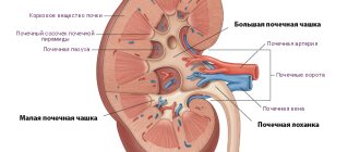
The kidney structures are distinguished by a fairly developed system of blood vessels, so the normal functioning of the organ depends on the functionality of the vascular network. Any disturbance of vascular circulation negatively affects renal functionality. Angiographic examination allows us to assess the functional and anatomical state of the renal vessels.
Progress of the procedure
If we talk about how kidney angiography is done, it will directly depend on which method the urologist has chosen.
There are two types of procedure:
- Translumbar aortography. During this procedure, the doctor performs a puncture of the aorta in the lumbar region of the patient.
- Transfemoral aortography. With it, contrast is supplied to the body through the femoral artery.
The doctor decides how best to conduct the study. At the same time, he necessarily takes into account the individual characteristics of the patient’s body.
Translumbar
In this case, angiography is performed in the following order:
- 20 minutes before the start, the patient is administered Morphine and Omnopon.
- The patient lies on his stomach and places his left hand along the body, and moves his right hand to the side. This is necessary so that his pulse and blood pressure are recorded during the study.
- Injection of a local anesthetic in the lower back. This is where the puncture needle will subsequently be inserted.
- The patient stops breathing for a while and at this moment the specialist pours contrast into the aorta through a puncture.
- Initially, doctors perform a nephrogram and an arteriogram.
- The procedure ends with a venogram.
- The patient rests for 5 minutes, after which an excretory urogram is performed.
- Novocaine (helps to avoid vasospasm) and Heparin (prevents blood thickening) are injected into the body with contrast.
After this procedure, the patient can get out of bed and move around after 24 hours.
Transfemoral
Transfemoral angiography is performed according to the following scheme:
- The patient lies on his back.
- The specialist gives him an injection of Novocaine.
- Contrast is injected into the femoral artery.
- After this, the patient undergoes a nephrogram, arteriogram and venogram. Next, the patient rests for 5 minutes.
- After the specified time has passed, he undergoes an excretory urogram.
After such a study, the patient needs to follow a pastel regime for 3 days.
The advantages of transfemoral angiography over translumbar one include the fact that there is no need for stitches after it.
Conducting a survey
Angiography scans use iodine-containing drugs administered through a catheter to fill blood vessels with contrast. The blood vessels themselves are transparent to X-rays. After filling with contrast, the circulatory system clearly appears on tomograms, allowing processing in 3D projection. Contrast in the kidneys after angiography is removed within 24 hours. For certain indications, the substance may be excreted for more than 3-4 days.
Injecting contrast through a catheter