Symptoms and complications
If everything is clear with the temporary form of pyeloectasia during pregnancy, then another type, occurring against the background of an infectious disease, poses a particular danger in such a difficult period.
The disease can cause difficult childbirth and a problematic postoperative period. If a pregnant woman is diagnosed with kidney stones, then surgery is prescribed immediately.
At the beginning of the disease, no symptoms may be observed at all. Only the bilateral form manifests itself immediately, stagnation of urine occurs, pain in the lumbar region, the outflow of urine almost stops, and the temperature rises.
In the case of pyeloectasia, it is not the disease itself that is dangerous, but the cause of its occurrence. When it is difficult to pass urine, stagnation occurs, which can lead to damage to the kidney tissue and serious pathology of acute renal failure. In addition, against the background of pyelectasis, a lot of inflammatory processes occur in the renal area.
Particular attention should be paid to urine, even without laboratory testing and ultrasound; when urinating, the urine will be cloudy and contain blood.
Complications of the disease include:
- Enlargement of the upper part of the kidney.
- Change in the width of the ureter.
- A sharp increase in pressure in the bladder.
- The process of urine returning back to the pelvis.
- Acute renal failure.
- The occurrence of an inflammatory process.
- Insertion of the ureter into the vagina.
In some severe cases of the disease and complications that arise, experts recommend termination of pregnancy to protect the woman’s life.
Methods of treatment for pregnant women
In case of already severe pyelectasis during pregnancy, it is recommended to place a urethral catheter, which is usually used to protect the child and preserve kidney function. The device is a thin plastic tube with a thickness of 3 to 4 millimeters. Thanks to it, urine can flow freely from the renal pelvis into the bladder.
Treatment should be carried out as early as possible to prevent infection that is dangerous for the child. In some cases, urgent insertion of a catheter is recommended. Possible complications after the procedure include urosepsis, increased inflammation, or severe renal colic.
It is recommended to limit the amount of protein consumed in your diet. Urologists and nephrologists allow eating salt, but in small doses (approximate figures: 2-3 g of sodium per day). For bilateral pyeloectasia, chloride and sodium intake should be reduced to 4-5 grams. Women are advised to take enough vegetables (tomatoes, cucumbers, eggplants, carrots) and fruits (kiwi, bananas, apples, pomegranates). Concentrated juices should be avoided, as they increase the risk of urolithiasis in the expectant mother.
Pyeelectasis of the kidney in a child
Pyeelectasis in the fetus, with the help of modern medicine, is diagnosed at the initial stages of the formation of the organs of the urinary system. In the second trimester, when screening studies are carried out, an ultrasound examination procedure is prescribed for pathologies of fetal development.
Of course, a diagnosis of pyeelectasis in a fetus can be made not only on the basis of ultrasound, since the child’s body is constantly changing, so it is customary to consider a disease only when the enlargement of the renal pelvis exceeds 10 mm, with the norm being 8 mm (it was established at 32 weeks). When conducting medical statistics, it was found that girls get sick almost a third less than boys. I consider the main cause of this pathology to be kidney disease in the mother and genetic heredity.
The main causes of intrauterine damage:
- Viral infection of the mother.
- Damage to the body of a pregnant woman by toxic substances.
- Irradiation.
- Taking medications.
- Severe toxicosis (especially in the last stages).
Pyeelectasis in the fetus also occurs due to perinatal damage to the nervous system, abnormal development of the urinary system, and disjointed formation of organs. It was found that renal pyeloectasia in the fetus is also associated with muscle hypertornus, which causes congestion in the ureter and causes the same process in the kidneys.
Those who have encountered dilation of the renal pelvis at least once in their life know what pyeloectasia is like in an infant. This is a disease that most often accompanies newly born babies or fetuses. The main feature of the disease is its hereditary nature.
The pathology develops on the right or left kidney, that is, it is unilateral. Depending on which kidney is affected, the diagnosis is made by the attending physician. If pathological signs are observed on two organs, then we can conclude that the disease is bilateral. If we turn to statistical data for help, we can conclude that pyeelectasis in an infant is observed more often in boys than in girls. The advantage is significant, because the number of diseases in males exceeds females by three to five times.
Causes of the disease
Parents are concerned about the question of why the disease develops in a newborn, and what are its symptoms? This is worth looking into. The clinical picture of the renal pelvis looks like a container that, using a certain level of pressure, accumulates urine in the kidneys. After this, urine follows into the ureter, then safely reaches the bladder.
Several reasons can contribute to pyelectasis in newborns:
- The location of the pelvis is behind the kidney. Such an atypical location is rare, but still occurs;
- a premature baby, one who was born prematurely, may feel general weakness in the body;
- neurological problems in premature babies;
- various types of inflammatory processes;
- high levels of bladder pressure;
- the reaction is the opposite, that is, the urine does not reach its destination, but returns;
- processes of narrowing of the ureter, clogged holes in it due to the formation of stones, pus, or tumors;
- there is a prolapse of one or two kidneys relative to their normal level of location;
- The baby does not urinate frequently, which causes the bladder to overflow with urine for a long time.
Abnormal development of a child, with pathologies of the urinary system, is the result of a congenital factor, as well as external stimuli on the body of the expectant mother and fetus.
It is worth highlighting the symptoms separately. Infants do not have it. The only characteristic symptom of pyeloectasia in children is inflammatory processes that begin to develop in the bladder due to the accumulation and non-excretion of a normal amount of urine. It is difficult to recognize the disease by external signs. Most often, parents learn about the disease only when serious complications are evident.
Pyeelectasis
A mild degree of pyelectasis requires only systematic observation and does not require drug therapy, since this is a natural disorder that goes away with age.
In severe cases and complications, surgery cannot be avoided; such operations are also performed for bilateral forms of the disease.
Involvement of both organs in the pathological process is observed in 30-35% of newborns with malformations of the urinary organs, and in older children it is considered a rare pathology.
Causes of bilateral damage:
- Chronic fetal hypoxia.
- Premature aging of the placenta (fetoplacental insufficiency).
- Pregnancy aggravated by alcohol and smoking.
- Serious illnesses of a pregnant woman.
- The impact of chemical and physical factors on the fetus.
- Intrauterine growth retardation.
- Malformations of the umbilical cord.
In older children, the causes of bilateral pathology are reduced to damage to the lower urinary tract:
- Bladder tumors.
- Injuries and subsequent cicatricial changes in the urethra.
- Anomalies of the structure of the urethra and bladder.
This pathology is more severe than a disorder only on the right or left. Hydronephrosis and renal failure are increasing rapidly. But, cases are recorded when such a disease is considered a diagnostic finding.
March 20, 2020
Pyeelectasia is a pathological expansion of the renal pelvis of an anatomical nature. Pyeelectasis of the kidney in a child is not a separate disease; this ailment refers only to indirect signs that the baby has impaired urine flow from the pelvis, which is often caused by the development of an infection or the presence of a congenital anomaly.
The disease has a congenital development; symptoms of the pathology are often detected in utero or immediately after birth. The disease is divided into several types:
- Unilateral - if the baby’s right kidney is affected - diagnosis: pyeloectasia of the right kidney. If a neighboring internal organ is also damaged, the baby is diagnosed with pyeloectasia of the left kidney;
- Bilateral - with expansion of both internal organs.
Pyeelectasis of the kidneys in infants, as a rule, is congenital and is associated with disturbances in the development of the body. In this option, the disease of the fetus is determined at the time of gestation, when a pregnant woman undergoes a routine ultrasound scan at 16-20 weeks of pregnancy.
Congenital pathology is often triggered by the negative impact of certain factors on the body of a pregnant woman.
The first 2 forms do not require therapeutic treatment; the pathology, as a rule, disappears on its own over time, when the newborn’s body matures and the urinary system begins to fully perform its main function.
A newborn who exhibits symptoms of pyelectasis should undergo regular ultrasound examinations. In the first year of life, ultrasound is performed every 3 months, in subsequent years - once every six months.
If the pathology begins to progress and a urinary infection joins the underlying disease, then the baby must undergo a urological examination, which includes intravenous urography and cystography.
After receiving all the results of the examination, the root cause of the pathology, its degree, as well as the level of disturbance of the outflow of urine are determined. Accurate identification of important medical fundamentals allows effective treatment to be prescribed.
Reasons for development
Often, pathology can occur even during intrauterine development of the fetus. That is why future and current mothers are interested in what the normal renal pelvis is for a child and what are the reasons for deviations from the mentioned values.
In the material below we will analyze in detail what the normal size of a child’s renal pelvis should be and what affects the change in their parameters.
Pyeelectasia in pregnant women: developmental features
Pyeloectasia of the kidney occurs in 1% of pregnant women, of which 20-30% of cases are caused by diseases of the genitourinary system. In 50%, the pathology also manifests itself in the form of hydronephrosis. Patients are at increased risk of pyelonephritis, renal failure, hypertension, and all-cause mortality. Ultrasound has made it easier to identify urological abnormalities. The degree of dilation of the pyelocaliceal system depends on the stage of pregnancy and the underlying etiology.
Antenatal hydronephrosis has received considerable attention, and ultrasonography has become a major screening tool. Much of the controversy surrounding the use of ultrasound stems from the difficulty in determining which lesions are obstructive and potentially harmful to fetal and maternal development. In general, patients with obstructive uropathy who pose a significant risk of neonatal death due to pulmonary hypoplasia may be considered candidates for prenatal treatment and evaluation.
Possible reasons
Pyeelectasis is quite common and affects pregnant women mainly in the third trimester. Hormonal changes and increased progesterone concentrations weaken the ureters. Dilatation allows urine to flow from the bladder back to the kidneys. However, not all people have hormones that expand the pelvicalyceal system.
If the renal pelvis is enlarged in a pregnant woman, this may indicate the following diseases:
- kidney infections;
- kidney cancer;
- obstruction of the urinary tract by a foreign body;
- congenital malformations of the pyelocaliceal system;
- cystitis;
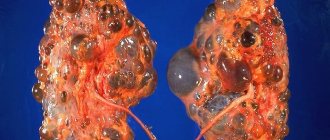
- cystic kidney disease;
- stones in the kidneys.
The baby in the womb grows steadily and takes up more and more available space. Fetal movements can disrupt the flow of urine. As a result, it can flow back into the bladder and then into the renal pelvis. Patients who suffered from bladder infections before pregnancy have a higher chance of developing pyelectasis. Fatigue, fever, and cold-like symptoms occur with pyelonephritis, an inflammation of the renal parenchyma and pelvis.
Other causes of pyeloectasia on the right side during pregnancy are various diseases and complications - stones, benign neoplasms of the bladder, urinary tract, colon or cervical cancer.
Development mechanism
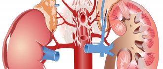
The kidneys are located to the left and right of the spine: in the back of the upper abdomen. They are protected by ribs and a thick fat capsule from external influences. The right kidney is approximately 2-3 cm lower than the left. If pain appears on the left or right, pathological changes occur in only one of the two kidneys. Sometimes back pain originating from nerve roots can be misinterpreted as kidney pain.
During pregnancy, the rapidly growing baby and the uterus require more space, so the two ureters are more or less subject to pressure, which can cause pyelectasis. Significantly enlarged renal calyx, pelvis and urinary tract occur in 3% of pregnant mothers.
The excretory organ must release pollutants and toxins and control electrolyte and fluid balance. However, with pyeloectasia of the right kidney, urinary function is impaired during pregnancy.
Toxic substances and pathogens damage mucous membranes. Patients complain only of nagging pain in the sides and back. If an infection has formed, it goes hand in hand with a fever. Symptoms of pyelectasis usually appear in the late trimester of pregnancy. To prevent this from happening, a woman should control her urination and consult a doctor in a timely manner.
Pyeelectasis can affect both kidneys. The left urinary tract is protected from damage by part of the intestine, but the right one is at significant risk of injury.
List of symptoms
Kidney pain is the first sign of complicated pyelectasis. The doctor detects problems with kidney function during routine blood and urine tests. Even back pain often indicates dilation of the renal pelvis during pregnancy.
Pain occurs on both the left and right side, depending on whether one or both kidneys are affected. During pregnancy, pain occurs when there is very high pressure on the urinary tract. Sometimes megacystis is observed - an increase in the size of the bladder. Normally, with minor pyelectasis, pain rarely occurs. Some patients experience pain and burning when urinating, high body temperature, chills, headache, fatigue or nausea.
Types of pyelectasis
According to the prevalence, pyelectasis is bilateral and unilateral. Both one and two kidneys can hurt at the same time. Women are more likely to suffer from kidney problems than men.
Based on etiology, primary and secondary forms of the disease are distinguished. The exact cause of the pathology cannot be established in all cases.
Risks for mother and fetus
Complications depend on the causative disease that caused pyelectasis. If the mother is diagnosed with urolithiasis, there is a possibility of renal colic and migration of bacteria into the systemic circulation. Sepsis threatens the life of both the pregnant woman and the fetus.
If you experience fever, kidney pain, dizziness or severe headache, you should urgently seek advice from a qualified medical professional to avoid possible complications.
Diagnostics
A method that can detect enlargement of the renal pelvis and the kidney itself, ultrasound, helps to make a diagnosis of renal pyeloectasia during pregnancy. The pregnancy period is associated with many restrictions on the diagnosis of a woman and a pregnant child, and also applies to various methods of irradiation, except for ultrasound. The doctor can only be guided by it and the collection of data on complaints of discomfort in the patient.
Since the disease has 2 ways of occurrence, diagnosis is carried out depending on the pathology.
Pyeloectasia of the fetal kidney is diagnosed during fetal development. Ultrasound shows the presence of pathology at 18-20 weeks of pregnancy.
In boys, deviations from the normal size of the renal pelvis can be detected at the 17th week. Detection of pathology does not mean that the disease is incurable. In most newborns, renal pyelectasis resolves without medical intervention.
After diagnosing pelvic dilatation in the fetus, the expectant mother is registered. A woman should regularly visit a doctor so that the specialist can see the full picture of the development of the pathology.
An enlarged pelvis can be detected before childbirth. This is also not a reason to panic. After birth, the baby’s body copes with the problem on its own without outside intervention.
The newborn is immediately examined by pediatricians with the assistance of a urologist and nephrologist. The child is monitored during the initial period of life, paying special attention to the genitourinary system.
The examination is carried out using ultrasound and urine tests. If the disease does not develop, you should visit a doctor once every six months. Otherwise, the doctor will schedule a visit.
The method for diagnosing pathology resulting from other diseases is the same as for congenital development. The disease is confirmed after studying urine tests and ultrasound data.
Kidney disease and cancer can lead to expansion of the pelvis. An increase in the size of the renal pelvis greater than the norm typical for a certain age indicates the appearance of pathology.
Prevention of pyelonephritis
Pyelonephritis is the process of stagnation of urine in the kidneys during pregnancy. In order to avoid this disease, first of all, you need to follow all the doctor’s instructions, drink a moderate amount of water, and lead a healthy lifestyle. If you are just planning to get pregnant, be sure to undergo a full medical examination, including examination of the genitourinary system, in order to cure all existing diseases, the presence of kidney infections, before pregnancy. Get treatment, and only then deal directly with planning the pregnancy itself.
Treatment
At the initial stage, no special therapy is required. The efforts of the newborn’s body are enough to cope with the problem on its own. Treatment of pyeloctasia in children begins when the disease progresses to a moderate and severe form.
At the second stage of the disease, the doctor prescribes medications appropriate to the age of the small patient. Not only the degree of damage is taken into account, but also the cause of the problem.
If the disease is the result of a kidney infection, appropriate medications are prescribed to eliminate the causative agents of the disease. A baby in this condition needs a special diet.
Severe cases of the disease require surgery. The reason for radical measures is complex unilateral pathologies. Surgical treatment is prescribed during the process of expansion of the pelvis of both kidneys.
The operation is even performed on infants if circumstances require it. The patency of the urinary tract is restored using an endoscope.
The muscle tissue is not cut with a scalpel; small incisions are made through which manipulation is carried out. The process causes minimal damage to the baby’s health.
To choose an effective treatment method, the doctor collects all the data so that he has a complete picture of the course of the disease. It was found that it is almost impossible to predict the development of pathology in a pregnant woman, as well as in a child. Whether the process will progress at birth is unknown. Bilateral renal pyeelectasis in the fetus is considered an absolute norm by specialists, due to the large volume of water in the mother’s body.
After birth, the child is thoroughly examined to determine whether the disease process continues. Statistics have revealed that the vast majority of cases go away on their own and do not require additional treatment. If the disease continues to progress, surgical intervention is possible.
Often, when pyeelectasis occurs, urolithiasis appears. If you have a stone, it is very important to rid your body of it as quickly as possible. For treatment purposes, both traditional methods (medicines) and surgical intervention are prescribed to remove stones from the kidneys.
Infections that enter the body and spread rapidly during congestive processes in the kidneys are treated with antibacterial drugs. All medications are strictly taken as prescribed by your doctor. Do not self-medicate, especially during such an important period as expecting a child.
If there is an accompanying wandering kidney, a nephrologist will recommend that the woman wear a specialized bandage to avoid the kidney getting into other areas.
Treatment of hydronephrosis
During pregnancy, pyeloectasia can occur due to hormonal imbalance. A woman's body experiences a release of hormones that can affect the frequency of contraction of the muscles of the urinary canal. In such cases, treatment is not prescribed, due to the fact that after childbirth the hormonal levels return to normal. If pyeloectasia appears as a result of infectious diseases, then drug therapy is mandatory. The attending physician prescribes additional tests that will help identify the pathogen and prescribe treatment.
In cases where the cause of dilation of the pelvis is a tumor on the internal organs or stones in the urine excretory tract, surgical intervention or, less commonly, termination of pregnancy is possible.
Prevention
Preventive measures to avoid the occurrence of pyelectasis:
- Timely consultation with a doctor, even with minimal discomfort during urination or the appearance of painful sensations. Conducting ongoing analyses.
- Conducting control ultrasound examinations at all stages of pregnancy.
- Avoid hypothermia of the body.
- Keep children away from very cold water.
- Do not hold back the process of urination for a long time. Teach your children to go to the toilet at the first request of the body.
- Periodically carry out sets of therapeutic and preventive exercises, especially if your work involves constant sitting. Such gymnastics will allow you to disperse urine through the ureters and avoid constrictions, kinks and stagnation.
- It is important to relieve all kinds of stress on the kidneys, adhere to a specialized diet, and consume the recommended amount of fluid.
To maintain the health of yourself and your child, you should listen to your body more often, because even with a minor failure, it gives signals of disaster.
Forms of the disease
In medicine, pyeloectasia is classified into several stages: mild, moderate and severe. After the birth of a child, it is impossible to determine the severity of the disease, since it has the ability to progress.
With mild and moderate degrees, the prognosis is favorable, since in this case the pathology disappears on its own. Or the disease will become less pronounced, which is associated with the rapid development of the urinary tract. However, the child should be examined by a doctor periodically. If after some time the disease progresses and kidney function deteriorates, the doctor prescribes surgery. If this is not done, there is a high probability of serious complications, including abnormal development of the renal pelvis.
Classification
Doctors divide the disease into types according to location and method of appearance. Depending on the location of the lesion, the pathology is:
- pyeelectasis of the left kidney (left-sided);
- pyeelectasis of the right kidney (right-sided);
- bilateral pyelectasis (both kidneys are affected).
The name of the disease depends on which anatomical structures are involved in the process:
- dilatation of the pyelocaliceal system;
- pyeloureterectasia;
- pyeloureterectasia;
- calicopyelectasia;
- ureteropyeloectasia.
The most common expansion of the pyelocaliceal system is on the right. This is explained by the physiology of the organ. Infants and adult men suffer from kidney pathology of this type. A severe form of the disease affects the calyces and ureters.
Depending on the cause of its appearance, the disease is divided into congenital and acquired. Each variety is either dynamic or organic.
Classification and treatment
There are degrees of severity of the disease. It is customary to distinguish the following varieties:
- mild – the right or left kidney of the fetus is affected; no special treatment is required; After birth, the child is registered with a nephrologist.
- medium - the renal pelvis is dilated in one of the organs; constant medical supervision is required through ultrasound examination (ultrasound); Drug therapy is prescribed depending on what triggered the disease.
- severe – bilateral pyelectasis. There are disturbances in the functioning of both organs; To prevent progression of the disease, urgent surgical intervention is required.
Despite the fact that the pathology itself is diagnosed during pregnancy, it is not possible to clarify the details and determine the severity of the disease in the fetus - this happens immediately after the birth of the baby.
After confirming the diagnosis, the expectant mother and baby should be observed by a doctor in order to avoid complications. Treatment of mild and moderate forms of renal pyelectasis is carried out through medical supervision and the prescription of medications. The program of therapeutic procedures is drawn up taking into account what exactly caused the pathology. First of all, it is necessary to eliminate the original source. Its elimination makes further treatment more effective. In most cases, pyeelectasis in the fetus is not considered a dangerous disease - the disease is moderate in nature and the chances that it will go away on its own before or soon after birth are very high. To normalize kidney function, the urinary system must form and mature completely.
Surgical measures for severe forms of the disease are carried out only after birth. There may be several reasons:
- the disease continues to progress;
- the performance of the urinary system and, in particular, the kidneys decreases;
- the size of the renal pelvis does not correspond to the age of the child.
Immediately before the operation, the baby is prescribed a course of antibiotics and safe drugs based on natural ingredients in order to prevent the risk of infectious and inflammatory processes. Surgical intervention is aimed at achieving the following tasks:
- identify the presence or absence of deviations;
- evaluate the functioning of the urinary system;
- correct defective areas;
- eliminate vesicoureteral reflux and restore normal outflow of physiological fluid.
Surgical measures in case of severe pyeloectasia should be applied as soon as possible - otherwise there is a risk of organ atrophy.
Recommended topic:
Pyeelectasia of the left kidney
After the operation, the baby requires mandatory observation by a pediatrician, urologist and neonatologist. It is recommended to take a three-month course of uroseptics - they eliminate the causative agents of the described pathological process. To avoid relapse of the disease, it is advisable to have the child’s urine tested every two weeks for six months. Several months after surgical treatment, some unfavorable changes may be observed in the results of examination of the baby’s urine: proteinuria (presence of protein in the urine), microhematuria (presence of leukocytes in urine; detection is possible only through microscopic examination), leukocyturia (pathogenic bacteria in areas of the urinary system; may be accompanied discharge of pus when urinating).
To exclude a possible relapse of the disease, observation by a doctor and strict adherence to the prescribed regimen are prescribed.
It is not recommended to treat pyeloectasia at home; constant visits to a neonatologist are required for a year after the birth of the child.
Reasons and features
The disease is not classified as independent. Pyeelectasis in an infant is a consequence of another pathology. Deviations in the functioning of the urinary system lead to the problem.
A congenital disorder is a consequence of the mother’s illness during gestation or uneven, from a physiological point of view, development of the child.
If pyeelectasis affects the right side, the child is diagnosed with a pathology of the right kidney, and on the opposite side - the left one. Rarely, dilatation of the pelvis of both kidneys occurs.
Bilateral pyelectasis in infants is dangerous because the body does not cope well with urine excretion. If you do not intervene in time, kidney atrophy will occur. Children over 5 years of age rarely suffer from this disease. Different periods of development are characterized by their own norms of renal pyelectasis in a child.
A newborn's kidneys form while in the mother's womb. Pyeelectasis in the perinatal period occurs when there are genetic abnormalities in the mother’s body.
Using an ultrasound, the doctor immediately determines what it is. Pyeelectasis of the left kidney in a child is rarely detected.
| Age | Kidney pelvis size |
| Fetus up to 32 weeks | 4-5 mm |
| 36 weeks - birth | 7-8 mm |
| Newborn - up to 1 month | No more than 7 mm |
| Baby up to 1 year | 6-7 mm |
Developmental disorders at an early stage are not considered dangerous to health. Due to the anatomical features of the body, boys suffer from the disease more often than girls. Other pathologies also lead to expansion of the pelvis.
Harmful external influences on the woman’s body lead to the appearance of problems during intrauterine development.
Stress and infections in the mother during pregnancy lead to illness in the child, which will be congenital in nature.
The acquired problem results from the following disorders:
- problems with urination in a child;
- weakened muscles (especially in premature babies), insufficient development of organs;
- pathology of the transition valve from the pelvis to the ureter;
- abnormal development of the child’s urinary system due to compression by other organs.
Forecast
The vast majority of cases (~96%) return to normal in the second trimester of pregnancy or early postpartum. The risk of postnatal renal pathology is increased by the absence of amniotic fluid or bilateral manifestations. Postpartum anomalies:
- primary vesicoureteral reflux;
- hydronephrosis;
- megaureter;
- doubling of the kidneys.
There is no guarantee that the disease, even with normal postpartum indicators, will not return at a later age. That is why the mother and the therapist must constantly monitor the baby’s health. The disease itself is not life-threatening, but the causes that caused its development are dangerous. If the normal outflow of urine from the kidney is obstructed, this leads to malfunctions of the entire urinary system. Decreased functional capacity can lead to atrophy of the renal structures and be life-threatening.
Video: The renal pelvis is dilated in a newborn. What is the reason?
Preventive actions
Prevention of the development of pyeloectasia in children consists of the expectant mother maintaining a correct lifestyle when planning and during pregnancy. She must monitor her health and take scheduled examinations responsibly. The risk of developing pyelectasis in children during the prenatal period increases with:
- the presence of harmful working conditions at work for a pregnant woman;
- history of spontaneous abortion;
- passive and active smoking;
- diseases of the urinary system;
- suffered from acute respiratory viral infection in the 2nd trimester of pregnancy;
- presence of cytomegalovirus infection.
Loading…
Share with friends!
Characteristics of disease diagnosis
Every person knows that during pregnancy it is impossible to undergo an x-ray examination under any circumstances. During the course of this disease, the main indication for its diagnosis is the results of an ultrasound examination; only thanks to them can one see how much the kidney is enlarged without harm to the child and the expectant mother. The bladder and abdominal cavity are checked. It is important to identify the disease in the early stages, which will help to begin treatment immediately and avoid serious consequences.
Symptoms of drug-induced kidney damage and treatment
Why is pyelectasis dangerous?
Pyeelectasis in a child leads to problems with urine output. This is especially dangerous if both kidneys are pathological in a child. The efforts of diseased organs are not enough to ensure a complete outflow of urine.
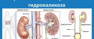
This threatens stagnation in the child’s kidneys. When the size of the pelvis is more than 10 mm, pyeloectasia turns into hydronephrosis.
Untreated renal pyelectasis in a child leads to pathological changes. Kidney diseases (urethrocele, pyelonephritis) and ureteral prolapse appear.
The pathology causes a urinary infection. There may be problems associated with the flow of urine back into the pelvis. This leads to inflammatory processes in the organ.
Main types of pyelectasis
With organic pyelectasis, the functioning of the urinary system is disrupted.
In modern medical practice, there are two main types of the disease:
- organic;
- functional.
In the first pathology, the functioning of the urinary system is disrupted, and in the second situation, the condition of the human body is directly disrupted.
Causes of kaliectonasia during pregnancy
Symptoms and complications
Pyeelectasis manifests itself in infants with the following symptoms:
- irritability;
- cry;
- poor appetite;
- temperature increase;
- vomiting and diarrhea.
- it is possible to detect pus in the urine.
In the initial stage, the disease does not manifest itself. Older children may complain of pain in the lower back and abdomen. In later stages of renal pyelectasis, the baby exhibits external symptoms.
Signs of the disease include swelling of the arms and legs, changes in the urine - cloudiness, the appearance of blood. Painful urination signals this disease.
If you discover any abnormalities in your child’s health, you should immediately consult a doctor.
Complications
In most cases, the prognosis for unilateral pyelectasis is favorable. However, if the disease continues to develop and there are no therapeutic measures, a number of complications may develop:
- hydronephrosis - expansion of the calyces and pelvis of the kidney;
- calicopyelectasia - an increase in the size of the pyelocaliceal system provokes compression of the renal tissues and leads to their atrophy and blockage of the urinary tubules;
- vesicoureteral syndrome - urine from the bladder into the ureter in the opposite direction;
- damage to the valve of the posterior wall of the urethra;
- kidney inflammation – pyelonephritis;
- megaureter – enlargement of the ureters due to frequent spasms;
- cystitis;
- pain when urinating;
- suppuration;
- complete atrophy of the organ.
There are often cases when even timely surgery does not guarantee protection against complications or relapse of the disease.
Prognosis and prevention
The detected pyeloectasia is a fairly serious pathology. Even after it has been successfully cured naturally or surgically, there cannot be an absolute guarantee that the disease will not return after some time. To prevent this, a set of rehabilitation measures is being carried out. The child must be registered with a dispensary and undergo regular examinations.
Doctors give a number of fairly simple recommendations that can be followed by any mother who wants to protect her child from possible kidney problems immediately after birth. First of all, this is the healthy state of the mother’s body itself - there should be no chronic kidney problems, no advanced diseases, no ongoing inflammatory processes. If, however, pyeelectasis in one form or another was detected in the fetus during routine examinations, mothers are advised to drink less liquid so that urine does not accumulate in the pelvis of the unborn child. Immediately after birth, careful medical supervision and detailed diagnostics are necessary.
How to diagnose intrauterine pyelectasis?
Normally, the size of the renal pelvis in the fetus in the womb is 4-5 mm 7 mm in the second . If there is a deviation from the specified dimensions, but not more than 2 mm, no treatment is required. In the future, the size of the kidneys is monitored in the dynamics of fetal growth.
Ultrasound diagnostics determines the size of the urinary organs, and if they exceed the norm and amount to 10 mm or more, then emergency therapy is indicated.
Therapy
Today, doctors cannot say for sure whether the pathology will progress after the baby is born. For example, bilateral pyelectasis is considered a physiological condition and develops due to excessive fluid intake by a woman and child. Therefore, it is important to undergo timely examinations by a doctor who can diagnose the disease and carry out therapy.
Usually, if the pathology does not go away over the years, then conservative therapy is prescribed. What treatment will be depends directly on the cause that led to the development of the pathological condition.
For example, if kidney disease is caused by urolithiasis, then the doctor prescribes medications that will dissolve solid formations and remove sand.
However, conservative therapy cannot always eliminate the disease; in some cases, surgical treatment is used - surgery. The decision about surgery is made by the attending physician after observation and examination. The operation will be indicated when kidney pathology leads to renal failure, dysfunction of the paired organ. In 25-40% of cases, the operation is performed in childhood.
Many methods for treating pathology have been developed. The operation is performed using endoscopic devices. Abdominal surgery is performed very rarely, in the most severe cases. Before the operation, the child must be prepared, preventive measures are carried out, and special therapy is prescribed with the help of medicinal plants.
Unfortunately, it is impossible to prevent the development of pathology. During the period of bearing a baby, a woman is recommended to lead a healthy lifestyle, eat right, and give up bad habits. It is also necessary to monitor your water balance and promptly treat your kidney pathologies.
According to doctors' forecasts, after surgical treatment the disease rarely returns. But such children should be observed by a doctor all the time, so that if the disease returns, it can be quickly cured, even in the initial stages.
Prevention of the pathological condition and prognosis
Most newborns with antenatal pyelectasis have an excellent prognosis. Determining mortality is difficult due to the significant number of stillbirths, terminated pregnancies, and missed diagnoses, all of which lead to underestimation of the true epidemiology. However, most studies indicate that morbidity and mortality are directly related to the underlying etiology of pyelectasis.
Knowledge of the natural history of each disorder that manifests as antenatal hydronephrosis provides a better understanding and assessment of morbidity and mortality than general examinations. Pyeelectasis does not always increase the risk of death in a fetus of either sex.
It is important to know! Obstructive processes that affect both kidneys are more dangerous than unilateral ones. The survival rate for unilateral renal obstruction approaches 100%, with only 15-25% of patients (as shown in the study table) requiring surgery surviving 4 years.
In the presence of a bilateral obstructive process, oligohydramnios is a predictor of an unfavorable outcome. Fetal urine is an important component of amniotic fluid. Maintaining a constant amount of water-salt balance is necessary for normal lung development. If oligohydramnios is present, pulmonary hypoplasia and skeletal compression can significantly affect the quality of life and survival of children.
The earlier the lesion develops, the more likely it is to affect the fetal kidneys, lungs and liver. In a series of 113 cases identified in the third trimester, the mortality rate was 13%. Detection in the second trimester is almost equivalent to death. The most vulnerable period for the development of the pulmonary artery is the sixth month of pregnancy; late oligohydramnios does not have an adverse effect on the formation of the bronchopulmonary system.
Types of pathology, complications
There are 2 types of violations:
- One-sided. In this case, pyeelectasis is observed on the left side of the fetus or on the right side. After birth, the problem mostly goes away on its own.
- Bilateral or physiological. This is a rare type of violation that can lead to negative consequences. If necessary, doctors decide to perform surgery.
If treatment of the pathology is not started in a timely manner, the child’s basic functional abilities of the urinary system organs are impaired in the first days of life, as a result of which the baby is at risk of developing pyelonephritis and other equally dangerous inflammatory complications. Therefore, immediately after birth, strict control is established over the baby, and his condition is regularly monitored, which will help to avoid the occurrence of other dangerous diseases.