Hydronephrosis during pregnancy is considered a cause of complications both during pregnancy and childbirth. Hydronephrosis in the fetus can be a transient disorder or a persistent abnormality leading to impaired renal function. Pathology of the pyelocaliceal system and kidneys is registered in 10% of women during pregnancy and in 20% of fetuses. The reasons for the development of dilation of the pelvis in a woman and the fetus are different, but the outcome is the same - decreased renal function and renal failure.
There are types of hydronephrosis in the fetus and pregnant women:
- Unilateral hydronephrosis on the right or left.
- Bilateral kidney damage.
Causes of hydronephrosis in the fetus
Congenital dilation of the renal pelvis in the fetus, leading to hydronephrosis, develops as a result of the following reasons:
- Fetal hypoxia during pregnancy, caused by a number of provoking factors:
- fetoplacental insufficiency caused by pathology of the blood coagulation system of a pregnant woman (folate cycle mutations, antiphospholipid syndrome, thrombophilia), preeclampsia;
- intrauterine infections in the fetus (cytomegalovirus infection, chlamydia, mycoplasmosis, ureaplasmosis, ARVI);
- premature aging of the placenta, accompanied by impaired blood flow in the vessels of the umbilical cord and fetus;
- anemia in a pregnant woman of moderate and severe severity.
- Malformations of the ureters and urinary organs:
- accessory vessel of the kidney;
- connective tissue dysplasia of the urinary tract, which leads to stretching of the pelvis;
- deviation or bending of the ureter;
- megaureter;
- ureterocele.
- Chromosomal pathology of the fetus. Hydronephrosis is most often recorded in Down syndrome, but the anomaly can also be present in other genetic disorders.
Causes of hydronephrosis such as hypoxia and disruption of the placenta most often initiate a one-sided process. Mutations and chromosomal abnormalities are the cause of congenital bilateral hydronephrosis in the fetus.
Dilatation of the pyelocaliceal system can be detected on ultrasound examination from the 17th week of gestation. If hydronephrosis develops as a result of intrauterine infection, then in addition to dilation of the pelvis, suspensions in the amniotic fluid, thickening of the placenta, oligohydramnios or polyhydramnios are determined.
With hypoxic damage to the fetus, intrauterine growth retardation is observed, as a rule, children are born low birth weight.
In the case of congenital hydronephrosis as a result of genetic pathology, other anomalies of organ development are simultaneously detected (cysts of the choroid plexus of the brain, swelling of the cervical fold, absence of nasal bones, hyperechoic intestine, enlarged bladder, heart defects and others).
Preventive measures
It is impossible to completely prevent the development of hydronephrosis in the fetus. There is only an opportunity to protect the baby’s body from the development of severe pathologies, various deteriorations and serious complications.
To do this, it is enough for the expectant mother to undergo regular examinations and ultrasound diagnostics of the child’s urinary system and her own, on the recommendations of her doctor. It will also be useful to adhere to a special diet, from which foods that increase urine output, vitamin and multivitamin preparations are excluded.
For better urine outflow, fruit drinks made from cranberries and lingonberries and special exercises in the knee-elbow position are recommended.
Early diagnosis of pathology allows not only to preserve the baby, his health and kidney functionality, but also to avoid serious consequences that can lead to a sad outcome. Hydronephrosis is not a reason to panic, but a stimulus to action for the sake of the unborn child.
Photo from the site mypochka.ru
Diagnosis and treatment of congenital hydronephrosis
Immediately after the birth of the child, an ultrasound examination is performed, which notes the size of the kidneys, cups and pelvis, and abnormalities of the ureters. A biochemical blood test is carried out (determining the level of electrolytes, urea, creatinine, glomerular filtration rate), urine tests with determination of protein and leukocytes. If the results are satisfactory, the next examinations are carried out at 3 months, six months, 9 months and a year.
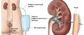
In case of bilateral pathology or with significant expansion of the cavities in the kidney on the right or left, percutaneous nephrostomy is performed and after a few days the functionality of the kidney is assessed. If the function is restored, a wait-and-see approach is chosen.
Depending on the results of repeated studies, surgical treatment is prescribed. It consists of plastic surgery of the ureteropelvic segment. The operation is most often performed laparoscopically.
How is it diagnosed?
The most reliable and suitable diagnostic method for identifying kidney pathologies - radiographic - is strictly prohibited during pregnancy; ultrasound is prescribed to expectant mothers. Since 1970, this instrumental method has been recognized as mandatory for a comprehensive examination of the fetus.
With its help, the doctor can assess:
- kidney size;
- echogenicity of the parenchyma;
- presence and amount of amniotic fluid;
- bladder fullness;
- posterior and anterior size of the pelvis (up to 33 weeks of pregnancy, their size is 4 mm, from 34 - 7 mm);
- the presence of neoplasms in the urinary system.
The information obtained in this way is enough to suspect a pathology or make an accurate diagnosis, and then begin timely treatment and prevent the occurrence of complications. Laboratory diagnostics are more difficult to perform.
In this case, it is necessary to collect urine from the unborn baby. The bladder is punctured, followed by material collection (vesicocentesis). Fetal urine in a normal state is characterized by its hypotonicity, its density is very low and even less than blood plasma.
With any pathological changes, a sharp change in the composition of the substance occurs. It becomes isotonic.
The content of chlorides, calcium and sodium is increased. Although this method is considered highly informative and makes it possible to make an accurate diagnosis, it is used only in the most difficult situations, when the child is in danger of losing an organ.
Another laboratory test is catheterization with contrast. In this case, the instrument (catheter) is inserted directly into the pelvis. Excess liquid is removed, and indigo carmine, a special safe colored liquid, is used instead. It is recognized by an ultrasound machine.
The liquid, like urine, must pass through all channels of the urinary system. In this case, doctors monitor the speed and characteristics of the passage of indigo carmine. The device allows you to identify the location of the problem.
Stagnation of urine is often accompanied by the development of bacteria, and then a complication of the situation. To identify them, a bacterial urine test is performed.
The most accurate and informative method for diagnosing hydronephrosis is ultrasound examination of the fetus, which is prescribed to assess developmental parameters and identify early pathologies:
- Ultrasound diagnostics allows you to assess the size of organs, their correspondence to the stage of development, functional characteristics, the presence of signs of inflammation, neoplasms and foreign elements.
- Ultrasound with a contrast agent is performed extremely rarely, since it requires technical equipment and poses a danger to the fetus. It is required to make a decision on further management of pregnancy and is the basis for surgical intervention.
- Laboratory diagnosis is complex and requires correct interpretation of results. Only a highly qualified specialist using high-quality equipment can take a urine test from the fetus. With the help of biochemical tests, parameters are determined that inevitably change with impaired renal function.
According to the diagnostic data, hydronephrosis of the right or left kidney can be detected, and the most unfavorable is the detection of bilateral pathology.
Consequences and prognosis of hydronephrosis in the fetus
The consequences of congenital renal hydronephrosis are determined depending on the degree of damage to the renal parenchyma, a decrease in their function, and concomitant pathology. Diagnosis of the functionality and other parameters of the child’s urinary system can only be determined after birth.
Compression of kidney tissue by expanded cavities filled with urine leads to atrophic processes in the parenchyma and persistent impairment of nephron function. The most unfavorable consequence of kidney hydronephrosis in the fetus is the development of chronic renal failure. And also against the background of the anomaly, secondary pyelonephritis often develops.
Unilateral hydronephrosis of the kidney during pregnancy on the right or left can be leveled out at the time of birth, as well as immediately after birth, which is often found in boys. Kidney damage on one side in the absence of other serious pathology in a child has a favorable prognosis. Kidney functions are preserved even if 90% of nephrons fail. Doctors choose observational tactics and repeat the study six months after the initial diagnosis. If kidney function is maintained or improved, then observation is continued. If the disease progresses, pelvisplasty is performed.
Bilateral congenital hydronephrosis usually remains after birth and requires careful diagnosis. Kidney damage on both sides has a poor prognosis and often requires surgery immediately after birth.
Hydronephrosis of the kidney in pregnant women - causes of development
The main reasons for the development of dilation of the pyelocaliceal system in a pregnant woman:
- Increase in the size of the uterus.
- Increased production of progesterone and prostacyclin, which leads to a decrease in the tone of the smooth muscles of the ureters and pelvis.
- Weakening of the ligamentous apparatus of the kidneys, which leads to their prolapse and disruption of the outflow of urine.
The topography and anatomy of the pregnant uterus determines changes in the work and functioning of the pelvic organs. The growing fetus puts pressure on the ureters passing behind the uterus, which leads to disruption of the passage of the resulting urine, expansion of the cups and pelvis. Hypotonia and dyskinesia of the ureters lead to the inability to compensate for compression from the outside, which is manifested by hydronephrosis.
The right kidney is most often affected, since it is located lower than the left and is in the area of direct pressure. A bilateral process is rare.
The risk of developing this pathology during pregnancy increases if a woman has urolithiasis and ureteral anomalies.
Symptoms and degrees of hydronephrosis in pregnant women
Symptoms of the disease in a pregnant woman often develop when an infection or exacerbation of urolithiasis occurs. In the vast majority of cases, the pathology is asymptomatic, which is where its danger lies. Unilateral lesions are often asymptomatic.
The first degree of hydronephrosis can be manifested by the following symptoms:
- periodic increase in temperature to low numbers;
- aching pain in the back.
A woman does not pay attention to such symptoms or associates them with pregnancy. Kidney function is practically not impaired, the organ is slightly enlarged. The prognosis for grade 1 is favorable.
The second degree is manifested by the following symptoms:
- renal colic;
- periodic dull and aching pain;
- increase in body temperature to subfebrile levels;
- periodic appearance of visible blood in the urine;
- blood pressure surges;
- deterioration of health associated with increasing anemia;
- swelling of the upper half of the body.
The kidney is enlarged by 20% of normal, its function is reduced by 30-40%.
The second degree of unilateral hydronephrosis also has a favorable prognosis, and with bilateral damage, the risk of developing gestosis, eclampsia and renal failure increases.
The third degree is considered the last stage of the disease. Symptoms of stage 3 hydronephrosis in pregnant women:
- constant high blood pressure and hypertensive crises;
- dizziness, sparkles in the eyes;
- pain in the lumbar region;
- swelling of the face, arms and legs;
- nausea and vomiting;
- itchy skin.
Kidney function is lost by 60-70%, its size is increased by 50%.
Unilateral grade 3 hydronephrosis with timely treatment and normalization of urine outflow has a favorable prognosis. Lack of help or a two-way process leads to end-stage renal failure. This consequence is rare.
Classification of the disease
There are 3 stages of hydronephrosis:
- Mild stage - slight stretching of the renal pelvis occurs under urine pressure. There are no malfunctions in the functioning of the organ.
- Middle stage - fluid pressure on the organ increases, leading to its enlargement and thinning of the walls of the renal pelvis. Kidney performance is significantly reduced.
- Severe stage - the size of the kidneys increases 1.5-2 times, a sharp expansion of the renal pelvis and calyces occurs. A significant increase in the size of the organ will lead to partial or complete disruption of their functioning, which can lead to the death of the fetus.
Why is hydronephrosis dangerous during pregnancy?
The danger of hydronephrosis for a pregnant woman is as follows:
- Development of gestational pyelonephritis.
- The likelihood of gestosis.
- Chronic renal failure.
- Eclampsia.
Preeclampsia and eclampsia are considered life-threatening conditions for women and children. In addition, with kidney failure, metabolic products accumulate. Azotemia negatively affects the development of the fetus.
Life-threatening situations rarely develop if a pregnant woman is registered with a antenatal clinic and under the supervision of doctors. Monitoring urine analysis and blood pressure allows you to identify kidney pathology at an early stage and take appropriate measures.
After childbirth, in almost all women, organ function is completely restored. Only with stage 3 disease do 20% of women experience a decrease in kidney function.