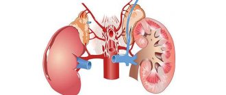
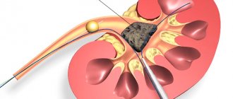
Kidney hypoplasia is a congenital structural disorder when the organ’s cellular structure is considered normal, but its size is far from normal. Apart from its inappropriate size, a small kidney is no different from a normal organ and can even function within its small dimensions.
The hypoplastic organ has layers standard for renal tissue and a narrow, thin-walled vessel.
Almost 50% of children with identified renal hypoplasia also have other abnormalities :
- doubling of a healthy kidney,
- bladder eversion,
- abnormal position of the urinary canal,
- narrowing of the kidney artery,
- cryptorchidism.
What are the causes of renal hypoplasia?
Kidney hypoplasia in children and adults develops for many reasons. They are usually divided into exogenous and endogenous (internal). External factors affecting kidney size:
- uncontrolled use of aggressive drugs;
- radiation exposure;
- smoking during pregnancy;
- alcohol abuse;
- infectious diseases;
- compression of the uterus during toxicosis.
Internal causes of reduced organs of the excretory system:
- violation of cell division during the intrauterine formation of organs of the genitourinary system;
- “sweet disease” - diabetes mellitus;
- liver diseases, which are fraught with intoxication of the body during pregnancy;
- pyelonephritis;
- secondary inflammation;
- low water;
Poor ecology affects the reduction in kidney size. - renal vein thrombosis;
- intrauterine development disorder.
The following can cause a kidney to shrink in size:
- bad ecology;
- bad habits, including poor nutrition;
- abdominal injury;
- taking medications.
How is hypoplasia diagnosed?
The examination reveals a kidney whose size is smaller than required, the number of calyces is no more than six, and the pelvis has a changed structure.
At the same time, the ureter may be of normal size or will also be reduced. In addition, it may
there will be difficulty in the flow of urine, expansion in the ureters, and the artery of the organ will definitely be underdeveloped.
The histological structure of the affected organ, provided there are no other complications, corresponds to the age norm. With damage on one side, deviations in the development of a normal kidney can be detected, such as its duplication, dysplasia, etc.
Underdevelopment of the kidney in an infant should be differentiated from secondary processes of organ pathology observed due to chronic inflammation and disorders :
- pyelonephritis,
- jade,
- renal artery stenosis,
- renal failure.
During the disease, the calyxes are not affected, but only their number and size decrease, and upon examination, overdevelopment in the second kidney is visible.
The disease is detected by:
- Ultrasound;
- Angiography – examination using the injection of a contrast agent into a large vessel;
- Urography - x-ray of the kidneys with a contrast agent;
- Ureteropyelography - studies by introducing a contrast agent through a catheter into the ureter;
- Nephroscintigraphy - examination of the functioning of an organ when searching for radioactive material;
- MRI combined with nephroscintigraphy;
- Radioisotope examination - examination by intravenous administration of a radioactive composition.
You will learn more about what angiography is and how it is performed in the video:
Clinically, with this defect, the condition of a normal kidney is of great importance, since its disease or injury can provoke renal failure.
Discriminating diagnosis of lesions of this type is carried out with a dwarf and wrinkled kidney . A biopsy in this case is not effective.
Symptoms of a reduced organ of the excretory system
Simple hypoplasia proceeds unnoticed, the organ is reduced on one side, all the main functions are taken over by the second kidney, which has the usual dimensions. Symptoms will begin to appear if one of the pair of organs weakens, this will provoke the development of pyelonephritis of the other. In this case, adults and children will experience high blood pressure. If the disease develops into chronic hypoplasia, the underdeveloped kidney is removed, since drug treatment will be ineffective.
Kidney hypoplasia is manifested by the following symptoms:
- retardation in physical development;
- dry mouth, constant thirst;
- increased blood pressure;
- swelling of the limbs;
- random loss of urine at night;
- specific smell of urine, color change;
- girdle pain in the back;
- pain when urinating;
- pallor and dry skin.
If you ignore the symptoms, the disease will later develop into a chronic form with subsequent complications.
Bilateral hypoplasia leads to the death of the child.
When bilateral hypoplasia is diagnosed, the prognosis is disappointing. During the first year of life, the child dies. Hypoplasia of the left kidney is more pronounced, this is due to structural features. The amygdala is located higher, large veins and many nerve clusters are attached to it. The reduced cavity on the left is accompanied by aching pain that radiates to the lower part of the body. If the disorder is not diagnosed in time, there is a high risk of “capture” of the second kidney.
Symptoms
Considering that the kidneys are a paired organ, in the presence of a unilateral lesion, a healthy kidney can compensate for the work of the underdeveloped organ. In this case, pronounced symptoms may not be observed, but at the same time, it must be taken into account that a person who has only one functioning kidney must be extremely careful, since the development of any infectious disease affecting a healthy kidney can have extremely adverse consequences.
At the same time, it is necessary to emphasize that a kidney reduced in size is predisposed to the development of pyelonephritis, that is, an inflammatory process due to the imperfection of its structures, which contributes to damage to organ tissues by pathogenic microflora. In this case, the person experiences a number of symptoms characteristic of pyelonephritis, including;
- chills;
- increased body temperature;
- severe lower back pain;
- urinary disorders.
In people with hypoplasia, pyelonephritis of a reduced kidney is usually common. If a person adheres to certain preventive measures, the number of cases of the development of an inflammatory process in the tissues of a reduced kidney can be minimized.
In the presence of frequent pyelonephritis in a reduced-sized kidney, healthy functional cells of the organ gradually die and are replaced by connective tissue, which leads to gradual shrinkage of the kidney and loss of its function. Among other things, with such an unfavorable course of the disease, gradual compression of the arterial artery is observed, which ultimately leads to a persistent increase in blood pressure.
If hypoplasia is observed in both kidneys, or if there is one kidney that is reduced in size and the second one has additional pathology, signs of damage to this paired organ will be noticeable from the very birth of the child. As a rule, with such an unfavorable option, the symptoms can be very intense. Manifestations of such an unfavorable course of hypoplasia include: a child’s retardation in mental and physical development;
- chronic diarrhea;
- swelling of the face and body;
- pale skin;
- low-grade fever;
- chronic renal failure;
- softening of bone tissue;
- parietal tubercles of the skull;
- protruding frontal tubercles of the skull;
- bowed legs;
- baldness;
- bloating;
- arterial hypertension;
- nausea and vomiting.
If there is bilateral hypoplasia in a child under the age of 1 year, the prognosis for the child’s life is usually unfavorable, since transplantation and drug treatment at this age are not possible. In this case, the symptomatic manifestations of renal failure increase quite rapidly, which leads to the rapid death of the child.
Pathological changes in a newborn
Hypoplasia of the paired organ in newborns is being diagnosed more and more often. Of all diseases related to the urinary system, this disorder accounts for 30%. After the birth of the child, renal dysfunction is detected using an ultrasound examination. If there are no difficulties with urine excretion, then this makes it difficult to suspect something is wrong. Hypoplasia of the right kidney in a child may not appear at all, this means that the neighboring organ performs a replacement function and copes with the load. If both kidneys are enlarged, then blood and urine are taken for analysis. A hypoplastic kidney is manifested by the following symptoms:
- frequent vomiting in a child;
- developmental arrest, proper weight gain is not observed;
- protective reflexes appear;
- elevated temperature;
- gastrointestinal disorder;
- rickets;
- sunken fontanel;
- soft bones.
Causes of the defect
In the fetus
Pathology begins at the moment when the child is still in the womb. The reasons causing this disorder are the improper development of cells in the body of the unborn child. The developmental defect is caused by external and internal factors.
External causes include processes such as a small amount of amniotic fluid, as well as deviations from the norm in the position of the fetus. These main reasons are supplemented by external pressure on the uterus (for example, tight clothing), bad habits of a pregnant woman, and exposure to radiation.
In addition, the use of potent drugs and previous infectious diseases are harmful.
Kidney hypoplasia in a child can be caused by a number of internal reasons:
- diseases of the mother's uterus;
- effect of fetal underdevelopment;
- pyelonephritis in the womb;
- renal vein thrombosis in a child;
- incorrect position of the embryo.
Nephrologists believe that most often the kidney can shrink due to internal pathologies.
Advanced diagnostic methods
In order to make a diagnosis, they resort to the method of differential diagnosis. Thus, other diseases of the urinary system are eliminated. Hypoplasia will be confirmed by the fact that the organ is reduced in size, but this does not affect its structure and integrity in any way. To identify pathology, use:
- Ultrasound. One of the most accessible and informative ways to detect changes at any age.
- MRI. It has maximum effectiveness, but the difficulty lies in the specifics of the diagnosis, so it is prescribed to children in extreme cases.
- Angiography, urography.
- Scintigraphy.
- Biopsy.
- Urine and blood tests for elevated white blood cells and protein.
Clinical picture and diagnosis.
With kidney dystopia, the clinical picture is due to the abnormal location of the organ. The leading symptom is pain that occurs when changing body position, physical stress, and flatulence. With cross dystopia, pain is usually localized in the iliac region and radiates to the groin area of the opposite side. Since damage to a dystopic kidney by a pathological process (hydronephrotic transformation, calculosis, pyelonephritis) occurs much more often compared to a normal kidney, the symptoms of these diseases are often associated. Intrathoracic dystopia can simulate a mediastinal tumor by clinical manifestations and radiographic findings.
With lumbar and iliac dystopia, the kidney is palpated in the form of a slightly painful, sedentary formation. Dystopia is usually detected with excretory urography, and in the case of a sharp decrease in kidney function - with retrograde pyelography. Characteristic signs of dystopia are noted: rotation and unusual localization of the kidney with limited mobility. Difficulties often arise in the differential diagnosis of lumbar, ileal dystopia and nephroptosis, especially in cases of so-called fixed nephroptosis, which, like a dystopic kidney, is characterized by low localization and low displacement of the kidney. However, on urograms with fixed nephroptosis, one can note the medial location of the pelvis and a long tortuous ureter. Sometimes only renal angiography helps to distinguish this condition, revealing a short vascular pedicle in dystopia and an elongated one in nephroptosis. Treatment. The attitude towards kidney dystopia is as conservative as possible. The operation is usually performed for dystopia complicated by hydronephrosis or calculosis. In cases of death of a dystopic kidney, nephrectomy is performed. Surgical relocation of the kidney is extremely difficult due to the scattered type of blood supply and the small caliber of the vessels.
Horseshoe kidney
Renal fusion accounts for about 13% of all renal anomalies. There are symmetrical and asymmetrical forms of fusion. The former include horseshoe- and biscuit-shaped buds, the latter - S-, L- and I-shaped buds.
With a horseshoe-shaped developmental anomaly, the kidneys grow together at the ends of the same name, the renal parenchyma has the shape of a horseshoe. The horseshoe kidney is located lower than usual, the pelvis of the fused kidneys is directed anteriorly or laterally. The blood supply is usually carried out by multiple arteries arising from the abdominal aorta or its branches.
In 98% of cases, the kidneys are fused at their lower ends. At the junction of the kidneys there is an isthmus, represented by connective tissue or full-fledged renal parenchyma, which often has a separate blood supply. The isthmus is located anterior to the abdominal aorta and inferior vena cava, but may be located between or behind them.
The anomaly occurs in newborns with a frequency of 1 in 400 to 1 in 500, with boys 2.5 times more likely than girls.
A horseshoe kidney is often combined with other anomalies and malformations. Dystopic location, poor mobility, abnormal discharge of the ureters and other factors contribute to the fact that the horseshoe kidney is easily exposed to traumatic influences.
Clinical picture and diagnosis
Most often, this malformation is manifested by abdominal pain, aggravated by straightening the torso, which is associated with compression of the vessels and aortic plexus by the isthmus of the kidney. Often, when urine passage is disrupted, a urinary infection is detected.
A horseshoe kidney can be identified by deep palpation of the abdomen in the form of a dense, sedentary formation.
The most reliable diagnostic method is ultrasound with Doppler ultrasound, which allows to detect the presence of an isthmus.
Radiologically, with good bowel preparation, the kidney has the appearance of a horseshoe, with its convexity facing downwards.
On excretory urograms, a horseshoe kidney is characterized by rotation of the pyelocaliceal system and a change in the angle formed by the longitudinal axes of the fused kidneys. If normally this angle is open downward, then with a horseshoe-shaped kidney it is open upward. The shadows of the ureters outline a “flower vase”: moving away from the pelvis, the ureters diverge to the sides, then gradually come closer together on the way to the bladder. The contours of the kidney are most clearly revealed during angiography in the nephrogram phase.
Treatment of hypoplasia using available methods
A small kidney is treated with different methods, it all depends on the patient’s condition and the degree of impairment of the filtration mechanism. For this use:
- Hemodialysis. This is a procedure for cleansing the blood of accumulated toxins that the kidney could not remove. Used in extreme cases where an organ has been reduced to its limit and removed. Thus, in order to maintain the patient’s vital functions, this procedure is carried out until the time of transplantation.
- Surgical intervention. The operation is performed if the organ has ceased to function normally, and pyelonephritis has developed against this background.
Surgical treatment is prescribed in cases where the organ has stopped functioning.
or chronic form of hypotension. If asymmetry of both kidneys is observed, then transplantation is indicated. - Drug treatment. A conservative method of therapy is indicated if only one cavity is reduced, but it can cope with the load. Therapy consists of prescribing a diet; the patient must lead a healthy lifestyle. Pregnancy in this case is an aggravating factor; maintenance or replacement therapy is prescribed. When the first disturbances appear, antibiotics, diuretics, antispasmodics and anti-inflammatory drugs are used.
Complications with hypoplasia
In some cases, if there is no treatment and a combination of other factors, the disease may become complicated. Characteristic signs of complications are swelling that appears in the morning, a change in skin tone to a paler one, a decrease in the amount of urine produced and general intoxication of the body. All this indicates the development of renal failure, which threatens the body with irreversible changes in many of its organs.
Due to the fact that an organ affected by hypoplasia is much more susceptible to various types of infections, inflammatory processes often begin. The infection can enter the kidney either through the blood (from other inflamed organs) or ascending through the urethra and bladder. All this increases the chances of developing pyelonephritis, timely treatment of which is necessary to avoid renal failure.
How to prevent the development of the disease?
If one kidney is smaller than the other, then it is not possible to get rid of this disease, but it is quite possible to avoid the development of complications and live a full life. To do this, it is enough to reduce the salt content in the diet and prevent dehydration of the body, since water is the key to proper metabolic processes. It is important to avoid hypothermia and injury to the area where the organ is located. If you feel worse, you should immediately inform your doctor. Various types of violations can be eliminated with timely treatment.
Prevention of kidney diseases
Modern medicine does not yet know how to grow a kidney to normal size, so the disease is considered incurable. However, this developmental anomaly does not always lead to serious consequences, leaving an adult the opportunity to live a full life without problems. It is enough to maintain a healthy lifestyle, ensure normal water metabolism in the body, as well as facilitated salt metabolism, and avoid hypothermia (especially in the lumbar region). At the slightest symptoms of the disease, you should immediately consult a doctor and avoid traditional methods of treatment.
Small buds
Leave a comment 3,138
One of the congenital defects of the excretory system is kidney hypoplasia. The pathology is characterized by reduced size of the right or left bean-shaped organ. At the same time, functionality is preserved, but against the background of 2 times less number of nephrons (specific kidney cells), the work of the diseased organ weakens. Dystopia may be observed, that is, a displacement of the location of the hypoplastic capsule. In most cases, the problem of excretory structures of different sizes is detected accidentally in adulthood and does not require treatment. It is more often diagnosed among the male half of the population.
Species diversity
Renal hypoplasia refers to congenital anomalies in the development of one of the paired excretory organs with normal cellular structure, but with weakened functionality and abnormally small sizes. The hypoplastic capsule of the kidney differs from the normal size of the organ, but at the same time it performs the job at the proper level, has a structure characteristic of renal tissue, and narrow thin-walled vessels.
The following types of hypoplasia of a paired organ are distinguished, requiring different approaches to treatment:
- Simple, when the left or right kidney is of different sizes. An underdeveloped structure is characterized by an insufficient number of nephrons and calyces. Underdeveloped formation of the body with oligonephronia, when there is a decrease in both kidneys with a small number of tubules and glomeruli. In this case, excessive formation of epithelium is noted. Hypoplasia with dysplasia, when organs are of different sizes and malformations of the tissue structure are noted, that is, cellular replacement.
In newborns with bilateral asymmetry, the presence of other developmental anomalies is noted, such as:
- doubling of a normal-sized kidney; eversion of the bladder bursa; shifted position of the ureter; narrowing of the renal artery; cryptorchidism in men (undescended testicle into the scrotum).
Return to contents
Diagnosis of hypoplastic kidney conditions
The main method for diagnosing hypoplastic conditions is ultrasound diagnostics (ultrasound). The scan reveals a decrease in the thickness and volume of the kidney.
To identify abnormalities in the structure of the urinary tract, excretory urography is performed - an x-ray diagnostic method. It involves examining the condition of the urinary tract after introducing a contrast agent, urografin, into the blood.
Excretory urography – wrinkled kidney
Immediately after entering the body, the contrast is excreted by the kidneys, which allows it to envelop the pelvis, ureter, bladder and urethra. X-rays are taken at the 7th, 15th and 22nd minutes. This time allows you to track the excretory function of the kidneys and identify abnormalities in the development of the organs of the urinary system.
Some urologists prescribe magnetic resonance imaging to quickly diagnose hypoplasia. The study allows not only to estimate the size, but also to obtain a 3D picture (three-dimensional image) of the organ. MRI is an expensive procedure and is therefore available only to a narrow range of medical institutions.
When diagnosing a primary contracted kidney in a newborn, only magnetic resonance scanning can detect the disease at an early stage.
Diagnosis of disorders at different stages
Differential diagnosis studies the child’s kidney as a whole, considering the primary task of searching for pathology of the organ structure - dysplasia and other serious abnormalities that impede the functioning of the kidneys. Hypoplasia is characterized by the preservation of normal anatomy and physiology, just in a reduced volume, which is confirmed by the diagnosis.
The following popular methods for determining disproportion are used:
- Ultrasound examination - affordable equipment and safety at any age ensure the popularity of diagnostics. Directly shows the changed dimensions of the kidney or indirectly, through analysis of the condition of the renal artery, calyces and pelvis. Magnetic resonance imaging and CT give the most unambiguous results, the problem is the availability of equipment; Angiography (introduction of marking reagents into the blood); Urography (a type of X-ray examination specifically of the kidneys); Scintigraphy (a study using radionuclide analysis with labeled isotopes) Biopsy (ineffective and practically not used)
For an older child, hypoplasia reveals itself in urine tests combined with high blood pressure. The high content of proteins in the analyzes serves as a significant cause for concern and the basis for complex diagnostics using the above methods in order to confirm or completely eliminate this problem.
Hypoplasia and pregnancy
A pregnant woman diagnosed with renal hypoplasia is able to give birth to a healthy child. This requires the absence of other pathologies of the urinary system. In addition, the expectant mother should undergo regular examinations in order to count on a successful pregnancy outcome.
In cases where an underdeveloped kidney often becomes inflamed and this leads to pyelonephritis, you must first undergo drug treatment. After the normal functioning of the reduced kidney and blood pressure levels are restored, pregnancy will proceed safely.
If renal failure develops as a result of kidney hypoplasia, then pregnancy is contraindicated.
The need and possibility of treatment
Unilateral kidney hypoplasia, which occurs without complications (inflammatory processes, increased pressure), does not require direct treatment (the debate among specialists on the issue does not subside, opinions are ambiguous). In standard situations, three constructive treatment options are possible:
When more than a third of the excretory capacity of a healthy organ is preserved and there are no deviations in the functioning of the second (left or right) kidney, they are limited to restraining drug therapy - antibiotics and uroseptics that prevent the development of inflammation, medications that lower blood pressure. If one kidney is healthy, and the hypoplasia of the second does not allow it to develop even a minimum third of the required capacity (or restraining treatment cannot prevent the development of complications), a surgical intervention called “nephrectomy” is performed - the underdeveloped organ is cut out. If both kidneys do not provide a healthy metabolism and chronic failure begins - the problem is temporarily eliminated by connecting to an artificial organ (hemodialysis), but this is a palliative - in such a severe case, only transplantation of a new kidney can put the patient back on his feet.
