Types of disease and symptoms
Therapy with medicinal plants and homeopathy is not able to rid a child of the disease.
If pyeloectasia in an infant does not go away on its own, you should listen to the doctors and take a course of antibiotics for prevention. If a severe form of the disease develops, do not refuse surgical intervention. The operation is safe and effective. This is the only treatment method for this situation.
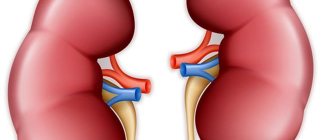
Since the kidneys are a paired organ, the disease can be unilateral or bilateral. The unilateral form is often represented by pyeelectasis of the left kidney. Pyeelectasis of the right kidney is 45% less common. Pathological expansion of the pelvis of both kidneys (bilateral form) is quite often characteristic of children. The one-sided form is also not uncommon in childhood, but is more common in adults.
There are three degrees of the disease, they are determined by the degree of damage: mild, moderate and severe. If not only the renal pelvis, but also the calyces of these organs (cavities) are dilated, then the disease is called calicopyelectasia.
The compensatory abilities of the child's body are incredibly high. Some signs that should be an “alarm bell” can be observed (but not necessarily!) only with bilateral pathology. At the same time, the likelihood of complications increases. And as soon as they begin, the child is taken to see a doctor, who prescribes an ultrasound of the kidneys and the fact of pyelectasis becomes obvious.
Most often, dilation of the pelvis causes:
- pyelonephritis;
- urethrocele;
- ureteral prolapse.
In order to avoid such and other equally serious diagnoses, at the first suspicion of problems with the kidneys, you should immediately take the child to the doctor. Parents should be alert to such signs as swelling of the arms and legs, face, especially in the late afternoon, cloudy urine, blood in the urine, frequent or infrequent urination, pain when emptying the bladder, deterioration in the child’s general well-being, frequent headaches, nagging pain in the lumbar region.
What causes the development of the disease, and what types are there?
Pyeelectasis in children often develops due to a genetic predisposition. If a mother is diagnosed with a disease, her unborn child is at risk. Doctors often observe the occurrence of pathology due to disorders that occur during the formation of the fetal urinary system.
The development of the disease in the last months of pregnancy is caused by the influence of x-rays, infection, or due to severe toxicosis in the mother. The development of the disease varies by gender: boys are more often affected by the disease, unlike girls.
Pyeelectasis in an infant is provoked due to the following factors:
- urinary retention, which does not correspond to age;
- weak muscles in newborns, especially premature babies;
- valve defect in the area of transition from the pelvis to the ureter;
- pathological formation of the urinary system as a result of its compression by other organs or large vessels;
- physiologically uneven maturation of the child.
The disease is divided into several varieties depending on the location of the disease. If the organ is damaged on one side, a diagnosis is made of pyeloectasia of the right kidney. When the tissue of a neighboring organ is affected, pyeloectasia of the left kidney develops. With abnormal expansion of two urinary organs, bilateral pyelectasis is diagnosed.
Most often, the disease can be diagnosed during pregnancy.
What are the consequences of renal pyelectasis in a child?
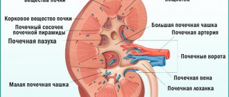
Pyeelectasis of the kidney in a child is a serious pathological process, during which the size of its internal element, the pelvis, increases. The normal size of the pelvis depends entirely on factors such as height, weight, age, body composition of the child or the size of the fetus during pregnancy. This element of the kidneys is necessary in order to temporarily accumulate filtered urine in the pelvis, which then enters the bladder.
Kidney pyeloectasia in children is not an independent pathological process, but is a sign of the presence of some other disease in the baby that disrupts the outflow of urea from the renal pelvis, which can be provoked as a result of exposure to infectious microorganisms or be a congenital anomaly.
This disease can be detected in the fetus both in the womb and in a newborn child, as a result of which it can be assumed that this anomaly is congenital. Pyeloectasia can be one-sided (that is, pyeloectasia of the right kidney and, in the case of damage to the adjacent renal pelvis, left-sided, a diagnosis of pyeloectasia of the left kidney is made) and bilateral - damage to both kidneys.
The bilateral form of the disease occurs somewhat less frequently in girls than in boys. Untimely provision of medical care can contribute to the formation of irreversible pathological changes in the affected kidney, which is reflected in disruption of its normal functioning.
This element of the kidneys is necessary in order to temporarily accumulate filtered urine in the pelvis, which then enters the bladder.
Causes
The causes of pyelectasis in children have been studied in detail, which makes their rapid detection possible.
The hereditary factor plays a major role in the occurrence of the disease: if the mother had such a diagnosis, then the likelihood of its occurrence in the baby increases sharply. The anatomy of the entire kidney may change, or the pelvis may extend beyond the renal structure.
Often provoked by prolonged presence of urine in the pelvis is its return from the bladder - vesicoureteral reflux. In normal physiology, this is prevented by a valve located at the entry point of the ureter into the bladder. When this valve does not close, urine during bladder contraction partially flows into the urethra and partially returns back to the kidneys.
Other causes of pyelectasis include:
- Inconsistency in the development of blood vessels and the ureter, when the blood vessels are more developed and create increased pressure on the ureter with weak tone.
- Excessive systematic intake of fluid into the body.
- The presence of mechanical obstructions in the urinary tract (pus, neoplasm, stone or sand, mucus).
- Urinary tract infections.
- Failure of urinary function. With a neurogenic manifestation, the child rarely pees, which causes frequent bladder overflow.
- Narrowing of any part of the urinary system located below the renal pelvis.
- Nephroptosis.
- A wandering kidney or its prolapse.
- Injuries to internal organs in the pelvis.
- High anatomical location of the ureter.
- Weakness of muscle tissue in premature or neonatal patients.
- Volley load on the excretory system after birth.
In boys, difficulty with urine output and subsequent stretching of the renal pelvis can occur against the background of phimosis or narrowing of the foreskin, which prevents the exit of the head of the penis and the normal process of urination.
Also provoking facts are an imbalance of the endocrine system with hormonal imbalance and poisoning, during which the kidneys are subjected to too high a load.
The video discusses in detail the reasons for the development of pyeloectasia, diagnosis and treatment of the disease.
A wandering kidney or its prolapse.
Complications and risks
Pyeelectasis can be complicated by very serious pathologies. The most dangerous of them:
- hydronephrosis – progressive expansion of the renal pelvis zone with the risk of atrophy of the kidney parenchyma;
- pyelonephritis – inflammation of the kidney tissue of varying volume and severity;
- megaureter - inherited or acquired lengthening and expansion of the ureters;
- renal failure.
Ultimately, pyeloectasia can result in nephropathy, a complex liver syndrome characterized by damage to two kidneys and renal failure.
Less often, pathology can be found in newborns, since ultrasound diagnostics is not included in medical examinations in the first month of a baby’s life.
Diagnostics
You can notice a pathological dilation of the renal pelvis in a child using an ultrasound examination, starting from the 18-20th week of the mother’s pregnancy. An attentive diagnostician is able to discern pyeloectasia in a male fetus already from the 17th week of pregnancy. Under no circumstances should the expectant mother panic if such a conclusion is made. The fact is that in many cases, the expansion of the renal pelvis can be physiological and will go away on its own.
Sometimes the problem is first detected in the fetus shortly before birth - at 34-36 weeks of pregnancy. In this case, there is also no need to be nervous.
After birth, the child must be examined by neonatologists with the assistance of a urologist and nephrologist. Observation is often continued until the child is one and a half years old. It is by this age that for many children the problem resolves itself. If this does not happen, the issue of treatment is decided.
Medical diagnostic monitoring is carried out for children with a mild degree of the disease once every six months - they do an ultrasound, evaluate the dynamic indicators of urine tests. The average degree of pathology requires diagnosis every three months. And only a severe form of the disease requires urgent medical measures and follow-up.
Sonographic signs of pathology are expansion of the size of the pelvis. Normally, the size of the pelvis in a fetus up to 31-32 weeks of pregnancy should not exceed 4 mm. At 36-37 weeks, the renal pelvis normally increases to 7 mm. If the expectant mother is told that the fetal renal pelvis exceeds 10 mm, this is an alarming signal that indicates the likely development of pyelectasis.
The normal size of the renal pelvis for children after birth is 6-7 mm; a slight excess of up to 8-9 mm can be considered an individual inherited feature. In children over 3 years old, the size of the pelvis can be within 8 mm. Exceeding the threshold of 10 mm at any age is grounds for visiting a nephrologist and urologist.
In newborns, to make or exclude a diagnosis of calicoectasia, it is necessary to conduct a complete examination of all organs of the urinary system and other nearby organs.
This involves carrying out the following diagnostic measures:
- blood tests (general and biochemical);
- general and bacteriological urine analysis;
- ultrasound examination of the kidneys;
- X-ray examination of the urinary tract;
- uroflowmetry;
- renal angiography;
- multislice computed tomography.
Based on the results of the examination, a conclusion is made about the nature of the pathological changes and the necessary treatment is prescribed.
Main reasons
Clinicians identify many reasons that lead to dilation of the renal pelvis. Considering that the primary factor in the development of pyeloectasia is stagnation of urine in the kidneys and difficulty in its normal outflow, the following diseases and conditions can contribute to the development of pathology:
- abnormal structure of the ureteropelvic apparatus;
- compression by vessels or internal organs of the lumen of the ureters;
- muscle weakness;
- rare urination;
- ureteral torsion;
- injuries;
- infectious kidney diseases – pyelonephritis, nephritis;
- autoimmune kidney pathologies – glomerulonephritis.
The fetus is diagnosed with pyeloectasia predominantly of the right kidney. Preeclampsia during pregnancy, kidney pathologies in the mother, genetic disorders, and bad habits of the mother during gestation can contribute to the development of pyeloectasia in the fetus and in infants in the first days of life. If the disease is hereditary, the pathology is diagnosed by ultrasound at 16–20 weeks of pregnancy.
In older children, pyeloectasia occurs when the organs of the genitourinary and urinary system become inflamed, as well as blockage of the ureters by mucous components, pus, and dead tissue.
With urolithiasis, the ureters are likely to be blocked by stones. In young children, against the background of calicopyelectasia, neurogenic bladder syndrome is observed, which leads to enuresis at an older age, constant pressure on the organs of the urinary system.
What symptoms should you pay attention to in children?
As a rule, the child complains of signs of a disease concomitant with pyelectasis. Parents need to be attentive to the volume of urine excreted in their newborn, especially if the diagnosis was made during pregnancy.
With pyeelectasis in newborns, the general condition worsens. They cry a lot, scream, and refuse to eat. Fever, diarrhea, and vomiting are possible. Older children talk about pain in the lumbar region, abdomen, and tingling.
If a child may have defects in the structure and development of the urinary system at the genetic level, the mother and father should consult a doctor at the first changes in his behavior. This is apathy, anxiety, poor urine output, and lack of appetite.
Recommendations for parents
If your child has mild or moderate pyeloectasia, do not panic. Doctors will ensure proper monitoring of the baby’s condition. On their own behalf, parents can only make sure that the load on the kidneys is reduced as much as possible. For this:
- the amount of liquid consumed should be limited, the amount drunk should not exceed the age norm;
- It is imperative to monitor how much the baby pees - ideally, the amount excreted is slightly less than or equal to the amount drunk;
- the child should not become overcooled or sit on cold surfaces;
- all infectious diseases (ARVI, influenza and others) must be treated under the supervision of a doctor, since the load on the kidneys during the period of illness increases, self-medication is completely excluded;
- Particular attention should be paid to taking medications. Many tablets and syrups are contraindicated for children with kidney problems or are dosed strictly individually.
To learn how the kidneys work, watch the following video.
What happens if the pathology is not treated?
If therapy is not carried out in a timely manner, pyeelectasis of the kidneys in a child will provoke compression of the organ, atrophy of its tissues, decreased performance and, as a result, complete death of the urinary organ.
In addition to disruption of urine passage, pyelonephritis develops. The infection reduces the level of kidney function, the structural condition deteriorates, which often leads to tissue sclerosis. There are a number of diseases that aggravate pyeelectasis. These are enlargement of the ureter, urethrocele, infection of the posterior urethral valves, and others.
Treatment
When renal pyeloectasia is detected in a fetus or newborn child, drug treatment is not always prescribed. If the pathology is asymptomatic, then parents are advised to follow the following rules:
- Regularly conduct follow-up ultrasounds and visit the child’s supervising doctor.
- Organize proper nutrition.
- Observe hygiene rules.
- Prevent the development of inflammatory diseases of the genitourinary organs.
If signs of progression of the pathology appear, which are detected on ultrasound, the child is prescribed courses of drug therapy aimed at ensuring normal urine outflow and eliminating emerging inflammatory processes. If pyeloectasia is provoked by urolithiasis, then the child is prescribed a diet that prevents the formation of stones, and appropriate treatment - conservative or surgical.
The need for corrective surgery for renal pyeelectasis is determined by the clinical picture and the presence of decreased renal function. According to statistics, surgical treatment of this pathology is prescribed in approximately 25-40% of cases. When performing such interventions, which can be performed using classical or endoscopic techniques, the surgeon eliminates factors that impede the normal outflow of urine (ureteral reflux, neoplasms, narrowing, etc.). After the operation, the child undergoes a full course of rehabilitation.
Classification
Calicoectasia is a congenital or acquired abnormality that can form on the right, left or both sides.
The right kidney is located slightly lower than the left and is more mobile, which leads to disruption of its blood supply, twisting and squeezing of the urinary canals.
This makes it difficult for urine to pass through the ducts of the right ureter into the bladder.
Calicopyelectasia, which develops in the left kidney, is much less common than in the right.
With left-sided caliectasis, identical changes occur. It is not possible to independently identify pathological changes in an organ.
An increase in the size of the renal pelvis in children up to 4.5 cm is considered calicopyelectasia and requires constant monitoring by a doctor.
Calicoectasia of both kidneys is a dangerous condition that requires hospitalization, as it can lead to complete cessation of their function. With such a lesion, a delay in the development of the child is possible.
Causes
Pyeelectasis in fetuses and newborns is detected quite rarely. The formation of an anatomical disorder usually occurs due to an increase in urine pressure in the kidneys due to difficulty in its outflow. In the fetus, right-sided pyelectasis is more often detected.
The main reasons for the expansion of the renal pelvis are the following factors:
- abnormal formation of the valve apparatus of the ureteropelvic junction;
- compression of the ureters by other organs or vessels due to anomalies in their structure;
- muscle weakness in newborns or premature babies;
- infrequent urination, in which the bladder is full of urine for a long time.
In the fetus, pyeelectasis can be detected by ultrasound at 16-20 weeks of pregnancy. Pathology can occur for the following reasons:
- genetic predisposition;
- preeclampsia and eclampsia during pregnancy;
- acute inflammatory kidney diseases suffered by the mother during pregnancy;
- pyeelectasis in the expectant mother.
In older children, pyeelectasis can be caused by the following diseases and conditions:
- pyelonephritis and other inflammatory processes in the kidneys lead to obstruction of the ureters with mucus, pus and dead tissue;
- urolithiasis causes blockage of the ureter with a stone;
- infections of the urinary system lead to the formation of scars in the ureters and renal pelvis;
- kinks or torsions of the ureters occur when the kidney prolapses;
- Excessive fluid intake leads to kidney overload;
- disruption of the innervation of the bladder causes a constant increase in pressure in the bladder.
How is pathology detected?
Often, pyeelectasis in a child is diagnosed in the womb during a routine ultrasound at 16-22 weeks or in the first year after birth. When the disease is detected in early pregnancy, constant monitoring studies are carried out using ultrasound. If the pathology progresses or the disease intensifies due to infection, a full examination is carried out, after which a complete picture of the disease is drawn up and a verdict is rendered.
Kidney pyeelectasis in a newborn can occur in three forms: mild, severe, moderate. The first two go away on their own over time, mostly without additional therapy. Severe forms are treated through surgery. When diagnosing a pathology in a newly swarming baby, it is necessary to carry out periodic ultrasound monitoring.
Causes
It has been studied that pyelectasolia in children is unevenly distributed in the boy-to-girl ratio. In boys, this pathology develops approximately 4 times more often. In girls, the genitourinary system is designed in such a way that narrowing of the lumen of the urinary tract occurs quite rarely, while in boys this is a fairly common occurrence. Although sometimes the narrowing of the renal pelvis is an individual factor, that is, a developmental norm determined by physiology.
The reasons for the appearance of this disease in a child may be different, but in 85% of cases it is heredity. There is also such a thing as calicopyelectasia. This is a pathology characterized by dilation of the calyces and pelvis of the kidneys, while calicopyelectasia is not a disease, but simply a deviation from the norm. As a rule, this is a congenital physiological abnormality, which nevertheless provokes problems with the kidneys and urinary system.
During intrauterine development at a period of 16 to 20 weeks, signs of pyelectasis can be detected using ultrasound. The most common causes of pathology are:
- genetics;
- late toxicosis and a sharp increase in blood pressure in the expectant mother;
- kidney diseases suffered during pregnancy.
Other reasons include:
- malpositioned renal pelvis;
- infection in the kidney cavity;
- inflammation of the genitourinary system;
- rare urination of the child, which leads to bladder overflow;
- change in pressure in the area of the ureters;
- blockage of the ureters with various purulent secretions;
- kidney prolapse;
- compression of the ureters by other organs due to their displacement or enlargement.
In children born before the due date, the muscles of the urinary system may be underdeveloped, which leads to pathology. In newborn babies, organs may grow unevenly, with the kidneys most often “lag behind” other organs, while simultaneously receiving significant stress. This leads to malfunctions, accumulation of excess fluid in the pelvis, and as a result, pyeelectasis of the kidneys develops in the infant.
It is known that the kidneys are a paired organ, so the disease can be either unilateral or bilateral. Unilateral pathology occurs more often, in most cases it is a disease of the left kidney. However, children are more likely to have a bilateral form.
What leads to enlargement of the renal pelvis is quite dangerous for the baby’s health. Infrequent urination that occurs with this pathology can lead to the development of pyelonephritis (chronic or acute). In addition, the complicated outflow of urine can cause compression of the kidney, which impedes its functioning and leads to tissue atrophy. Over time, this can lead to kidney dysfunction and atrophy.
Pyeelectasis in an infant has the ability to “hide.” When diagnosing this disease, parents should provide their child with a complete urological examination. This must be done to identify the causes and severity of the pathology.
The following forms of pyelectasis are distinguished:
- mild - at this stage the child is not prescribed medication, but at the same time the dynamics of the course of the disease and the child’s development are constantly monitored, since over time the genitourinary system develops and strengthens, which leads to the self-elimination of pyelectasis;
- medium - with this form, taking appropriate medications is necessary. The frequency and amount of the medication course is determined by the doctor according to the dynamics of the disease and based on test results;
- severe - in addition to intensive drug therapy, surgical intervention is used.
Children under one year of age (during intensive growth) fall into the risk category for the most common diseases.
Although many experts are inclined to say that kidney pyeloectasia in a newborn often goes away on its own, if signs of pathology are detected, the child must be constantly monitored by a doctor for several years. This will allow you to prevent complications in time and begin the necessary treatment.
Possible complications
In the absence of timely treatment, serious complications may occur in the form of:
- chronic pyelonephritis;
- pyonephrosis (purulent process in the kidney);
- hydronephrosis (expansion of the renal pelvis and calyces, leading to atrophy of the kidney tissue);
- glomerulonephritis (inflammation of the renal glomeruli and changes in the structure of the tubules);
- kidney stones (stones and sand appear);
- renal failure (impairment of all organ functions);
- urosepsis (spread of infection).
Negative changes are provoked by prolonged stagnation of urine and impaired circulation in the vessels of the organ.
In some cases, such phenomena can be aggravated by the development of infection and lead to disability, and if both kidneys are affected, to death.
Prevention and prognosis
There is no specific prevention of pyeloectasia, but the risk of developing pathology can be reduced even at the stage of pregnancy. Women are advised to control the volume of daily fluid, avoid exposure to harmful factors, and monitor the condition of the kidneys in case of a complicated nephrourological history.
The prognosis for renal pyeloectasis in a child varies greatly, depending on the causes of the pathology, combination with other diseases, and symptoms. In case of persistent functional disorders of the kidneys, appropriate treatment is carried out. With the development of chronic renal failure, an adequate diet, long-term drug therapy to prevent or treat complications, and a kidney transplant for end-stage chronic renal failure are required.
Humanity has diseases that can be hidden and asymptomatic. They are often discovered by chance, during the examination of other obvious ailments. Such “hidden” and asymptomatic pathologies include renal pyelectasis. This is an enlargement of the renal pelvis (sometimes calyces) in a person, which leads to difficulty urinating. When the pelvis enlarges, the urine accumulated there has difficulty passing through the ureter. This in itself is not dangerous, but a malfunction of the urinary system and difficulty urinating can lead to inflammatory processes in the kidneys and genitourinary system.
Renal pyeloectasia is detected less frequently in infants than in adults. In the case of children, this pathology can be congenital and be an anomaly in the development of the fetus. This deviation can be detected at the stage of intrauterine development by ultrasound examination.
How to get rid of pathology in a baby?
Treatment for enlarged pelvis in infants depends on the etiology of the disease and the severity of development. Mild degrees of pyeloectasia do not require special therapy, and the disease goes away with age. Moderate pathology requires treatment, most of which is aimed at getting rid of the bacteria that caused the infection. Severe forms of the pathology are treated through surgery. Surgery is performed if bilateral pyelectasis develops.
Surgical intervention solves the problem of obstructed urine outflow and eliminates reflux. No open incisions are made. The operation is endoscopic. To protect the baby from complications after manipulation, doctors prescribe gentle antiviral drugs. The dangerous age for re-exacerbation of the disease is 6-7 years. After all, during this period the baby is actively growing. Next time, be extremely careful during your child’s puberty.
Treatment tactics
After diagnostic measures and determination of the severity of pyeloectasia, therapy is prescribed. The treatment regimen varies greatly and depends on many factors: the causes of urinary stagnation, concomitant diseases and complications, and the clinical picture. Drug therapy is based on the following groups of drugs:
- anti-inflammatory and antibacterial agents;
- uroantiseptics;
- anti-stone formation drugs;
- means for reducing creatinine, urea, restoring the electrolyte composition of blood plasma;
- diuretics.
Preventive actions
For pyeloectasia, there are no effective exercises, diets, etc. Doctors recommend not to stop taking antiviral drugs, which are harmless to the baby, during the recovery period after surgery. To ensure that the child does not experience complications, the following conditions must be met:
- do not overcool;
- be examined regularly;
- cure colds and other infections on time;
- warm up daily to prevent urine stagnation;
- control urination;
- Healthy food;
- maintain personal hygiene.
What is pyelectasia? This is an expansion of the collecting system due to the accumulation of urine. The diagnosis terrifies expectant or new parents. In fact, a timely diagnosed disease that occurs under the supervision of specialists does not pose a threat to the baby’s life. You should unquestioningly follow the recommendations of doctors and not refuse surgical intervention if treatment requires it.
Nutrition for pyeelectasis
If pyeelectasis is detected in children or adults, the specialist prescribes a diet that must be followed. The main principle of dietary nutrition for this disease is to reduce the amount of protein consumed (no more than 60 grams per day), as well as increase the consumption of fats and carbohydrates. An important point of the diet is to limit the intake of salt, pasta, canned foods, pickles, chocolate, and mushrooms. Meals should consist of lean meats and fish, steamed or boiled.
Also, with pyelectasis, control over the amount of fluid consumed is necessary. The daily dose of liquid is calculated by the formula: 30 ml per 1 kilogram of the patient’s weight.