Few people know what the term “anechoic formation in the kidney” means - sometimes medical words mislead people and cause fear of the diagnosis. In fact, everything is explained simply; you just need to have a general understanding of this pathology.
First, it’s worth understanding the concept of “echogenicity.” An echogenic formation is a change in the structure of the kidney. For example, the density of an organ can be increased or, conversely, decreased. Different echogenicity of the kidney can lead to the development of the following types of formations:
- anechoic;
- hyperechoic;
- isoechoic;
- hypoechoic.
What is echogenicity
The essence of the ultrasound method is the different ability of tissues and organs to reflect ultrasound waves. The ultrasound source sends ultrasound waves to the area of the body of interest to the doctor, and then receives the signal reflected from the anatomical structures. This research method is also called echolocation. The ability of organs to reflect ultrasound waves is called echogenicity. This is the main characteristic of fluids and tissues in diagnostics using ultrasound.
On the device monitor this parameter is visualized in the form of a black and white picture. The intensity of dark and white colors depends on the density of the structures: the denser the fabric, the better it reflects ultrasonic waves and the more of them return to the sensor. High-density formations will appear light on the monitor. Low-density liquids and formations absorb ultrasound rather than reflect it. Therefore, they appear dark on the scanner screen.
Types of echogenicity
In ultrasound diagnostics, when describing the results of the study, several types of echogenicity of structures are distinguished:
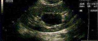
- Anechoic objects . Completely absorb ultrasonic signal. Visualized on ultrasound as a black spot on the kidney. Also called echo-negative formations.
- Hyperechoic formations . They have a high density and are displayed on the monitor in light gray and white colors. In the kidneys, calcifications, stones, and areas of inflamed tissue can look like this.
- Isoechogenic structures . They have a density characteristic of normal organs and tissues. Visualized in gray color of medium intensity. An isoechoic formation in the kidney is a possible sign of a tumor.
- Hypoechoic formations . They have low density and absorb most of the ultrasonic waves. On the scanner screen they appear as dark areas, almost black. This is what kidney cysts, abscesses, kidney tuberculosis, and hematomas look like.
Types of echogenic formations
Echogenicity is one of the main parameters and means the ability of tissues to reflect ultrasound waves during ultrasound. The echogenicity of pathological formations varies:
- Hypoechoic - does not reflect sound well, is a tissue material with a structure such as pus, blood clots, and less commonly tumors and cysts.
- Hyperechoic – has a dense structure, reflects ultrasound well (stones, dense tumors, inflamed areas).
- Isoechogenic – close in structure to healthy tissues, represents benign tumors or congenital anomalies.
- Anechoic - does not respond to ultrasound, is a liquid medium.
Anechoicity is not a disease, but only a diagnostic sign. This is a blind area from which waves are not reflected, so it is visible on the echogram as a dark spot. The appearance of an anechoic formation on the kidney means that in this area there is not normal dense tissue, but a cavity with a liquid center.
Anechoic formation in the kidney - what is it?
Anechoic (echonegative) kidney formations are areas in the organ that completely absorb ultrasonic waves. On the ultrasound scanner monitor they appear as spots of rich black color. Echonegativity is characteristic of cystic formations with liquid contents that do not have an internal structure.
Hypoechoic formation in the kidney - what is it?
These are areas with reduced echogenicity. Detection of hypoechoic structures is possible with abscesses and kidney tumors.
Cyst formations are classified according to the following criteria:
- quantity: multiple and single;
- structure: simple (do not have internal partitions) and complex (represent multi-capsule compartments filled with liquid contents);
- localization: cysts can be located in the pyelocaliceal system, in the medulla or in the renal cortex.
Treatment and possible consequences
As mentioned above, anechoic renal formation is not an independent disease. Therefore, treatment will be carried out depending on the cause of the pathology. If an ultrasound reveals a hemorrhage or hematoma in the kidney, mandatory surgery is prescribed. In case of suppuration - drug treatment. Removal of the cyst is necessary if there is a threat of its rupture.
If anechoicity appears due to cysts in the kidneys, then the following circumstances may result:
- Penetration of infection - it can be determined by high fever and severe pain in the lower back and abdomen.
- Rupture of the cyst - this is indicated by pain that becomes unbearable; it is localized in the back or abdomen at the site of the rupture.
- Hydronephrosis is swelling of the kidneys. In this case, difficulties arise with the passage of urine.
- Kidney atrophy - partial or complete. In this case, kidney failure may develop.
Manifestations of anechoic formation in the kidneys
Cysts are often an incidental finding on ultrasound, since in more than 60% of cases their presence does not cause any symptoms. If the cystoid formation has reached a large size, then it affects the functioning of the organ and puts pressure on its connective tissue capsule. This is manifested by the following symptoms:
Aching dull pain in the lumbar region. Irradiation of pain into the groin area, side, and abdomen is possible. The reason is the pressure of the cyst on the organ capsule and irritation of the sensitive nerve endings located in it.
Prolonged increase in body temperature to 37.3–37.5 0C. Temperatures can rise to higher numbers. The cause of fever is a violation of the outflow of urine due to deformation of the cyst of the pyelocaliceal system with subsequent inflammation of the renal parenchyma, as well as infection of the cystoid formation and the development of a kidney abscess.
Hematuria is the appearance of red blood cells in the urine. The number of red blood cells can vary: from an increase, determined only by urine analysis, to “meat slop-colored urine” noticeable to the naked eye. The cause of hematuria is damage to the renal vessels, as well as the involvement of the organ parenchyma in the pathological process, leading to an increase in the permeability of the renal membranes to red blood cells.
Impaired excretion of formed urine. Develops when a cyst obstructs the renal pelvis and the mouth of the ureter. The development of hydronephrosis is dangerous - a significant expansion of the renal pelvis due to the large volume of accumulated fluid. Leads to damage and necrosis of the renal parenchyma.
Complications
Typically occurring without symptoms, kidney cysts can in rare cases cause complications:
- Cyst rupture. Accompanied by intense pain in the side and lower back.
- Infection of education. Manifested by increased body temperature and increased pain.
- Development of hydronephrosis. It manifests itself as aching pain in the lumbar region, dysfunction of the organ, and the development of renal failure.
- An increase in the number of cysts up to polycystic kidney disease. The complication leads to atrophy of the organ parenchyma and the development of chronic renal failure.
Diagnostic measures
Diagnosis of the disease depends on the circumstances under which the abnormalities are detected. This could be a routine examination, diagnosis of another disease, or examination of the kidney itself. First of all, an ultrasound scan of the organ and anamnesis are required.
Laboratory tests also play an important role:
- A blood test can determine the number of white blood cells. If there are a lot of them, then this indicates the presence of inflammation in the body. The level of red blood cells is detected.
- Urinalysis - it determines the presence of blood, protein, urea. A urine test can reveal how well your kidneys are working. To do this, compare the amount of water drunk and urine excreted.
If an ultrasound was performed, but due to the location of the organ, the results are not so reliable, then additional research methods will be needed:
- CT or MRI - these methods allow you to study the kidneys from all sides.
- Dopplerography of the renal vessels.
- Puncture biopsy - a puncture is made at the site of the organ and tissue is taken for laboratory testing.
- Differential diagnosis. It is important, since there are several reasons for the appearance of anechoic formations. In order not to confuse the disease with other similar kidney lesions, differential diagnosis is carried out. It is especially important if there is a suspicion of cancer.
Diagnosis of anechoic formations
The primary detection of cystoid formations occurs, as a rule, during a standard ultrasound examination. To identify the nature and make an accurate diagnosis, additional studies are carried out.
This:
- General urine analysis . Allows you to identify gross disturbances in the functioning of the urinary system.
- General and biochemical blood test . In the presence of a chronic inflammatory process, changes in the leukocyte formula will be detected. Renal failure is detected by an increase in the concentration of urea and creatinine in the blood.
- Ultrasound Dopplerography of renal vessels . Using this method, you can determine the presence or absence of blood flow in the formation. The absence of blood vessels in a cyst is an important sign that it is benign.
- Nephroscintiography . It is carried out to determine the functional activity of the kidneys. Allows detection of renal failure.
- Needle biopsy. Collection of biomaterial followed by histological analysis of the sample is an opportunity to perform a differential diagnosis of malignant and benign tumors.
What diagnostic options are optimal?
You should start with simple and minimally invasive techniques.
Ultrasonography
Echography allows you to quickly, painlessly, without trauma to tissues and with high reliability detect any of the tumor-like neoplasms. Main criteria for ultrasound diagnostics:
- organ size;
- echogenicity (structures reflect the ultrasound signal differently);
- condition of renal tissues and changes in blood flow.
If an ultrasound scan reveals an isoechoic structure of the tumor, then it may be a benign neoplasm, which is almost no different from normal renal tissue. If the doctor identifies a hypoechoic formation, then the presence of a cyst can be assumed. Hyperechogenicity is characteristic of solid tumors without fluid inclusions. In addition to the hypoechoic option, cysts and tumors with cavities may have an anechoic appearance on ultrasound scanning.
A significant disadvantage of ultrasound is the likelihood of not noticing small neoplasms and difficulties during examination against the background of obesity or intestines distended by gases. In addition, you need to take into account the accuracy of ultrasound diagnostics, which is no more than 70%.
X-ray examination
The most informative are x-ray techniques, in which a special contrast agent is injected into the urinary tract and subsequent images are taken. These include:
- excretory urography;
- retrograde pyelography;
- nephrotomography;
- renal arteriography and venocavography.
The disadvantage of any x-ray examination is the radiation exposure and the need for traumatic intervention in the urinary system. Whenever possible, computed tomography or magnetic resonance imaging should be preferred for renal tumors.
As additional methods, according to indications, the doctor may prescribe endoscopic and radioisotope examination options.
If a hypoechoic renal formation is detected on ultrasound, the diagnosis must be continued: a safe and non-traumatic method of echography reveals a tumor, but does not allow making a correct diagnosis. X-ray techniques help confirm the assumption, clarify the location of the large tumor in the right or left kidney and make a decision on treatment tactics.
Causes of development of benign bladder tumors
The causes of the development of bladder tumors have not been reliably elucidated. Great importance in matters of etiology is given to the effects of industrial hazards, in particular, aromatic amines (benzidine, naphthylamine, etc.), since a high percentage of neoplasms are diagnosed in workers employed in the paint, paper, rubber, and chemical industries.
Prolonged stasis of urine can provoke the formation of tumors. Orthoaminophenols contained in urine (products of the final metabolism of the amino acid tryptophan) cause proliferation of the epithelium (urothelium) lining the urinary tract. The longer urine is retained in the bladder, and the higher its concentration, the more pronounced the tumorigenic effect of the chemical compounds it contains on the urothelium. Therefore, in the bladder, where urine is stored for a relatively long time, various types of tumors develop more often than in the kidneys or ureters.
In men, due to the anatomical features of the structure of the genitourinary tract, diseases that interfere with the outflow of urine quite often occur (prostatitis, strictures and diverticula of the urethra, prostate adenoma, prostate cancer, urolithiasis) and there is a high probability of developing bladder tumors. In some cases, the occurrence of tumors in the bladder is promoted by cystitis of viral etiology, trophic, ulcerative lesions, and parasitic infections (schistosomiasis).
Manifestation of symptoms
In most cases, inflammatory processes in the kidneys are discovered accidentally during a routine examination. This is due to the fact that during the appearance of a cyst in the body, symptoms do not appear and the patient does not complain of disturbances.
If an inflammatory process develops in the form of an abscess, tumor or hematoma, which are associated with injury, then the signs appear quite pronounced.
Basically, the patient has a low-grade fever. In some cases, the temperature can rise to higher levels.
Symptoms and diagnosis of bladder cancer
One of the most common symptoms of early bladder cancer is bloody urine, but this may also indicate that the kidneys are simply shutting down. So you should not panic ahead of time and should immediately go to the nearest hospital. When hematuria appears, the patient may feel excellent, he feels great. If the cancer is advanced, it can grow deeper into the tissues and walls of the organ, which causes pain when urinating. As the disease progresses, the pain becomes chronic, the pain is transmitted to the perineum, anus, scrotum, the pain is hellish and spreads to the nearby area. Often the tumor is accompanied by an infection, which also leads to cystitis.
The most reliable way to examine bladder tumors is to examine them using an endoscope, that is, visually through a special device. It works like this: a hollow metal device is inserted into the urethra, having previously been lubricated with a lubricant for the best insertion. Then the doctor proceeds to directly examine the bladder. Urea sediments are also taken for samples, and sometimes a biopsy is done. A biopsy is the removal of a piece of biological material, such as tissue, for detailed examination in a laboratory under a microscope. The modern method is radiography. In this case, the patient is injected with a special contrast and immersed in a special apparatus, which takes pictures of the internal organs. The contrast will highlight the tumor.
When treating such problems, the best solution is surgical intervention.
In advanced or severe cases, as well as to certainly kill the disease, radiation therapy and courses of chemotherapy are used. If the tumors are small and benign, they can be removed using an endoscope with minimal intervention in the patient's body. If it is not possible to remove it in this way, then a more complex operation is performed. Several small incisions are made in the area slightly above the pubic area and removal is performed through these holes. If the cancer has reached the last stage, the bladder can be removed, either completely or partially. If the bladder is removed completely, then the ureters are sutured into the abdominal septum and care is carried out using medical bags that are attached to the area where the ureters come out.
Isoechogenic changes in the structure of the uterus
The similarity of such formations with the structure of the uterus makes it difficult to detect on an ultrasound examination. They are detected in cases where the inclusion has a capsule, or when there is a feeling of compression of the developing formation. For a long time the process does not manifest itself with external symptoms, the patient is not bothered by anything.
If, after an ultrasound, a focus was discovered in the uterus that had an acoustic density similar to that in the surrounding tissues, then this indicates the development of the following processes:
- Uterine cancer is the presence of an adenomatous node in the mucous membrane of the organ, which appears and takes root on the myometrium.
- Presence of myomatous node.
- Presence of an adenomatous node.
- Rokitansky syndrome.
To clarify the diagnosis, you need to undergo additional research methods. Ultrasound helps to detect the presence of a tumor. Echodensity depends on the stage and degree of development of the pathology. In the initial stage, they are all visualized as isoechoic. As the process progresses (enlarges), malignant neoplasms are characterized by the appearance of hypoechoic, hyperechoic, and anechoic inclusions in the lesion.
What else can ultrasound images record?
A dark highlight near the kidneys, which has regular contours, is a possible consequence of hemorrhage, which indicates an anechoic formation in the kidney parenchyma. When bleeding occurs, the area becomes larger. When blood clots, the neoplasm does not reflect the sound wave.
Dark outlines may be a sign of a prolonged inflammatory process, which is accompanied by the accumulation of pus.
This can lead to complications such as:
- cyst rupture with extensive bleeding;
- compression of the kidney parenchyma, which impairs the functionality of the organ;
- nephropathy.
Treatment of tumor processes
Hypoechoic formations
If a congenital pathology was hidden under a hypoechoic formation, the doctor decides to carefully monitor the patient. When the problem threatens the normal functioning of the organ, surgical elimination of the anomaly is performed using kidney plastic surgery. If the patient has suffered an injury and a hematoma has formed in the kidney cavity, it does not require therapy, since it soon goes away on its own. Serious measures are taken when blood accumulation negatively affects the kidneys. Medicines are used to treat suppuration in the kidneys. Surgery is required only in extreme cases. And cysts do not require radical treatment. Only a doctor can make a decision on how to treat a patient, based on the results of tests and studies performed.
Echogenic focal formations
The choice of treatment method directly depends on the type of formation and its size. When the patient has no symptoms and the formations are small, constant medical supervision is necessary. If there is no growth and nothing interferes with a person’s life, therapy is not applied. Changes that lead to kidney dysfunction and active growth of the formation are considered signals for surgical intervention. Echo-dense benign lesions are removed surgically, and doctors are usually able to save the kidney. Malignant tumors require radical therapy, so complete removal of the damaged kidney is performed.
In addition to surgical treatment, chemotherapy and radiation are used. They are necessary in the presence of cancerous tumors. Most often they act as an auxiliary therapy to suppress the development of malignant tumors. Used before and after surgery to remove a tumor. They can be the basis of therapy if it is impossible to excise the tumor.
Types and forms
“Blind” areas on ultrasound images may have a number of differences:
- By organ. Anechoic formations can occur both in the right and left kidneys separately, and on both organs at once.
- By location on the kidney. They can be located in the sinus of the kidney or in its parenchyma.
- According to the form. Can be round or oval.
- By structure. They may give a “shadow” from the contour, have partitions, “hairs”, and fatty inclusions.
- In count. Education can be either one or several.
The cysts themselves are also classified:
- Simple. Shape: round or oval, possibly with partitions. The formation itself can give a “shadow” from its contour.
- Echinococcal. They are round in shape and contain calcenite inclusions inside. The walls of the area are echogenic.
When the hepatic vein expands, anechoic formations will connect with its branches. Doppler in such a situation will not show normal blood flow.
The simplest option is an avascular anechoic formation. In this case, blood vessels are not displayed in the area, and there is no (not visible) blood flow.
Reasons for appearance
An anechoic renal object indicates the presence of a cyst in the organ. It is for this type that the readings of blind zones during examination are characteristic. If the cysts are presented in the form of separate round cavities-capsules, then during an echogram they resemble distant tissue structures.
If an ultrasound examination reveals a renal cyst in the human body, it is characterized as an anechoic avascular formation. This indicates the absence of blood vessels and blood circulation in this area.
Until today, medicine has not determined the exact origin of cysts. Experts (nephrologists and urologists) associate congenital growths in the kidneys, called multilocular, with anomalies that occur during intrauterine development.
The appearance of anechoic objects can also be caused by inflammatory processes localized in the renal pelvis. The presence of stones in the organ can also provoke such a disease.
Neoplasms in the urethral area
Urethral polyp. This is a type in which the connective tissue grows in large quantities and causes severe focal inflammation. Among the male half, the polyp prefers places closer to the prostatic area. In women, this is most often the urinary tract.
Urethral carbuncle. Girls and women are most susceptible to this disease. Most often, part of the mucous membrane falls out of the urethra, which leads to inflammation and the possibility of catching an infection, and blood circulation throughout the entire area is also disrupted.
Urethral papilloma is a common formation with many villi that can be located along the entire length of the urethra in both men and women.
If the tumor is benign and small, a person can live 80 years and not know that he has something similar. Since such a tumor does not show itself in any way and does not manifest itself in any way. As the formation grows, there may be discomfort, burning, and bleeding. In cases where the tumor appears externally, doctors perform simple resection and electrocoagulation. If the location is the inner surface, then doctors resort to urethroscopes and the TUR method.
High density formations
The kidney parenchyma is isoechoic, but on a gray background there may be lighter areas that indicate a dense structure. For example, kidney stones or calcifications resulting from tissue necrosis and calcification. Unlike stones, calcifications rarely have a regular shape and clear boundaries.
A neoplasm in the kidney may also have an isogenic structure, in which case it is indicated by a change in shape, volume, and renal highlights. To find out what it is, additional studies are carried out, for example, radiography or computed tomography with the introduction of a contrast agent.
Depending on the blood supply, the contrast accumulates unevenly in the tissues. Thus, a hypervascular formation is usually a malignant tumor that accumulates contrast agent faster and its structure appears denser on images. Therefore, when examining with contrast, terms such as hyperdense or hypodense are used.
The concept of hyperdense means increased density to the surrounding tissues and indicates good vascularization or a hematoma due to bleeding, and cysts appear hypodense in the image. Therefore, computed tomography with the introduction of a contrast agent helps in the differential diagnosis of echogenic formations of the kidney.
If common diagnoses are more or less clear to most people, then some formulations can confuse the majority of patients. A striking example is an anechoic formation. Hearing this, many will probably be scared, immediately imagining some serious pathology.