Hypodense liver lesions appear for various reasons - these can be tumors, cysts, metastases and malignant formations. When a hypodense formation exceeds 4 cm, it is necessary to perform a biopsy of the lesion, take tests for markers of hepatitis A, B, C, and take tests for tumor markers to exclude oncological formations.
Hypodense formations in the liver are focal neoplasms of different nature and scale.
Patient examination
During intraoperative examination of the abdominal cavity for diagnostics in Germany. The liver is changed, dark greenish in color, slightly enlarged. There is an infiltrative process in the subhepatic space and the projection of the head of the pancreas. The wall of the gallbladder is thickened. In the liver parenchyma, there are multiple dense whitish foci from 3 mm to 15 mm intraparenchymatous and with exit under the capsule. One of them at the edge of the NW was taken for histological examination. Under ultrasound control, puncture drainage of the cystic formation was performed. CA 19-9 - 259 units/ml. There are several hypodense foci in the liver - presumably metastases.
Blood chemistry. Total bilirubin 23.2 (up to 21 µmol/l); Total protein 75 (67 - 87 g/l); Albumin 36 (34 - 50 g/l); Blood glucose 4.9 (3.3 - 6.1 mmol/l); AST 49 (up to 40 U/l); ALT 44 (up to 40 U/l); Creatinine 82 (62 - 132 µmol/l).
General blood analysis. Red blood cells 4.47 (3.9 – 5.6 * 10”12/l); Hemoglobin 133 (124 - 172 g/l); Hematocrit 41.3%; ESR 40mm/hour); Platelets 295 (180 - 320 * 10"9/l); Leukocytes 6.9 (4.0 - 8.8 * 10"9/l); Band 5 (1 - 5%); Segmented 62 (40 - 70%); Lymphocytes 21 (23 - 40%); Monocytes 7 (3 - 8%). Blood type: A (II) Rh factor: negative.
Conclusion
According to the results of a diagnostic examination in Munich: diffuse changes in the liver, hypodense foci in the right lobe of the liver, probably of secondary origin, zones of transient arterial hyperdensity in the right lobe of the liver. The posterior and left semicircles of the pancreas are involved in the tumor process. Along the upper and lower edges of the pancreas, in the mesentery of the small intestine along the middle colic artery, in the hepatoduodenal ligament there are multiple enlarged soft-elastic lymph nodes from 10 mm to 20 mm.
Histological examination: the result of an urgent biopsy was confirmed. A metastasis of poorly differentiated adenocarcinoma was detected in the liver.
Surgical treatment was performed in Germany. Atypical resection of the liver. Cholecystectomy. The bile duct is crossed proximal to the cystic duct. The wall is up to 3 mm thick, the total diameter is up to 20 mm, there is transparent bile in the lumen, the stent is located in the distal stump, removed. The distal stump is sutured. The distal part of the stomach is mobilized along the greater curvature with the intersection of the lesser omentum. A hepaticojejunostomy was formed on an isolated Roux loop of the small intestine using separate interrupted sutures. A gastroenteroanastomosis was performed 20 cm distally with a continuous suture.
Postoperative recovery was satisfactory. Observation after treatment in Munich showed positive dynamics. Recommended. Consultation to decide on the advisability of chemotherapy, observation by an oncologist at the place of residence, strict adherence to the regimen and diet, limiting stress.
ger-doc.de
Malignant neoplasms
A sign of a malignant formation is intoxication syndrome, which manifests itself in loss of strength and unmotivated weakness.
The reasons why renal cell cancer develops are described in general in various sources. These often include:
- various unfavorable external factors;
- changes in the body that are associated with internal pathologies;
- genetic predisposition;
- malfunctions of the immune system.
There are a number of signs by which one can suspect that a neoplasm in the kidney is malignant. Among them are the following:
- sudden weight loss with proper nutrition;
- intoxication syndrome, which manifests itself in loss of strength and unmotivated weakness;
- hematuria, the cause of which cannot be determined;
- feverish condition;
- hypertension of a malignant nature;
- swelling of the legs;
- enlarged lymph nodes;
- in the later stages of tumor development, pain appears.
The listed clinical signs of malignant tumors are more typical for the later stages of their development, when the size of the tumor is significant. While the tumor is small, there may be no clinical manifestations, that is, the course of the disease is asymptomatic.
Causes of prolapsed kidney: symptoms and treatment
The biggest problem with malignant tumors is their tendency to metastasize. Sometimes metastases to other organs signal the development of a malignant process much faster than the tumor itself, localized on the kidney.
Attention: if a patient has gross hematuria without symptoms of renal colic, then he urgently needs to be examined for malignant renal neoplasms.
Hypodense and hypervascular lesions
Different pathologies are visualized differently on a tomogram. When a hypodense formation appears in the liver, the image shows an area that is darker in color than the surrounding area. The structure of such tissue is heterogeneous and is characterized by a decrease in density. The image may reveal lighter areas of compacted tissue than the surrounding ones, with a uniform structure. In this case, they speak of a hypervascular (hyperdense) formation with tissue compaction.
Changes in structures on the tomogram indicate the presence of liver disease.
Diseases associated with hypodense formations in the liver
With hypodense compactions, hemangioma, adenoma, cholangicarcinoma, and fatty liver are often diagnosed.
A hypodense lesion can be a consequence of many diseases, both benign and malignant:
- Hemangioma is a benign formation that appears as a result of dilation of blood vessels, ranging in size from 2 to 13 cm. The lesion develops asymptomatically, when it reaches a large size, it manifests itself as dull pain in the right hypochondrium, increased pressure. Possible degeneration into a malignant tumor.
- Liver adenoma is a tumor of the epithelial cells of the organ, fenced off from the surrounding tissues by a capsule. It is more often diagnosed in women who have taken oral contraceptives. Symptoms of adenoma are pale skin, pain in the right hypochondrium, thirst.
- Fatty degeneration is a dysfunction of the fatty tissue of the liver. Characterized by impaired intestinal function, nausea, loss of appetite.
- Hemangiosarcoma of the liver is a malignant formation that replaces all liver tissue. It is more common in men who abuse alcohol.
- Metastasis is a secondary focus of a malignant tumor of any location. Metastases in the liver may indicate cancer of the stomach, pancreas, lungs, and breast. Most often, metastases appear on the surface of the left lobe, since this area is located closest to the pancreas.
- Cholangiocarcinoma is a malignant tumor that affects the bile ducts and liver. Smokers, patients with Crohn's syndrome, HIV-infected people, and people with hepatitis C are at risk of getting sick.
Diseases associated with hyperdense formations
Hyperdensity formation, such as hyperplasia, consists of connective tissue and is characterized by increased cell proliferation. This compaction is the opposite of hypodense formations, as it is visualized as a lighter spot. Compaction characterizes several types of hyperplasia - classical and non-classical hyperplasia. The first type occurs in 70% of clinical cases, has an abnormal structure, and a central scar is detected. The non-classical form has hyperplasia of the bile ducts and changes in blood vessels. It is more often observed in women aged 35 to 50 years. The onset of the disease may occur without visible symptoms.
Adrenal masses
The adrenal glands are paired endocrine glands of the human body. The main function of which is the production of biologically active substances - hormones. The gland is divided into the cortex and medulla.
The formation of glucocorticoid and mineralocorticoid hormones occurs in the cortex. The formation of dopamine, adrenaline, and norepinephrine occurs in the medulla of the gland. The cortex is divided into the zona reticularis, zona glomerulosa and zona fasciculata.
This clear division into zones depends on the corresponding production of hormonal substances in each of these areas.
The shape of the left adrenal gland is different from the right. The left one is shaped like a semicircle, the right one resembles the top of a mountain.
Unfortunately, mass formation of the adrenal gland is not uncommon. In the adrenal glands, as in other endocrine glands, tumors are formed that have a benign and malignant course.
Neoplasms of the adrenal medulla
One of the most common and poorly studied brain tumors is pheochromocytoma. The tumor secretes biological substances - catecholamines (adrenaline, norepinephrine, dopamine).
These substances mobilize the body and help it survive stress, therefore, normally, they are in an active state for a short time, then disintegrate. In the presence of a tumor, active substances enter the blood in unlimited quantities.
Which provokes the corresponding clinical manifestations.
Pheochromocytoma – tumor of the adrenal medulla
It is characterized by spontaneously occurring sympathoadrenal crises, during which a rise in blood pressure to high units, profuse sweating, palpitations and a feeling of fear are recorded.
In its development it has three forms of flow: constant, paroxysmal and mixed. Long-term exposure to catecholamines causes organic changes in almost all organs and systems of the body.
The attacks occur suddenly and also end suddenly. The levels of catecholamines in the blood at the time of the attack and after it are different. They become almost normal immediately after the attack. The course of the disease can be benign or malignant.
What is adrenal incidentaloma
When a tumor acquires a malignant course, metastases form, which spread to the lymphatic vessels and tissues of other organs, where they grow and create foci for the production of catecholamines. In this case, even after surgical removal of the lesion in the adrenal gland, sympatho-adrenal crises will not stop, since the release of catecholamines will be carried out by metastatic lesions.
The tumor can form in both the left and right adrenal glands. Treatment is often surgical; tumor removal occurs after careful preoperative preparation, which involves taking medications that stabilize blood pressure and suppress catecholamines.
Ganglioneuroma is a tumor of the adrenal medulla consisting of nervous tissue. Symptoms are not expressed or absent at all, more often it has a benign course, and does not reach large sizes in its development. Often, ganglioneuroma can develop in both adrenal glands at once; in this case, the symptoms of the process will depend on which organ has the larger tumor.
Diagnostic methods
Hypodense formation in the liver is determined by such methods as magnetic resonance imaging, X-ray computed tomography, positron emission tomography. Magnetic resonance imaging (MRI) is a safe diagnostic method that identifies tumors and metastases of internal organs, allows you to determine the exact location of the tumor and determine its size. X-ray computed tomography (CT) is an accurate method that helps doctors quickly and correctly make a diagnosis. Positron emission tomography makes it possible to see how deep the tumor grows, its exact boundaries, and size. Some tumors can be diagnosed using ultrasound. Ultrasound makes it possible to see focal lesions of the liver parenchyma.
To accurately determine the benignity or malignancy of a tumor, it is necessary to select material for a biopsy and conduct laboratory diagnostics.
How to do CT and MRI correctly
In order for a study using CT and MRI methods to be informative, you need to know what these studies are, as well as how to properly prepare for them.
Computed tomography is a highly accurate diagnostic method that uses X-rays. For this reason, strict indications are necessary for performing a CT scan so as not to be exposed to radiation unless absolutely necessary.
The big advantage of CT compared, for example, with ultrasound, is the high standardization of the method. This means that the CT slice images will be of high quality, regardless of the qualifications of the doctor who performed the study. The radiation dose that a patient receives during a CT scan is very small and cannot cause any harm to the body.
Preparing for CT
You should not eat food several hours before the test. Before the procedure, remove all metal objects from the body - rings, crosses, hairpins. If you have a pacemaker, be sure to inform your doctor about it! If you previously had an allergy to a contrast agent, this must also be mentioned before the procedure.
Magnetic resonance imaging is used to examine the liver using nuclear magnetic resonance. Performed according to strict instructions. Just like CT, it is a highly accurate standardized research method. The difference is that when examined using MRI, the patient is not irradiated.
Preparing for an MRI
The study must be carried out on an empty stomach. The last meal should be no later than 5 hours before the procedure. Before starting the procedure, you should remove all metal objects from the body.
Any of these research methods is prescribed by the attending physician if indicated.
The information is provided for informational purposes only and does not constitute a guide to action. If any of these symptoms occur, consult a doctor.
Treatment of hypodense formations
Focal lesions with hypoattenuating visualization on diagnostic images should be treated immediately after detection and an accurate diagnosis. This will help avoid complications and transformation of benign tumors into malignant ones. Surgical intervention is necessary when the formation is large in size and is unsafe for the normal functioning of the liver; there is a threat of tumor rupture. It is impossible to remove a formation that affects two lobes of the liver, since a person cannot live without the organ.
After surgical treatment, you must additionally follow a diet. Be sure to avoid alcohol, which is toxic to the liver. It is important to limit the intake of fatty, fried foods, smoked and peppered foods. Compliance with such rules will help reduce postoperative risk to a minimum. If signs of illness are detected, consult a doctor; the sooner treatment is started, the lower the risk to health will be.
infopechen.ru
What is tissue hypodensity?
The term hypodense comes from the Latin hypo- “lowered” and the English density “density”, so from the name it is clear that it means a decrease in density.
Each tissue of the body has a certain density, which is determined by the intensity of color on X-ray and tomographic images. If an organ is diseased, on the tomogram film its structure will be of a non-uniform color, with areas of darkening.
The hypodense formation is darker in color than the surrounding tissue. This indicates that the density in this area is lower than in other parts of the organ.
The detection of such a low-density focus always indicates a pathological process in the organ.
Such changes in the density of liver tissue can be caused by any disease, from benign neoplasms (cysts and hemangiomas) to malignant tumors with metastases. In some cases, this picture may be due to congenital pathologies.
Benign liver tumors
Classification of benign tumors of the liver and intrahepatic bile ducts (According to Hamilton, 2000) Epithelial tumors:
- hepatocellular adenoma;
- focal nodular hyperplasia;
- adenoma of the intrahepatic bile ducts;
- cystadenoma of the intrahepatic bile ducts;
- papillomatosis of the intrahepatic bile ducts.
Nonepithelial tumors:
- hemangioma;
- infantile hemangioendoteleoma;
- angiomyolipoma;
- lymphangioma and lymphangiomatosis.
Tumors of mixed structure:
- solitary fibrous tumor;
- benign teratoma.
Mixed changes:
- mesenchymal hamartoma;
- nodal transformation;
- inflammatory pseudotumor.
The most common benign liver tumors are hemangioma, hepatocellular adenoma, and focal nodular hyperplasia. Other forms of tumors are extremely rare.
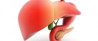
Pancreas and its role
The organ for the most part belongs to the exocrine type. Here the synthesis of enzymes that promote digestion occurs - they participate in the production of gastric juice, which is then sent to the duodenum. Inflammatory processes in this area often take a chronic form. This leads to the appearance of a focal formation of the pancreas provoked by pancreatitis.
The area of the gland where hormones are produced that regulate metabolic processes is of the endocrine type. These hormones include insulin, glucagon, somatostatin, pancreatic polypeptide, and ghrelin. They participate in glucose metabolic processes, regulate the production of glands, and influence the feeling of need for food. If pathology manifests itself in this area, the carbohydrate balance is disturbed, and tumors provoke intensive growth or suppression of hormone production.
Important. The pancreas structurally includes several parts - the head, neck, body and tail. It is in the first part that most formations arise.
General features of pathology
Benign liver tumors, when small in size, have no clinical manifestations and are detected by chance during ultrasound. Complaints usually appear when tumors are large. Benign tumors should be differentiated from primary liver cancer, as well as from metastatic lesions. The diagnostic algorithm includes the use of ultrasound, CT, MRI, and blood testing for tumor markers (AFP, CEA, CA19-9). In difficult differential diagnostic situations, videolaparoscopy, angiographic examination, and fine-needle puncture biopsy are performed.
Do you still think that it is difficult to cure the liver and glandular ducts?
Judging by the fact that you are reading these lines now, victory in the fight against liver disease is not yet on your side.
Have you already thought about surgery? This is understandable, because the liver is a very important organ, and its proper functioning is the key to health and well-being. Nausea and vomiting, yellowish or grayish skin tone, bitter taste in the mouth, dark urine and diarrhea. All these symptoms are familiar to you firsthand.
But perhaps it would be more correct to treat not the effect, but the cause? We recommend reading the story of Olga Krichevskaya, how she cured her liver and cleansed her gall bladder. Read the article >>
Hypodense formations in the liver occur for various reasons, these can be metastases, cysts, tumors, and malignant neoplasms. If the lesion exceeds a diameter of 4 centimeters, a biopsy and blood tests for the presence of hepatitis markers are indicated to exclude cancer.
Hemangiomas
Hemangioma of the liver (ICD-10 code - D18.0) is the most common benign tumor of the liver, accounting for 85% of all benign neoplasms of this organ. The ratio of women to men in terms of incidence is 5:1. It is most common in patients aged 44-55 years.
According to modern concepts, liver hemangiomas are dysontogenetic formations, that is, they are considered as a malformation of the vascular system during embryogenesis. Hemangiomas never become malignant
, but in childhood they must be differentiated from hemangioendotheliomas, which in a large percentage of cases undergo malignant transformation. The size of hemangiomas varies from a few millimeters to 30-40 cm or more. The tumor can affect part of a segment, an anatomical lobe of the liver, or be more extensive, in some cases occupying almost the entire abdominal cavity.
R. Virchow described hemangiomas with special properties, characterized by signs of uncontrolled infiltrative growth, and called them “devouring”.
He identified 3 types of liver hemangiomas:
- simple, or capillary;
- cavernous, or cavernous;
- membranous.
BC Shapkin (1970) proposed to distinguish the following types of liver hemangiomas:
- cavernous hemangioma;
- dense hemangioma with significantly pronounced fibrosis and calcification;
- liver hemangiomatosis without cirrhosis and with cirrhosis;
- mixed form of hemangiomas.
Macroscopically, hemangioma is usually dark cherry or dark red in color. It has a soft-elastic consistency and, when cut, has the appearance of a fine-mesh sponge. A characteristic feature is hyalinosis in the center of the tumor (an irregularly shaped gray or white area). Microscopically, the tumor is represented by clusters of vascular lacunae filled with blood, connected by multiple vascular anastomoses of various sizes
. Clusters of lacunae are surrounded by unchanged endothelial cells.
Small hemangiomas (up to 5.0 cm in diameter) do not appear clinically, but large ones (>10 cm)
and
giant (>15 cm)
tumor sizes, patients experience pain, signs of compression of neighboring organs, and hypocoagulation syndrome associated with hypercoagulation in the hemangioma.
Complications of liver hemangiomas:
- spontaneous tumor rupture;
- tumor necrosis;
- hemobilia;
- sharp twisting of the tumor;
- thrombocytopenia (Kasabach-Merritt syndrome);
- hemangiomatous liver degeneration;
- cardiovascular failure.
Complicated course of the disease is observed in 5-15% of patients. The most common and most dangerous complication that can be provoked by abdominal trauma is tumor rupture with intra-abdominal bleeding.
. The mortality rate for this complication reaches 75-85%, the frequency is 5% for large and giant hemangiomas, but cases of ruptures and small hemangiomas not exceeding 5 cm in diameter have been described. When a hemangioma ruptures with the development of intra-abdominal bleeding, the clinical picture is characterized by an acute onset, tachycardia, pale skin, a drop in hemoglobin levels, arterial hypotension, and signs of the presence of free fluid in the abdominal cavity.
Hemobilia
may manifest as profuse gastrointestinal bleeding or recurrent low-intensity hemorrhage with the presence of melena or coffee-ground vomiting.
Kasabach-Merritt syndrome
characterized by severe thrombocytopenia, massive pinpoint hemorrhages on the skin. In this case, the coagulogram reveals signs characteristic of DIC syndrome.
Heart failure
develops in the presence of massive arteriovenous shunts in large hemangiomas.
Diagnostics
The ultrasound picture
of hemangiomas in most cases is represented by a formation with clear uneven contours, a heterogeneous (mainly hyperechoic) structure, much less often - in the form of a homogeneous hyperechoic formation with clear, even contours. Only in some cases (5% of cases) there are no clear contours of the hemangioma, and the internal structure is mixed. Most cavernous hemangiomas belong to low-vascular formations, and only in some cases an arterial spectrum of blood flow is recorded inside the hemangiomas in the presence of afferent vascularization around (Fig. 59-1).
Rice. 59-1. Ultrasound for liver hemangioma: B-mode image (a): TUMOR - tumor, VHD - right hepatic vein, VHM - middle hepatic vein, IVC - inferior vena cava; in color duplex scanning mode (b), the arrow indicates the tumor.
Characteristic signs of hemangiomas on CT in the native phase are clear contours, homogeneity of the structure of small formations with a density of 38 to 43 units on the Hounsfield scale, as well as the presence of a hypodense area in the center (hyalinosis), which is more common in large and giant hemangiomas (Fig. 59 -2).
Rice. 59-2. Computed tomogram: giant hemangioma of the right lobe of the liver in the arterial phase of contrast.
In the arterial phase, a pathognomonic sign is the accumulation of a contrast agent along the periphery of the tumor like “tongues of flame.” In the delayed phase, the hemangioma becomes iso- or hyperdense in relation to the unaffected parenchyma. In malignant tumors, during this phase of the study the formation becomes hypodense, maintaining its heterogeneous structure.
During angiographic examination (celiacography)
Hemangiomas are characterized by rapid accumulation of contrast agent in vascular lacunae in the form of snowflakes and the absence of pathological vessels and clear tumor boundaries.
Performing a needle biopsy
with liver hemangiomas, especially those located superficially, it is dangerous due to the possibility of developing intra-abdominal bleeding.
Treatment
Indications for surgical treatment arise when the hemangioma is large (>10 cm). With such sizes, pain occurs as a result of compression of neighboring organs. The issue of the need for surgical treatment of large asymptomatic hemangiomas is still being discussed.
Surgical treatment is indicated in the following cases:
- hemangioma size is more than 10 cm in diameter;
- the presence of distinct clinical manifestations;
- the impossibility of reliably excluding a malignant process before surgery.
The validity of the indications for surgical treatment of large and giant hemangiomas is determined by the presence of constant pain syndrome in patients, a decrease in the functionally active mass of the hepatic parenchyma, the development of its dystrophic changes due to steal syndrome, disorders of the blood coagulation system, as well as the danger of hemangioma rupture.
If there are indications for surgical treatment for other diseases of the abdominal organs, it is advisable to simultaneously remove easily accessible smaller hemangiomas.
Of course, with such a complication as tumor rupture, emergency surgery is necessary.
In case of violation of the integrity of hemangiomas, manifested as hemobilia, angiographic examination and X-ray endovascular occlusion of the branches of the hepatic artery
, feeding the tumor, which allows in most cases to achieve effective hemostasis.
Surgical treatment in most cases is performed in the scope of peritumoral resection, since, given the benign nature of the tumor, one should strive for maximum preservation of healthy liver parenchyma
. Extensive anatomical resections of the liver are performed for giant hemangiomas, which almost completely replace the volume of the anatomical lobe.
If the risk of liver resection is high, they resort to ligation of the hepatic artery or cryodestruction of the hemangioma
. However, ligation of the hepatic artery is associated with the possibility of developing liver necrosis, and cryodestruction can be effective only for small tumor sizes. If it is impossible to remove the hemangioma or there are contraindications to surgery, as well as in order to prevent possible complications, a method of selective x-ray endovascular occlusion of the arteries feeding the tumor has been developed and used (Fig. 59-3).
Rice. 59.3. X-ray contrast celiacogram for giant hemangioma of the right lobe of the liver: a - initial study; b — after X-ray endovascular occlusion of the hepatic artery (no contrast enhancement of the tumor is noted).
V.D. Fedorov, V.A. Vishnevsky, N.A. Nazarenko
medbe.ru
Characteristics of hepatic lesions
All volumetric neoplasms determined during computed tomography are divided depending on a number of parameters: density, structure, shape, contours, size, localization.
Density is the main characteristic of any tissue, measured on computed tomography in Hounsfield units. Based on density, foci can be hypodense, hyperdense, or isodense. Based on the density indicator, there is reason to assume what is in the structure of the lesion, whether there is fluid, blood, or soft tissue components.
Cysts are divided into multi-chamber and single-chamber, they have or do not have visible walls, and contain bile. A foreign body or parasite, soft tissue or cystic component is determined inside.
With necrosis, there may be a heterogeneous structure; the duration of the pathological process is indicated by the identification of calcifications and lime.
Shape and contours
The shape of the formations is close to an elongated, irregular ball. Circuit:
- uneven or smooth;
- unclear or clear;
- visible along the entire perimeter or only in a limited area.
The formation is measured on an axial section; if a control study is necessary, a so-called marker lesion is selected. The dynamics of changes in the size of the lesion are assessed over time.
In order for the doctor to see the whole picture, the description of the computed tomography must indicate the location of the pathology. The outbreak may be located:
- deep in the liver;
- about with veins;
- near the gallbladder and ducts.
Such information suggests the etiology of the problem.
The number of pathological areas may vary. A solitary lesion in the liver is always single; with cancer there are many such altered areas. If one metastasis is detected, the doctor assigns stage M1 (according to the TNM measurement system).
But it must be remembered that multiple focal formations are not always metastases. The doctor will also need to conduct a differential diagnosis, carefully comparing all the available signs.
Features of contrast agent accumulation
The higher the accumulation of contrast agent, the better the tissue is supplied with blood. And vice versa, the slower the parenchyma accumulates contrast, the less developed the vascular network.
The more actively the tissue density decreases after completion of the administration of the substance, the more intense the blood flow in the affected areas of the liver.
Technology of diagnostic activities
When performing computed tomography and magnetic resonance imaging, it is important to pay attention to preliminary preparation. For example, if it is necessary to examine the left lobe of the liver, the patient should be correctly positioned on the couch under the manipulators of the device. The organ is partially covered by the ribs, sternum, lungs and other systems.
Therefore, incorrect body positioning may result in false images on the monitor. Patients should lie still. This is problematic with mentally ill people and children. Particular care is needed when handling critically ill patients. Therefore, some mental and physical characteristics are a contraindication to these studies.
What are these formations?
A hypodense lesion is a localized area in the image of the liver on a tomograph monitor. It is colored darker, which reflects the low density. Hyperdense areas, on the contrary, are lighter, which indicates tissue compaction in these places. Low density is caused by liquid media or cells that contain a lot of cytoplasm.
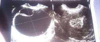
Clinical manifestations
Regardless of the nature of the neoplasm, the symptoms at the initial stages of disease development are the same:
- Aching or nagging pain in the lower abdomen, on the side where the ovarian mass is localized, or in the entire abdominal cavity with a bilateral course.
- Pain during sexual intercourse in the area of the appendages, which radiates to the thigh, leg, and lower back.
- Inability to get pregnant for a long time with regular sex life and full health of the partner.
- Menstrual irregularities. In this case, the regularity of menstruation can be either low or frequent.
- Frequent urge to urinate and defecate due to compression of the bladder and intestines. This symptom occurs when the formation has reached an impressive size.
- Bloating, feeling of heaviness in the abdominal cavity.
If a formation on the ovary causes a hormonal imbalance, then the following symptoms appear:
- weight gain;
- change in hair growth (increased hair growth);
- a sharp decrease/enlargement of the mammary glands;
- the appearance of acne;
- deepening of the voice;
- amenorrhea (lack of menstruation);
- the occurrence of Itsenko-Cushing syndrome.