Instrumental diagnostics
Bladder catheterization - this method is carried out using a special catheter, which differs by gender - for men, women and children. The use of such equipment is subject to strict rules of antiseptics and asepsis.
Catheterization is not performed in case of acute inflammation of the urinary system to prevent the spread of the infectious process up the urinary canals.
Basically, catheterization is organized for patients with benign enlargement of the prostate gland with urinary retention. In some cases, catheterization is required to determine the volume of residual urine.
Bougienage of the urinary canal - the method is carried out with the aim of expanding the narrowed passages of the urinary canal, to establish the degree of narrowing and clarify the localization of the stone in the canal. To organize diagnostics, bougies of increasing sizes are used, which are introduced at the very beginning of the study. Before starting the diagnosis, a gel is injected into the urinary canal to provide local anesthesia.
Needle biopsy - it can be open, which is performed during surgery, or closed, which is an instrumental diagnosis of the condition of the kidneys. A closed biopsy is necessary in a situation where other methods have not given the expected results. Such diagnostics are carried out only in hospital conditions under ultrasound control.
Cystomanometry - an examination is carried out to measure pressure in the bladder. It makes it possible to determine the condition of the bladder wall and its functionality.
After the bladder is completely emptied, 50 ml of warm liquid or gas of a specific predetermined volume is injected into it through a catheter. Using the same catheter, the pressure in the bladder is measured during the first urge to urinate. Also, pressure is measured during urination, which helps to recognize the patency of the urination channel and the sphincters in it.
Uroflowmetry - this examination is carried out to determine the functioning of the muscle that pushes urine out of the bladder. At the same time, the volumetric flow rate of urine through the urination channel is measured, which is recorded on a separate graph depending on the time interval. In patients who are characterized by urinary incontinence, the level of maximum pressure is markedly reduced compared to normal values.
Endoscopic examination - thanks to the use of an endoscope, the doctor can examine the inner surface of the organ being studied. An endoscope can be soft or hard, and in modern medicine, with the help of endoscopes, not only the examination of the internal surface of an organ is carried out, but also minor surgical interventions, the volume of which is constantly increasing as endoscopic devices progress and become more complex.
In urology, endoscopic examination is carried out to study the condition of the urinary canal, bladder and renal collecting system.
Instrumental methods for examining the kidneys make it possible to identify an increase in the size of the organ and the main damaging factors in it.
Kidney tuberculosis: symptoms and signs
There are no specific symptoms in the clinical picture of renal tuberculosis. In the early stages of the disease, there may be a slight malaise, rarely a low-grade fever, and in about a third of cases no symptoms are observed at all. As the process progresses, the following may appear:
The pain is characterized by an aching, dull nature, but the destructive process in the kidney with impaired urine outflow leads to an increase in symptoms up to the manifestations of renal colic.
In 1% of cases, arterial hypertension is observed in the early stages of the disease, which in advanced cases occurs in 20%.
Functional diagnostics
Renal function testing methods play an important role in the diagnosis of kidney pathologies because they help recognize and predict the outcome of treatment for a particular renal pathology.
There are a large number of methods for functional examination of the kidneys, but the most popular are the following:
- Concentration and water tests, which together constitute an accommodation test.
- Detection of the presence of residual nitrogen or urea in the blood.
- Simultaneous detection of the presence of residual nitrogen and urea in the blood and subsequent comparison of the obtained indicators.
It is important to remember that even the simplest functional study of the kidneys, which consists of monitoring urination, the specific gravity of urine excreted per day with the patient’s usual diet and in his usual environment, helps to get an approximate idea of the quality of kidney function.
Causes and mechanism of development
There are a number of main reasons leading to certain diseases:
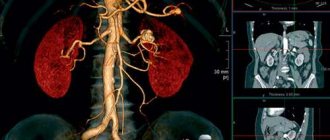
X-ray examination
X-ray examination in urology plays an important and sometimes even decisive role. To organize this study, the patient undergoes preliminary preparation. A thorough bowel movement is required, so a few days before the diagnosis the patient is prescribed a special diet with reduced carbohydrate intake, and on the evening before the diagnosis a cleansing enema is performed.
In some cases, it is necessary to take activated carbon throughout the day and laxatives the evening before the examination. The patient comes for diagnostics on an empty stomach, but in order to prevent excessive formation of gases in the morning, it is recommended to drink strong tea with crackers, since gases make it difficult to see the clear outlines of the kidneys.
Symptoms of kidney disease
Manifestations of kidney diseases can be divided into local and general. Local symptoms include those whose occurrence is directly related to the kidneys:
Radioisotope survey
Radioisotope study of kidney function is the simplest, fastest and least traumatic diagnostic method, which makes it possible to study the function of each kidney separately.
Before the examination, the patient is injected intravenously with a special substance, which is absorbed by the kidneys and subsequently quickly excreted in the urine. The patient takes a sitting or lying position, after which equally calibrated sensors are fixed over both kidneys, and a third sensor is installed in the heart area. The location of the kidneys is determined by palpation or x-ray. The resulting radiographs are recorded on special devices.
Treatment of kidney tuberculosis
Therapy of renal tuberculosis involves an individual approach with the use of certain anti-tuberculosis drugs.
There are first-line anti-tuberculosis drugs (primary) and reserve drugs. The main drugs include isoprinosine and other drugs based on isonicotinic acid hydrazides, ethambutol, rifampicin, streptomycin. Reserve drugs include prothionamide, ethionamide, cycloserine, kanamycin, aminosalicylic acid, etc. The use of fluoroquinolones (lomefloxacin) also seems promising.
When deciding how to treat kidney tuberculosis, it is necessary to rely on complex drug therapy, taking into account the stage and type of process, the severity of intoxication, individual dosage, the condition of the patient himself, as well as other systems and organs of his body. During treatment, it is important to remember the likelihood of impaired renal and liver function, the development of severe dysbacteriosis, allergic and other side effects.
Treatment of the disease requires the use of nonspecific anti-inflammatory drugs and angioprotectors and is long-term from half to one year. Treatment with folk remedies can only be carried out as maintenance therapy and requires prior consultation with a doctor.
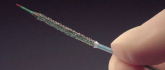
If symptoms of impaired urine outflow appear, its restoration through nephrostomy or installation of a ureteral catheter-stent is indicated.
The destructive process requires surgical treatment (nephrectomy) with preliminary anti-tuberculosis therapy for 2-4 weeks to prevent damage to the remaining kidney.
The local destructive process does not involve removal of the entire kidney, but only sanitation (cavernotomy) or resection of the lesion (cavernectomy).
Timely detection and successful conservative therapy are the key to a favorable prognosis.
Dispensary registration
Treatment of kidney tuberculosis is carried out in a specialized anti-tuberculosis institution. Patients who have suffered a pulmonary form of the disease are placed in a dispensary after treatment and undergo periodic examinations.
Source: https://www.pro-urology.ru/bolezni/pochki/tuberkulez-pochek.html
Radionuclide survey
Radionuclide examination has become firmly established in the medical practice of urology and nephrology. Such diagnostics helps to identify kidney dysfunctions in the early stages of their development, which is difficult to diagnose using other methods.
This method is relatively simple to implement and makes it possible to re-examine during treatment without risks to the patient’s health. Also, radionuclide compounds can be used for patients with high sensitivity to various radiocontrast agents. Based on the objectives of the upcoming diagnosis, the doctor selects a suitable radionuclide tracer.
The advantages of this diagnostic method lie in the possibility of simultaneous assessment of the total functioning of the kidneys and the functioning of each kidney separately. The method is characterized by low radiation exposure to the body and does not cause discomfort to the patient; it can be implemented many times, and this is important for monitoring throughout the course of treatment.
The disadvantage of such diagnostics is the impossibility of identifying the cause of detected violations, and errors may also occur due to incorrect installation of sensors.
Classification
Renal tuberculosis is classified based on clinical and radiological signs: