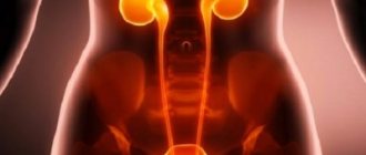
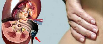
Renal colic syndrome
Finding a calculus in the renal pelvis or ureter (irritation of the mucosa Þ spasm, urinary retention Þ stretching of the ureter, pelvis above the site of obstruction)
F : - acute pain ( characteristic
)
- dysuria
- hematuria
OO : - positive symptom of tapping on the affected side
LIMO : - Rg (review: stones, excretory urography, ureteral catheterization)
— Ultrasound of the kidneys (calculus, dilation of the pelvis)
Modern diagnostic methods
The diagnosis of “acute nephritis” is made on the basis of a comprehensive clinical, laboratory and instrumental examination. At the first stage of diagnosis, the doctor takes into account the patient’s complaints and medical history, paying special attention to possible causes of the disease - previous infection, medication, occupational hazards, concomitant diseases.
Clinical diagnosis of the disease includes examination of the lumbar region, palpation of the kidneys and checking for renal symptoms - effleurage (pain when lightly tapping the lumbar region with a fist), Pasternatsky (sharp pain in the back with a sudden shift of weight from the toes to the heel). The standard examination plan for patients with suspected nephritis includes:
- general clinical blood and urine tests;
- urine analysis according to Nechiporenko;
- urine analysis according to Zimnitsky;
- ultrasound examination of the kidneys;
- R-graphy (urography) of the kidneys with contrast;
- CT, MRI;
- fine-needle biopsy with puncture and subsequent morphological examination of renal tissue.
Nephrotic syndrome
The most common syndrome that occurs in acute and chronic kidney damage, associated with degenerative changes not only in the tubular epithelium, but also in the glomeruli.
F : - pronounced “renal” edema
OO : - pronounced “renal” swelling, pale skin. AH is not typical.
LIMO : — in used an.kr.: ↑ total cholesterol, hypo- and disproteinemia,
— in urine: significant proteinuria (> 3 g/l), hematuria, waxy, granular and hyaline casts are not typical
Classification of jades
The kidneys are an important organ of the human body, responsible not only for the formation and excretion of urine, but also for maintaining chemical, electrolyte and osmotic homeostasis - the constancy of the internal environment. The structure of the kidneys is complex and includes several anatomical and physiological formations:
- The nephron is the main functioning apparatus of the kidney, which consists of the renal corpuscle, the glomerulus (glomerulus) and the capsule. It is a network of convoluted vessels in which the processes of filtration and reabsorption occur, i.e. primary urine is formed.
- Interstitium is the tissue surrounding the nephrons.
- The collecting apparatus is a system responsible for the collection and further excretion of urine.
Damage to any of these structures leads to serious consequences and gross disruptions to the functioning of the organ. Depending on the level at which the pathological process develops, three forms of kidney inflammation are distinguished:
- glomerulonephritis – damage to the glomeruli;
- pyelonephritis – inflammation of the collecting apparatus;
- Tubulointerstitial nephritis is an inflammatory disease of the kidney tubules and interstitial tissue.
Acute renal failure (ARF)
Rapidly developing insufficiency of excretory function of the kidneys (excretion of protein metabolism products and acid-base regulation); usually associated with acute impairment of renal blood flow, glomerular filtration, and tubular reabsorption. 4 periods: initial, oligoanuric (up to 200 ml/day), restoration of diuresis (initially polyuria), recovery. 3 etiological options: prerenal (marked decrease in renal blood flow), renal (ischemia or damage to the glomeruli or tubules), postrenal (acute urinary retention).
F : - “renal” edema
OO : - “renal” edema, pale skin
- possibly a systolic murmur over the renal artery
- renal hypertension syndrome
LIMO : — in used an.kr.: ↑ creatinine, ↑ urea, ↑ K+, ↓ Na+, acidosis
- in urine: anuria, oliguria or normuria; proteinuria (<3 g/l), hematuria (usually microhematuria), granular and hyaline casts, with chronic during possible ↓ urine density
— Ultrasound: normal or enlarged kidney size
Main clinical syndromes in kidney diseases
1. Urinary syndrome is a laboratory concept that includes moderate proteinuria, hematuria, leukocyturia and cylindruria. It is the most important and most constant sign of kidney and bladder damage, in some cases it is the only sign. The presence, nature and severity of urinary syndrome should be judged by the results of a general urinalysis, quantitative methods of urine testing according to Nechiporenko, Kakovsky-Addis, Amburge, and the determination of daily proteinuria.
1.1. Proteinuria (excretion of protein in the urine) can be of renal or non-renal origin. Organic renal proteinuria is caused by damage to nephrons and structural disorganization of their basement membrane, which leads to increased permeability of the glomerular capillary wall to plasma proteins (glomerulonephritis, amyloidosis, pyelonephritis). Violation of the permeability of glomeruli occurs as a “molecular sieve”, that is, first of all, low molecular weight proteins are lost - albuminuria, and as the process progresses, the loss of large molecular proteins also begins. Renal proteinuria is always combined with cylindruria.
Functional prerenal proteinuria is associated with an increase in the concentration in the blood of low molecular weight proteins that are easily filtered in the kidneys: in blood diseases, hemolysis, myeloma (Bence-Jones protein), extensive muscle injuries (myoglobinuria), burns. With postrenal proteinuria, proteins enter the urine from the urinary and genital tracts (admixture of inflammatory exudate, cells of disintegrating tumors).
The persistence and massiveness of proteinuria are of diagnostic importance. Persistent proteinuria always indicates kidney disease. Massiveness is determined by daily protein loss: up to 1 g/day – moderate proteinuria; 1-3 g/day – moderate; more than 3 g/day – massive proteinuria.
1.2. Hematuria is the release of more than 1-3 red blood cells in the urine in the field of view. Depending on the intensity of erythrocyte excretion, a distinction is made between microhematuria - the color of urine is not changed, the number of erythrocytes is up to 100 in the urine, and macrohematuria - urine the color of “meat slop”, the number of erythrocytes cannot be counted. Red blood cells can originate either from the kidneys or from the urinary tract. Renal hematuria is observed with glomerulonephritis, kidney infarction, kidney tumors, extra-renal - with urolithiasis, bladder tumors, cystitis, false hematuria - menstruation. With renal hematuria, as a rule, the urine simultaneously contains a large amount of protein, the red blood cells are leached (do not contain hemoglobin). Detection of the so-called protein-erythrocyte dissociation, that is, hematuria with minor proteinuria, often indicates the extrarenal origin of hematuria associated with the pathology of the urinary tract. Another sign of non-renal hematuria is its intermittent nature (large fluctuations in intensity). Finally, to clarify the localization of the source of hematuria, the so-called three-glass Thompson test is used: when the patient empties the bladder, he secretes urine sequentially into three vessels. When bleeding from the urethra, hematuria is greatest in the first portion (initial hematuria), when bleeding from the bladder - in the last portion (terminal hematuria); with renal hematuria, red blood cells are distributed evenly in all three portions (total hematuria).
1.3. Leukocyturia - excretion of more than 6-8 leukocytes in the urine in the urine, according to Nechiporenko - more than 4000/ml. Leukocyturia of renal origin is observed with pyelonephritis, pyelitis, tuberculosis, abscess or carbuncle of the kidney. To determine the source of leukocyturia, the Thompson test is also used :
When urinating in the morning, the very initial portion of urine is released into the first glass, the rest of the urine into the second, and its remainder into the third. The predominance of leukocytes in the first portion indicates urethritis or prostatitis, and in the third - a disease of the bladder. A uniform distribution of leukocytes in all portions may indicate kidney damage.
2. Nephrotic syndrome is a clinical and laboratory concept that includes massive proteinuria, which is the key condition for the development of all other signs of this syndrome: hypoproteinemia (total serum protein below 60 g/l), dysproteinemia, hyperlipidemia, severe edema. Nephrotic syndrome develops as a result of secondary immune-inflammatory damage to the membranes of the glomerular tubules, which leads to a sharp increase in their permeability to plasma proteins and causes massive proteinuria. With excessive filtration of proteins through the membranes of glomerular capillaries, secondary proteinuric damage to the tubular apparatus and renal stroma → nephrosclerosis develops. Developing hypoproteinemia leads to a decrease in plasma oncotic pressure and contributes to the occurrence of edema. Hypoproteinemia is combined with dysproteinemia → hypoalbuminemia and hyper-a2-globulinemia. Hypoalbuminemia increases the formation of lipoproteins (cholesterol, lipids and phospholipids) → hyperlipidemia.
Nephrotic syndrome in adult patients in the vast majority of cases is not an independent disease, but only accompanies many primary and secondary kidney diseases (glomerulonephritis, amyloidosis). The main and often the only complaint of patients is persistent swelling, which often reaches the level of anasarca. During the period of increasing edema, diuresis decreases, urine has a high relative density (1030-1040) and contains a large amount of protein (more than 3.5 g/day). The urine sediment contains a large number of casts and renal epithelial cells. In peripheral blood, the most constant sign is an increase in ESR to 60-85 mm/hour.
3. Arterial (renal) hypertension syndrome (AH) is one of the common signs of kidney disease. There are: parenchymal renal hypertension, which develops with diffuse lesions of the renal parenchyma: glomerulonephritis, pyelonephritis, nephropathy of pregnancy, diabetic glomerulosclerosis, renal amyloidosis; renovascular hypertension, which occurs when the renal vessels are narrowed (congenital or atherosclerotic). Diffuse parenchymal diseases of the kidneys or narrowing of the renal vessels lead to the development of renal ischemia, more precisely to a decrease in perfusion pressure in the renal vessels and a decrease in pulse fluctuations of the afferent arteriole, to which the JGA baroreceptors react. As a result, the renin-angiotensin-aldosterone system is activated, resulting in an increase in the tone of peripheral arterioles and an increase in total peripheral resistance, as well as sodium and water retention in the body, leading to an increase in cardiac output and circulating blood volume.
The clinical picture of renal hypertension is practically no different from the symptoms of hypertensive syndrome of other origin. However, in contrast to hypertension, renal hypertension is characterized by:
1) a higher level of diastolic blood pressure (more than 110-120 mm Hg), which persists throughout the day, even during sleep;
2) relatively common (in 20-25% of cases) malignant course of hypertension: the rise in blood pressure rapidly progresses, the level of diastolic blood pressure exceeds 120-130 mm Hg. Art., severe complications from the vessels of the brain, heart, aorta quickly develop, renal failure progresses;
3) a crisis course is quite rare.
4. Renal failure syndrome is a pathological condition that develops as a result of a violation of the basic functions of the kidneys and is characterized by azotemia, disturbances in the electrolyte-water balance and acid-base state. Kidney failure can be acute or chronic. Acute renal failure (ARF) occurs suddenly, is most often reversible (temporary ischemia of the cortex), can be intrarenal (acute glomerulonephritis or pyelonephritis, thrombosis of renal vessels) and extrarenal (shock, collapse, hemolysis, burns, intoxication, dehydration). OPN Clinic
: after exposure to the etiological factor, a period of oligo-anuria develops, patients complain of severe weakness, persistent nausea and vomiting, drowsiness and confusion, the smell of ammonia from the breath, dry, pale skin, muscle twitching, bloating. Particularly dangerous during this period are hyperkalemia (possible cardiac arrest), acute gastrointestinal bleeding, and blood clotting disorders. The period of recovery of diuresis is characterized by the gradual development of polyuria, which leads to the restoration of basic renal functions within 3-4 weeks.
Chronic renal failure (CRF) is a gradually progressive deterioration of glomerular and tubular renal function, reflecting the irreversible loss of functioning parenchyma. Initial signs of chronic renal failure appear when the mass of active nephrons decreases to 50-30% of the initial number. The manifestations of chronic renal failure are based on azotemia, severe water-salt imbalance and metabolic acidosis.
Azotemia (uremia)
– excessive concentration in the blood of nitrogen-containing products of protein metabolism - urea, ammonia, creatinine, uric acid, etc. The degree of renal azotemia reflects the degree of reduction in the mass of active nephrons and corresponds to the degree of chronic renal failure. Signs of uremic intoxication: fatigue, headache, itching, sleep disturbance, vomiting, hypothermia, bleeding of mucous membranes, decreased glucose tolerance. A characteristic feature of uremia is the phenomenon of transmineralization: in the cells the content of sodium, calcium, chlorine and water increases, and outside the cells - potassium, magnesium, and inorganic phosphates. Often, water-salt imbalance is accompanied by a picture of potassium intoxication (muscle weakness, adynamia, apathy, cardiac dysfunction) or magnesium (central nervous system depression, drowsiness, stupor and coma, osteodystrophy). Hypocalcemia is manifested by seizures and heart rhythm disturbances. Metabolic acidosis develops due to severe loss of bicarbonates in the urine.
In the clinical course of chronic renal failure, four stages of development are distinguished: latent, compensated, intermittent and terminal. The first three stages are conservative stages of chronic renal failure, when the decrease in glomerular filtration does not reach the threshold level of 10-15 ml/min, therefore diuresis is preserved, patients often report satisfactory health and performance. In the latent stage
diuresis is within normal limits, the Zimnitsky test reveals a slight decrease in the relative density of urine (hyposthenuria), the concentration of electrolytes in the blood serum is within normal limits, a decrease in GFR to 40-59 ml/min, creatinine concentration 0.1-0.18 mmol/l, urea 8.3-8.8 mmol/l.
In the compensated stage
, patients develop asthenia, dyspeptic symptoms (decreased appetite, unpleasant taste and dry mouth), moderate polyuria and nocturia, and hypoplastic anemia develops (usually when GFR decreases below 25 ml/min).
In the Zimnitsky test - isosthenuria, GFR 30-40 ml/min, in the blood - hyperkalemia, hypocalcemia, serum creatinine 0.2-0.28 mmol/l, urea - 8.8-10.0 mmol/l. In the intermittent stage
of chronic renal failure, severe azotemic intoxication and metabolic acidosis develop;
complaints from the gastrointestinal tract (unpleasant taste in the mouth, thirst, nausea, vomiting, hiccups, heartburn, stomatitis); the skin is dry, flabby, pale, muscle tone is reduced, often arterial hypertension. In the Zimnitsky test - hypoisosthenuria, GFR 20-30 ml/min, serum creatinine 0.3-0.6 mmol/l, urea 10.1-19.0 mmol/l, electrolyte imbalance is aggravated. The terminal stage of chronic
renal failure is characterized by an increase in dystrophy (renal cachexia), worsening anemia, arterial hypertension, left ventricular heart failure, and encephalopathy. GFR decreases to less than 15 ml/min, oliguria increases, serum creatinine is more than 1.0 mmol/l, urea is up to 25-35 mmol/l. Uremic polyneuropathy is manifested by hyper- and paresthesia in the form of burning, crawling, itching, and numbness. When examining the respiratory organs - hyperhydration of the lungs, moist rales. From the gastrointestinal tract - ulcerative lesions of the mucous membrane, uremic colitis, pancreatitis, peritonitis. Total heart failure develops, possibly uremic pericarditis. A severe complication of chronic renal failure is uremic coma: it develops gradually and is preceded by severe general weakness, headache, painful nausea, periodic vomiting, itching, insomnia, blurred vision, motor restlessness, which is replaced by apathy, drowsiness and the development of a coma. The skin is pale, dry, with traces of scratching; ammonia odor from skin and mouth; repeated vomiting, often mixed with blood; breathing is initially deep, noisy (Kussmaul type), then shallow and irregular (Cheyne-Stokes type); fibrillary muscle twitching, high hypertension, hypothermia, friction sounds of the pericardium, pleura, and peritoneum (“the death knell of the uremic”) are heard.
some issues of specific pathology of the kidneys and bladder
1. Acute glomerulonephritis (AGN) is an acute immune-inflammatory disease with predominant damage to the glomerular apparatus of both kidneys. It develops most often after a streptococcal infection (sore throat, scarlet fever, exacerbation of chronic tonsillitis, erysipelas, pneumonia). AGN can also occur with other antigenic influences: administration of vaccines, serums, medications (non-steroidal anti-inflammatory drugs, sulfonamides), after a viral infection (hepatitis B, measles, rubella, cytomegalovirus, herpes simplex). The development of the disease is facilitated by hypothermia (especially wet cold), operations, injuries, and physical activity. The immune complexes formed as a result of antigenic exposure are deposited on the glomerular basement membrane, damage it and cause a complex immune inflammatory process.
The most vivid clinical picture is presented in the classic triad form of AGN. 10-12 days after exposure, swelling, change in urine color (“meat slop”), lower back pain (acute kidney swelling), headache, oliguria, nausea, vomiting, possible convulsions (eclampsia), severe weakness, thirst appear. Swelling appears quickly (overnight) and is accompanied by pale and dry skin. Edema is one of the earliest and most common signs of AGN (70-90% of patients), mainly localized on the face, in combination with pallor of the skin and swelling of the neck veins forming facies nephritica. Due to significant fluid retention in the body, acute left ventricular failure (pulmonary edema) and cerebral edema (convulsive syndrome) may develop. Blood pressure is in the range of 140-160/90-110 mmHg and decreases relatively quickly. High and stable hypertension often indicates a transition to a chronic form. Kidneys with AGN are symmetrically enlarged and painful. In urine tests: oliguria, renal hematuria, massive proteinuria, cylindruria, decreased GFR. Blood test: leukocytosis, increased ESR, appearance of antistreptococcal antibodies, increased content of α2- and γ-globulins. Prognosis of AGN: recovery occurs in 70% of patients. A transition to a chronic form is possible (preservation of any extrarenal sign and proteinuria for a year), the reasons for which are the individual characteristics of the body, late diagnosis, and inadequate therapy.
2. Chronic glomerulonephritis (CGN) is a chronic immune-inflammatory disease of both kidneys with predominant damage to the glomerular apparatus, the outcome of which is the development of nephroangiosclerosis (secondary shrinkage of the kidney) and chronic renal failure. The etiology and pathogenesis are similar to those of AGN, but, in addition, the mechanisms of self-progression of CGN play an important role. Chronic inflammation is caused by the persistence of the etiological factor (antigenic exposure), insufficiency of local phagocytosis, unbalanced immune response, damage to the renal tubules by prolonged proteinuria, and sclerosis of the glomeruli.
The following clinical forms of CGN are distinguished:
· Latent
- the most common form of CGN (about 40-45% of all patients), manifested by isolated urinary syndrome, state of health is satisfactory, extrarenal syndromes (edema, hypertension, fundus changes) are absent, urine analysis shows proteinuria less than 1-2 g/day, microhematuria , slight cylindruria, sufficient specific gravity. The course is benign.
· Nephrotic
- occurs in 20-25% of patients, characterized by weakness, lack of appetite, decreased diuresis, significantly pronounced edema, including abdominal edema, unstable hypertension, massive proteinuria (more than 4-5 g/day), cylindruria, microhematuria, hypoproteinemia, disproteinemia, hypoplastic anemia, increased ESR. In the period between relapses of nephrotic syndrome, moderate urinary syndrome persists. Nephrotic GN is characterized by the development of so-called nephrotic crises: intense abdominal pain, peritonitis-like syndrome, increased body temperature, erysipelas-like skin changes, intravascular hemocoagulation.
· Hypertensive
- occurs in 20% of patients, characterized by intense headaches, dizziness, decreased visual acuity, “fog” before the eyes, pain in the heart, shortness of breath, palpitations, severe stable hypertension, expansion of the borders of the heart to the left, on the ECG - signs of LV hypertrophy. Hypertension is well tolerated by patients, even at significant levels (SBP > 200 mm Hg), but can be complicated by cerebral crises. Fundus - narrowing and tortuosity of the arteries, the phenomenon of “crossover”, “silver” or “copper wire”, hemorrhages, swelling of the optic nerve nipple. Urinalysis: moderate proteinuria, microhematuria, decreased urine density, early decrease in GFR. Often complicated by acute LV failure, the course is slowly progressive with the obligatory transition to chronic renal failure.
· Hematuric
- gross hematuria predominates, proteinuria is insignificant, blood pressure is normal, there is no edema (Bourget's disease - hematuric glomerulonephritis). A rare form (6% of patients), the course is favorable, chronic renal failure rarely develops.
· Mixed
form - combines the signs of nephrotic and hypertensive forms. This is classic Bright's nephritis, currently rare (7%), characterized by a steadily progressive course, 10-year survival rate 30%.
During CGN, a remission phase is distinguished (minor hematuria, blood pressure stabilization, minimal proteinuria) and an exacerbation phase with three degrees of activity. In addition, the course is divided into slowly progressive (benign) and rapidly progressive (malignant).
3. Pyelonephritis (PN) is an infectious nonspecific inflammatory process of the collecting system and renal tubules. From a clinical point of view, the classification schemes of pyelonephritis include the concept of the nature of the process (acute or chronic) and its prevalence (unilateral or bilateral). From an epidemiological point of view, three population groups are clearly defined as being most at risk of developing pyelonephritis:
ü First peak
incidence occurs in early childhood (up to 3 years), girls predominate (8:1), which is associated primarily with the structural features of the female genitourinary sphere (short length with a relatively large diameter of the urethra, proximity to the vagina and anus). The route of infection into the kidneys in childhood is almost always urogenic; one of its stages is the development of cystitis. PN in childhood, as a rule, takes a latent form, worsening during puberty, at the beginning of sexual activity, during pregnancy or after childbirth.
ü Second peak
The incidence of PN occurs during the most active reproductive age (18-30 years), the predominance of women remains (7:1). Most PN diseases at this age are associated with pregnancy and childbirth, as well as defloration (cystitis and “honeymoon” PN). Particular importance in the development of gestational renal failure is given to the increase in the content of estrogen in the blood during pregnancy (causing hypotension and dyskinesia of the pyelocaliceal system and ureters, decreasing the tone of the bladder), as well as the mechanical effect of the pregnant uterus on the urinary tract (impaired urine outflow).
ü Third peak
The incidence of PN occurs in old and senile age and is characterized by a progressive increase in the incidence of men. This is due, first of all, to a progressive decrease in the activity of the prostate gland (the secretion of “anti-infectious” factors decreases), as well as to the increase in hypertrophic and tumor processes in the prostate, leading to hemodynamic disturbances.
3.1 Acute pyelonephritis is associated with the penetration of gram-negative flora into the kidney parenchyma by urogenic or hematogenous routes, especially in the presence of occlusion of the upper urinary tract or vesicoureteral reflux. The clinical picture is characterized by febrile or pyretic fever, shaking chills, profuse sweating, and symptoms of general intoxication. Typical pain is in the lumbar region, there is a forced position of the patient (bending and bringing the leg towards the body on the affected side), muscle tension in the lower back and anterior abdominal wall, pain on palpation and tapping in the kidney area. General urine analysis: leukocyturia, bacteriuria. Complete blood count: leukocytosis, increased ESR.
3.2 Chronic pyelonephritis is most often caused by Escherichia coli, mycoplasma, Proteus vulgaris, staphylococci, and Pseudomonas aeruginosa. The exacerbation and development of the disease is facilitated by cooling, impaired urodynamics, urolithiasis, prostate adenoma, diabetes mellitus, and acute pyelonephritis. Infection can enter the kidney through hematogenous, urogenic, and lymphogenous routes. Clinical symptoms: general weakness, headache, loss of appetite, lower back pain (often unilateral), painful frequent urination, chills (chills with severe exacerbation), increased body temperature (usually low-grade), pallor of the skin, facial pastiness, pain when palpated and tapping of the lumbar region, increased blood pressure, displacement of the left border of the ostium. Tofilo's symptom
: in the supine position, the patient bends the leg at the hip joint and presses the thigh to the stomach - in the presence of pyelonephritis, pain in the lumbar region increases. Blood test: leukocytosis, toxic granularity of neutrophils, increased ESR. Urinalysis: urine is cloudy, alkaline, reduced density, severe leukocyturia, bacteriuria, moderate proteinuria, rarely microhematuria. Nechiporenko test - predominance of leukocyturia over erythrocyturia. Zimnitsky test - hyposthenuria. The disease usually progresses slowly with the development of chronic renal failure.
Causes of pathology
Kidney disease can occur for various reasons. Most often, the disease results from:
- Autoimmune process. As a result of a malfunction of the immune system, antibodies against kidney cells begin to be produced. This process leads to inflammation - glomerulonephritis.
- Bacterial type infection. When bacteria enter the kidneys, they begin to multiply, causing inflammation. It leads to pyelonephritis. If kidney disease is not treated, kidney abscesses can form. Infectious pathologies lead to disruption of the outflow of urine.
- Intoxication. In any pathology caused by pathogenic microorganisms, toxic substances and waste products of viruses and bacteria accumulate in the kidneys. They settle in the kidneys, getting there from the blood. As a result, toxic nephropathy develops. This is a reversible process that occurs after exposure to toxins.
- Violation of the metabolism and chemical composition of urine is the main cause of urolithiasis. Stones form in the renal pelvis, disrupting the flow of urine and injuring the mucous membrane of the kidneys and ureters. This process is joined by a secondary infection, resulting in pyelonephritis.
Signs of illness
It is not always possible to diagnose the disease yourself, since the symptoms of nephritis are easily confused with manifestations of another disease. However, there are characteristic signs that help identify the development of kidney nephritis, these are:
- intense headache;
- weakness;
- polydipsia (constant thirst);
- oliguria (decreased daily urine volume);
- hyporexia (decreased appetite);
- nausea, vomiting, diarrhea;
- renal colic (acute lower back pain);
- paresthesia (tingling or numbness);
- involuntary muscle contractions due to micronutrient deficiency;
- difficulty breathing due to cardiac or thoracic hydrops;
- paleness, peeling of the skin;
- decreased ability to work;
- temperature drop to 35°;
- thinning and brittle nails;
- weakening of hair.
In addition, with nephritis, the body swells, the face is more susceptible.
The main symptoms of the disease in acute form:
- feeling cold;
- muscle tremors;
- body temperature above 37 degrees;
- excessive sweating.
Signs of nephritis characteristic of a chronic course:
- yellowing of the skin;
- frequent emptying of the bladder;
- cloudy urine.
Glomerulonephritis is manifested by paroxysmal pain in the lower back, swelling, and decreased urine volume. In addition, blood pressure increases, and blood is observed in the urine.
Symptoms of purulent nephritis:
- acute pain in the kidney area;
- migraine;
- blood clots in urine;
- frequent, painful urination;
- general weakness;
- the temperature rises.
Tubulointerstitial nephritis is manifested by fever, swelling, and blood in the urine.
With Alport syndrome, severe myopia, hearing impairment occurs, and the concentration of leukocytes in the body increases.
With pustular nephritis, when the kidneys begin to become inflamed, the body temperature rises to 41°, severe pain in the head, nausea, and bouts of vomiting appear. This disease is characterized by excessive sweating, increased fatigue, and hypotension (low blood pressure).
Folk recipes
Treatment of jade with folk remedies is carried out only after consultation with a specialist.
To eliminate kidney inflammation, you can use the following recipes:
- Brew 25 g of bearberry with boiling water (200 ml), cover and leave. After 30 minutes, strain, take 30 ml 4 times a day half an hour before meals. Used for nephritis in acute or chronic form.
- Pour 10 g of orthosiphon (kidney tea) into 200 ml of boiling water, steam for 15 minutes, then cool and filter. Daily dose – 100 ml twice. The therapeutic course lasts from 4 to 6 months.
- Pour 20 g of herbal mixture (birch leaves, strawberries, stinging nettle, flax fruits) with 200 ml of boiling water, place in a water bath for 15 minutes. Cool, filter and consume 200-400 ml once or twice 15 minutes before meals. Used for glomerulonephritis.
- Take 20 g of dry raw materials (parsley roots, celery, asparagus, fennel seeds), fill it with filtered cold water, leave for 6 hours. Then simmer over low heat for 10 minutes. Take 100 ml three times a day. Eliminates inflammation in the kidneys and urinary tract.
- Pour boiling water (200 ml) over 5 g of anise seeds, cover and leave for 30 minutes. Then strain and consume 50 ml three or four times a day, half an hour before meals. Used for chronic nephritis.
All these recipes can be used at home after doctor's approval. Folk remedies are used as part of complex therapy: medications, diet, healthy lifestyle.
Drug and surgical therapy
Many patients ask the question: “How to treat nephritis?” The disease in its acute form is treated in a hospital setting. The patient should adhere to bed rest until the inflammation activity decreases. Kidney treatment must be comprehensive, and therefore it is necessary to adhere to a diet.
Medicines are used to eliminate the causes of the inflammatory process. If the disease was provoked by a drug, the doctor cancels it, and an antidote is introduced into the patient’s body, which binds the remaining toxin in the blood. In case of severe poisoning and accumulation of hazardous substances, hemosorption and plasmapheresis are prescribed.
If nephritis occurs due to infection of the kidneys by pathogens, then antibacterial and antimicrobial drugs are used for treatment.
But it is also necessary to influence the mechanisms of nephritis development. For this purpose, antihistamines and hormonal drugs are used, which improve blood flow in the kidneys.
In addition, symptomatic treatment of nephritis is carried out. With the help of medicinal solutions that are injected into a vein, water-electrolyte and other disorders are corrected. It is necessary to control the volume of urine excreted and, if necessary, take diuretics. In case of kidney dysfunction, extrarenal blood filtration is prescribed - hemodialysis.
Kidney disease is treated using the following medications:
- Azathioprine – has immunosuppressive and cytostatic effects.
- Prednisolone is a glucocorticoid drug with antihistamine and anti-inflammatory effects.
- Heparin is a medication with an anticoagulant effect that is allowed to be used only by inpatients.
- Dipyridamole normalizes blood circulation and restores immunity.
Treatment of kidney inflammation is a long process that takes at least 1.5 months, and in some cases can last for several years. The most suitable drugs are selected by the doctor, who determines the treatment regimen.
Principles of treatment
After making a diagnosis and determining the type of nephritis, the attending physician (urologist, nephrologist or therapist) selects a treatment plan, including recommendations for adjusting lifestyle and nutrition, and taking medications. The acute course of the disease is an indication for hospitalization of the patient.
Correction of lifestyle and nutrition
In the first days of any form of nephritis, treatment consists of bed rest, which can be expanded only after the patient’s condition improves and positive dynamics in laboratory tests. Therapeutic nutrition consists of following a “renal diet” (table No. 7, 7a, 7b according to Pevzner). Basic principles of the diet:
- The diet should be varied, balanced and high in calories (approximately 3500 kcal/day).
- Food is taken in small portions, 4-6 times a day.
- To avoid the formation of edema, fluid is limited to 1-1.25 liters per day.
- For the same reason, the salt content in food is reduced to 2-3 grams/day. If you have high blood pressure, it is better not to add salt to your food at all.
- Since protein foods are difficult to process, and the products of its metabolism can “load” the kidneys with unnecessary work, the content of this nutrient in the daily diet is reduced to 0.8-1 g/day.
Other reasons
Kidney disease may be caused by hemolysis. As a result of the destruction of red blood cells and the release of hemoglobin, it settles in the glomeruli of the kidneys. This blockage makes work difficult. As a result, acute renal failure develops. The following can also lead to illness:
- Impaired blood flow. With atherosclerotic plaques, a change in the diameter of the vessel occurs, which leads to insufficient blood flow to the cells of the organ. The result of this pathology is metabolic nephropathy. It can last for years without any symptoms.
- Low blood pressure. As a result of low blood pressure (systolic below 70 mmHg), blood filtration in the glomeruli stops. As a result, they die (necrosis appears).
- Injuries. Kidney disease can occur as a result of injury. This could be a bruise or an incised wound, in which the kidney tissue is damaged and the function of the organ is lost.
If the formation of the fetus in the womb is disrupted, an abnormality in the development of the kidneys may occur. They may be forked or drooping. Sometimes children have an extra kidney.